. 2022 Sep 18;23(18):10912.
doi: 10.3390/ijms231810912.

Skeletal Muscle Dysfunction in Experimental Pulmonary Hypertension

Affiliations

Skeletal Muscle Dysfunction in Experimental Pulmonary Hypertension

Kosmas Kosmas et al. Int J Mol Sci. .

Abstract

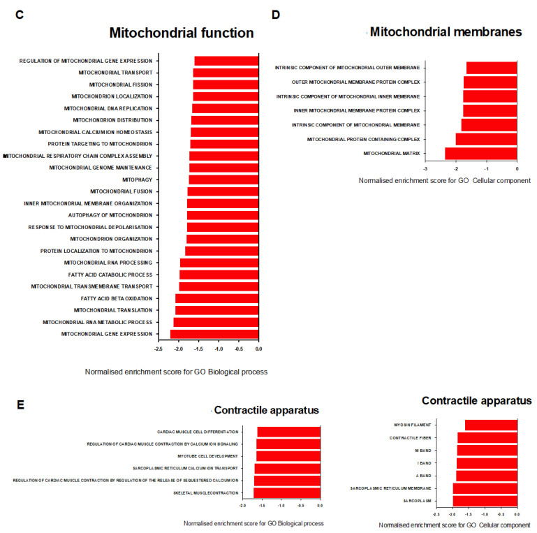
Pulmonary arterial hypertension (PAH) is a serious, progressive, and often fatal disease that is in urgent need of improved therapies that treat it. One of the remaining therapeutic challenges is the increasingly recognized skeletal muscle dysfunction that interferes with exercise tolerance. Here we report that in the adult rat Sugen/hypoxia (SU/Hx) model of severe pulmonary hypertension (PH), there is highly significant, almost 50%, decrease in exercise endurance, and this is associated with a 25% increase in the abundance of type II muscle fiber markers, thick sarcomeric aggregates and an increase in the levels of FoxO1 in the soleus (a predominantly type I fiber muscle), with additional alterations in the transcriptomic profiles of the diaphragm (a mixed fiber muscle) and the extensor digitorum longus (a predominantly Type II fiber muscle). In addition, soleus atrophy may contribute to impaired exercise endurance. Studies in L6 rat myoblasts have showed that myotube differentiation is associated with increased FoxO1 levels and type II fiber markers, while the inhibition of FoxO1 leads to increased type I fiber markers. We conclude that the formation of aggregates and a FoxO1-mediated shift in the skeletal muscle fiber-type specification may underlie skeletal muscle dysfunction in an experimental study of PH.

Keywords: FoxO1; PAH; skeletal muscle; type II fibers.


Conflict of interest statement

The authors declare no conflict of interest.

Figures

Figure 1
Figure 1
(A) Increased ventricular systolic pressure (RVSP) in SU/Hx-induced PH rats is seen, and (B) no significant difference on left ventricular systolic pressure (LVSP) can be seen when they are compared to those of the controls. (C) Right ventricular hypertrophy was assessed by Fulton’s Index (ratio of right ventricular weight to left ventricular and septal weight) in SU/Hx-induced PH rats and this is compared to that of the controls. (D) Decreased endurance for SU/Hx animals in treadmill exercise testing. Average time running for SU/Hx-induced PH rats is compared to that of the controls. Values are expressed as the mean ± SEM for six animals per group. Statistical analysis is conducted by a Student’s t-test, *** p < 0.001, **** p < 0.0001, ns: not significant.
Figure 2
Figure 2
(A) Decreased abundance of Type I markers (Actn2 and Tnni1) and markers of mitochondrial biogenesis (Tfam and PGC-1α) in diaphragms from animals with SU/Hx-induced PH when compared to those of the controls. (B) Decreased abundance of Type I markers (Actn2 and Tnni1) and markers of mitochondrial biogenesis (Tfam, PGC-1α, SIRT-1) and increased abundance of Type II markers (Myh1 and Myh4) in soleus muscles from animals with SU/Hx-induced PH when compared to those of the controls. (C) No difference in abundance of Type I markers (Actn2 and Tnni1), markers of mitochondrial biogenesis (Tfam, PGC-1α, SIRT-1), and Type II markers (Myh1 and Myh4) in EDL muscles from animals with SU/Hx-induced PH when compared to those of the controls. Expression is normalized to Nup133. Values are expressed as the mean ± SEM. Statistical analysis is conducted by a Student’s t-test, * p < 0.05, ** p < 0.01, ns: not significant. (D) In the soleus muscle from the control animals, there is less than 1% type II muscle fibers as compared to the 25% proportion that is seen when it was stained with Myh1/2/4/6 (arrows) in the soleus muscle from animals with SU/Hx-induced PH. Immunofluorescence microscopy of soleus cross sections from experimental animals (n = 3 animals per group). Scale bar, 100 μm. (E) Abundance of the Type II markers MYH1/2/4/6 in soleus from SU/Hx animals when compared to that of the controls. Immunoblot with GAPDH are used as a loading control and (F) quantitative analysis of MYH1/2/4/6 protein (n = 9 animals per group). Values are expressed as the mean ± SEM. Statistical analysis is conducted by a Student’s t-test, ns: not significant.
Figure 3
Figure 3
(A) Paraffin-embedded longitudinal sections of soleus muscle that is stained with desmin (Z-disc) and troponin-I specific antibodies showing a compromised and disorganized sarcomeric organization with aggregate-like structures (arrows) in SU/Hx-induced PH rats when they are compared to those of the controls. (B) Thick transverse desmin aggregates along the sarcolemma (arrows) and an assembly of troponin components along the sarcolemma (arrows) in transverse cryosections of soleus from animals with SU/Hx-induced PH is seen, and these are compared to those of the controls. Immunofluorescence microscopy with desmin and troponin-I specific antibodies, respectively. Nuclei are visualized after DAPI staining. Scale bar, 20 μm. n = 3 animals per group.
Figure 4
Figure 4
(A) No difference is seen in atrophy markers (MuRf1, Atrogin) in diaphragm and soleus muscle from SU/Hx-induced PH rats when they are compared to the controls. Expression normalized to Nup 133. (B) No difference is seen in average cross-sectional area and in the number of soleus muscle fibers per field for SU/Hx-induced PH rats when these are compared to those of the controls. Representative images of troponin I-stained soleus sections from SU/Hx-induced PH rats and controls. Scale bar, 20 μm. (C) Decreased ratio is seen in the soleus weight-to-tibia length from SU/Hx-induced PH rats when compared to that of the controls. Values are expressed as the mean ± SEM. Statistical analysis is conducted by a Student’s t-test, * p < 0.05, ** p < 0.01, ns: not significant.
Figure 5
Figure 5
(A) L6 cells are differentiated in 2% fetal bovine serum media for 5 days. Myotubes structures are indicated with arrows. (B) Decreased RNA levels of Glut1 and increased RNA levels of Glut4 and Myh4 in differentiated L6 cells (Day 7) are found when these are compared to the undifferentiated cells. Expression is normalized to GAPDH. Values are expressed as the mean ± SEM. Statistical analysis is conducted by a Student’s t-test, * p < 0.05, ** p < 0.01, **** p < 0.0001. (C) Immunoblot results are showing increased levels of Type II marker MYH1/2/4/6 and Glut4, decreased levels of Glut1 and unchanged levels of type I marker Hexokinase II in L6 lysates from differentiated L6 cells (Day 7) when these are compared to those of the untreated undifferentiated cells. Beta-actin was used as a loading control.
Figure 6
Figure 6
(A) There is an increased expression of FoxO1 mRNA in the EDL (fast twitch) when this is compared to the soleus (slow twitch) from the control animals. (B) A trend for the upregulation of FoxO1 mRNA in soleus of SU/Hx-induced PH is compared to that of the control animals. (C) A trend for the upregulation of FoxO1 mRNA in diaphragm of SU/Hx-induced PH is compared to that of the control animals. Expression is normalized to Nup 133. Values are expressed as the mean ± SEM. Statistical analysis is conducted by a Student’s t-test, ** p < 0.01, ns: not significant. (D) There is an increased expression of myogenin and FoxO1 mRNA in the whole cell lysates from differentiated L6 cells (Day 7) when this is compared to the whole cell lysates from undifferentiated cells. Expression is normalized to GAPDH. Values are expressed as the mean ± SEM. Statistical analysis was conducted by a Student’s t-test, ** p < 0.01. (E) Immunoblots and quantitative analysis showing that there was a cellular fractionation of the L6 myoblasts and myotube lysates. There was predominantly nuclear FoxO1 localization in myoblasts. There was cytosolic p-FoxO1 localization in myoblasts and myotubes. GAPDH was used as a marker for cytosolic fraction, and H3 was used as a marker for nuclear fraction. Values are expressed as the mean ± SEM. Statistical analysis was conducted by a Student’s t-test, * p < 0.05. (F) L6 myotube structure formation (indicated with arrows) after FoxO1 inhibitor treatment (AS1842856, 100 nM, 24 h). Immunoblots showing that there were unchanged levels of type II marker MYH1/2/4/6 and increased levels of type I marker Hexokinase in L6 myotube lysates after FoxO1 inhibitor treatment.
Figure 7
Figure 7
(A) Venn diagram showing the number of differentially expressed genes (DEGs) in the SU/Hx vs. control groups in three tissues: soleus, EDL, and diaphragm. (B) Hierarchical clustering analysis of the top 10 most significantly upregulated genes for soleus, EDL, and diaphragm, the two most significantly downregulated differential expressed genes that were identified in soleus, and the 10 most downregulated differential expressed genes in EDL and diaphragm between SU/Hx and control groups. (C) Volcano plot of the mRNA transcripts of soleus, EDL, and diaphragm in the SU/Hx vs. control groups. Significantly downregulated genes are in red, significantly upregulated genes are in green, nonsignificant genes are in gray. n = 6 individual animals per group.
Figure 8
Figure 8
Gene ontology enrichment analysis of biological processes, cellular components, and molecular functions in diaphragm transcripts between SU/Hx and control groups. (A) The most significantly upregulated biological processes, interleukin production-related terms, cytokine regulation-related terms, and the most significantly upregulated molecular functions, cytokine-related terms, immunity-related terms, and the most significantly upregulated cellular components and immunity-related terms; (B) the most significantly upregulated biological processes, collagen metabolism-related terms, and the most significantly upregulated cellular components and collagen-related terms; (C) the most significantly downregulated biological processes and mitochondrial function-related terms; (D) the most significantly downregulated cellular components and mitochondrial membrane-related terms; (E) the most significantly downregulated biological processes, contractile apparatus-related terms, and the most significantly downregulated cellular components and contractile apparatus-related terms by false discovery rate (FDR).
Figure 8
Figure 8
Gene ontology enrichment analysis of biological processes, cellular components, and molecular functions in diaphragm transcripts between SU/Hx and control groups. (A) The most significantly upregulated biological processes, interleukin production-related terms, cytokine regulation-related terms, and the most significantly upregulated molecular functions, cytokine-related terms, immunity-related terms, and the most significantly upregulated cellular components and immunity-related terms; (B) the most significantly upregulated biological processes, collagen metabolism-related terms, and the most significantly upregulated cellular components and collagen-related terms; (C) the most significantly downregulated biological processes and mitochondrial function-related terms; (D) the most significantly downregulated cellular components and mitochondrial membrane-related terms; (E) the most significantly downregulated biological processes, contractile apparatus-related terms, and the most significantly downregulated cellular components and contractile apparatus-related terms by false discovery rate (FDR).

Similar articles

Cited by

References

    1. Batt J., Ahmed S.S., Correa J., Bain A., Granton J. Skeletal muscle dysfunction in idiopathic pulmonary arterial hypertension. Am. J. Respir. Cell Mol. Biol. 2014;50:74–86. doi: 10.1165/rcmb.2012-0506OC. - DOI - PubMed
    1. Enache I., Charles A.-L., Bouitbir J., Favret F., Zoll J., Metzger D., Oswald-Mammosser M., Geny B., Charloux A. Skeletal muscle mitochondrial dysfunction precedes right ventricular impairment in experimental pulmonary hypertension. Mol. Cell. Biochem. 2013;373:161–170. doi: 10.1007/s11010-012-1485-6. - DOI - PubMed
    1. Malenfant S., Potus F., Fournier F., Breuils-Bonnet S., Pflieger A., Bourassa S., Tremblay E., Nehmé B., Droit A., Bonnet S., et al. Skeletal muscle proteomic signature and metabolic impairment in pulmonary hypertension. J. Mol. Med. 2015;93:573–584. doi: 10.1007/s00109-014-1244-0. - DOI - PubMed
    1. Gonçalves D., Padrão A.I., Ferreira R., Justino J., Neuparth M.J., Vitorino R., Fonseca H., Silva A.F., Duarte J.A., Leite-Moreira A., et al. Signaling pathways underlying skeletal muscle wasting in experimental pulmonary arterial hypertension. Biochim. Biophys. Acta. 2015;1852:2722–2731. doi: 10.1016/j.bbadis.2015.10.002. - DOI - PubMed
    1. de Man F.S., Van Hees H.W., Handoko M.L., Niessen H.W., Schalij I., Humbert M., Dorfmüller P., Mercier O., Bogaard H.J., Postmus P.E., et al. Diaphragm muscle fiber weakness in pulmonary hypertension. Am. J. Respir. Crit. Care Med. 2011;183:1411–1418. doi: 10.1164/rccm.201003-0354OC. - DOI - PubMed