

Effects of Vitamin D Supplementation on Adipose Tissue Inflammation and NF-κB/AMPK Activation in Obese Mice Fed a High-Fat Diet
- PMID: 36142842
- PMCID: PMC9506068
- DOI: 10.3390/ijms231810915
Effects of Vitamin D Supplementation on Adipose Tissue Inflammation and NF-κB/AMPK Activation in Obese Mice Fed a High-Fat Diet
Abstract
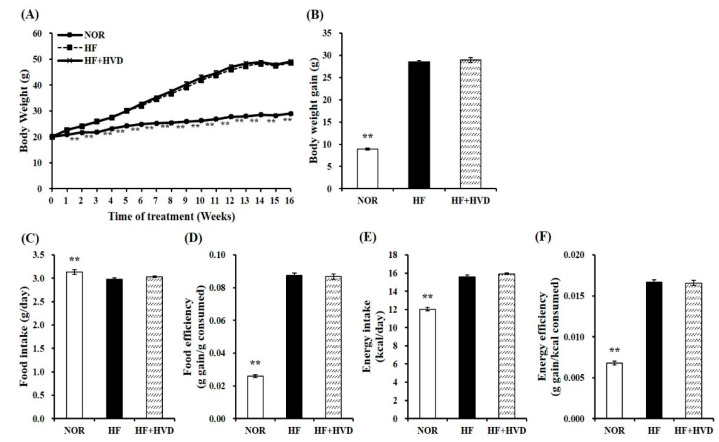
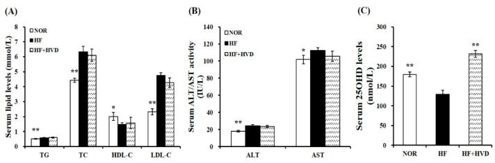
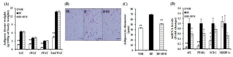
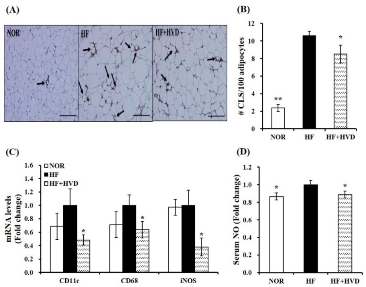
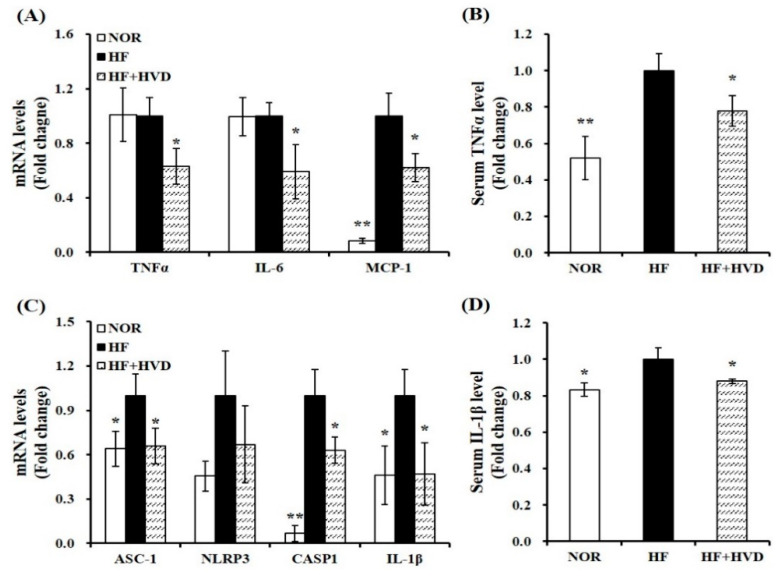
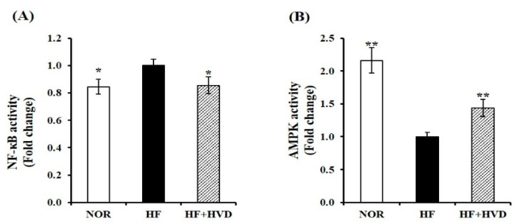
Adipose tissue expansion is strongly associated with increased adipose macrophage infiltration and adipocyte-derived pro-inflammatory cytokines, contributing to obesity-associated low-grade inflammation. Individuals with vitamin D deficiency have an increased prevalence of obesity and increased circulating inflammatory cytokines. However, the effect of vitamin D supplementation on obesity-induced inflammation remains controversial. Male C57BL/6J mice received a low-fat (10% fat) or high-fat (HF, 60% fat diet) containing 1000 IU vitamin D/kg diet, or HF supplemented with 10,000 IU vitamin D/kg diet for 16 weeks (n = 9/group). Vitamin D supplementation did not decrease HF-increased body weight but attenuated obesity-induced adipose hypertrophy and macrophage recruitment as demonstrated by the number of crown-like structures. Vitamin D supplementation significantly reduced the mRNA expression of CD11c, CD68, and iNOS, specific for inflammatory M1-like macrophages, and decreased serum levels of NO. In addition, significant reductions in pro-inflammatory gene expression of IL-6, MCP-1, and TNFα and mRNA levels of ASC-1, CASP1, and IL-1β involved in NLRP3 inflammasome were found in obese mice supplemented with vitamin D. Vitamin D supplementation significantly increased obesity-decreased AMPK activity and suppressed HF-increased NF-κB phosphorylation in adipose tissue from obese mice. These observed beneficial effects of vitamin D supplementation on adipose tissue expansion, macrophage recruitment, and inflammation might be related to AMPK/NF-κB signaling.
Keywords: NOD-, LRR- and pyrin domain-containing protein 3 (NLRP3); adenosine monophosphate-activated protein kinase (AMPK); adipose tissue; inflammation; nuclear factor-kappa B (NF-κB); obesity; vitamin D.
Conflict of interest statement
The author declares no conflict of interest.
Figures
Similar articles
-
Vitamin D Insufficiency Exacerbates Adipose Tissue Macrophage Infiltration and Decreases AMPK/SIRT1 Activity in Obese Rats.Nutrients. 2017 Mar 29;9(4):338. doi: 10.3390/nu9040338. Nutrients. 2017. PMID: 28353634 Free PMC article.
-
14-Deoxygarcinol improves insulin sensitivity in high-fat diet-induced obese mice via mitigating NF-κB/Sirtuin 2-NLRP3-mediated adipose tissue remodeling.Acta Pharmacol Sin. 2023 Feb;44(2):434-445. doi: 10.1038/s41401-022-00958-8. Epub 2022 Aug 9. Acta Pharmacol Sin. 2023. PMID: 35945312 Free PMC article.
-
Chikusetsu saponin IVa ameliorates high fat diet-induced inflammation in adipose tissue of mice through inhibition of NLRP3 inflammasome activation and NF-κB signaling.Oncotarget. 2017 May 9;8(19):31023-31040. doi: 10.18632/oncotarget.16052. Oncotarget. 2017. PMID: 28415686 Free PMC article.
-
Reappraisal of Adipose Tissue Inflammation in Obesity.Adv Exp Med Biol. 2024;1460:297-327. doi: 10.1007/978-3-031-63657-8_10. Adv Exp Med Biol. 2024. PMID: 39287856 Review.
-
Message Transmission Between Adipocyte and Macrophage in Obesity.Adv Exp Med Biol. 2024;1460:273-295. doi: 10.1007/978-3-031-63657-8_9. Adv Exp Med Biol. 2024. PMID: 39287855 Review.
Cited by
-
Omega-3 Fatty Acid and Vitamin D Supplementations Partially Reversed Metabolic Disorders and Restored Gut Microbiota in Obese Wistar Rats.Biology (Basel). 2024 Dec 20;13(12):1070. doi: 10.3390/biology13121070. Biology (Basel). 2024. PMID: 39765737 Free PMC article.
-
The correlation between serum vitamin D status and the occurrence of diabetic foot ulcers: a comprehensive systematic review and meta-analysis.Sci Rep. 2024 Sep 20;14(1):21932. doi: 10.1038/s41598-024-73133-0. Sci Rep. 2024. PMID: 39304728 Free PMC article.
-
From the Sun to the Cell: Examining Obesity through the Lens of Vitamin D and Inflammation.Metabolites. 2023 Dec 20;14(1):4. doi: 10.3390/metabo14010004. Metabolites. 2023. PMID: 38276294 Free PMC article. Review.
-
Effects of an in vitro vitamin D treatment on the inflammatory responses in visceral adipose tissue from Ldlr-/- mice.Nutr Res Pract. 2024 Feb;18(1):19-32. doi: 10.4162/nrp.2024.18.1.19. Epub 2023 Dec 11. Nutr Res Pract. 2024. PMID: 38352213 Free PMC article.
-
The Interplay between Liver and Adipose Tissue in the Onset of Liver Diseases: Exploring the Role of Vitamin Deficiency.Cells. 2024 Sep 30;13(19):1631. doi: 10.3390/cells13191631. Cells. 2024. PMID: 39404394 Free PMC article. Review.
References
-
- Xu H., Barnes G.T., Yang Q., Tan G., Yang D., Chou C.J., Sole J., Nichols A., Ross J.S., Tartaglia L.A., et al. Chronic inflammation in fat plays a crucial role in the development of obesity-related insulin resistance. J. Clin. Investig. 2003;112:1821–1830. doi: 10.1172/JCI200319451. - DOI - PMC - PubMed
MeSH terms
Substances
LinkOut - more resources
Full Text Sources
Research Materials
Miscellaneous
