

Correlation between Type I Interferon Associated Factors and COVID-19 Severity
- PMID: 36142877
- PMCID: PMC9506204
- DOI: 10.3390/ijms231810968
Correlation between Type I Interferon Associated Factors and COVID-19 Severity
Abstract
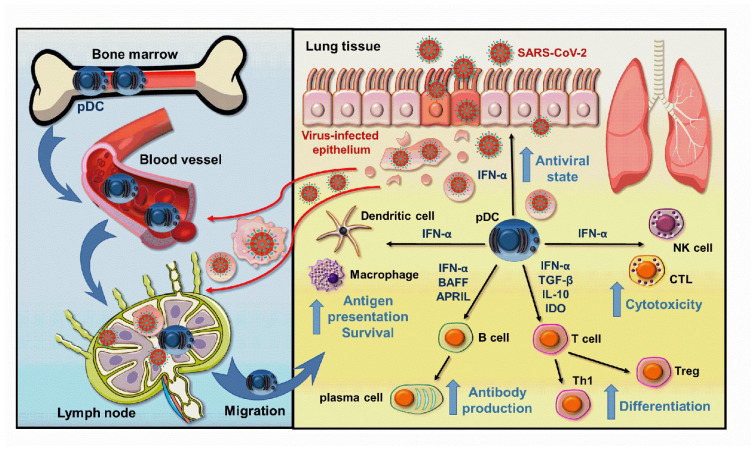
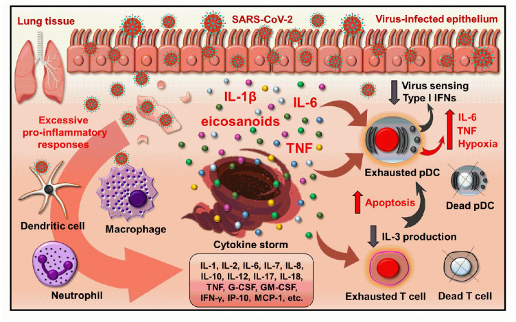
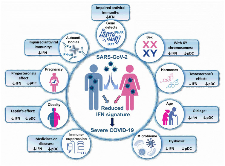
Antiviral type I interferons (IFN) produced in the early phase of viral infections effectively inhibit viral replication, prevent virus-mediated tissue damages and promote innate and adaptive immune responses that are all essential to the successful elimination of viruses. As professional type I IFN producing cells, plasmacytoid dendritic cells (pDC) have the ability to rapidly produce waste amounts of type I IFNs. Therefore, their low frequency, dysfunction or decreased capacity to produce type I IFNs might increase the risk of severe viral infections. In accordance with that, declined pDC numbers and delayed or inadequate type I IFN responses could be observed in patients with severe coronavirus disease (COVID-19) caused by the severe acute respiratory syndrome coronavirus 2 (SARS-CoV-2), as compared to individuals with mild or no symptoms. Thus, besides chronic diseases, all those conditions, which negatively affect the antiviral IFN responses lengthen the list of risk factors for severe COVID-19. In the current review, we would like to briefly discuss the role and dysregulation of pDC/type I IFN axis in COVID-19, and introduce those type I IFN-dependent factors, which account for an increased risk of COVID-19 severity and thus are responsible for the different magnitude of individual immune responses to SARS-CoV-2.
Keywords: COVID-19; IFN signature; SARS-CoV-2; antiviral response; plasmacytoid dendritic cell 1; risk factor; type I interferon.
Conflict of interest statement
The authors declare no conflict of interest.
Figures
Similar articles
-
Differential plasmacytoid dendritic cell phenotype and type I Interferon response in asymptomatic and severe COVID-19 infection.PLoS Pathog. 2021 Sep 2;17(9):e1009878. doi: 10.1371/journal.ppat.1009878. eCollection 2021 Sep. PLoS Pathog. 2021. PMID: 34473805 Free PMC article.
-
SARS-CoV-2 Evasion of the Interferon System: Can We Restore Its Effectiveness?Int J Mol Sci. 2023 May 27;24(11):9353. doi: 10.3390/ijms24119353. Int J Mol Sci. 2023. PMID: 37298304 Free PMC article. Review.
-
Type I IFNs: A Blessing in Disguise or Partner in Crime in MERS-CoV-, SARS-CoV-, and SARS-CoV-2-Induced Pathology and Potential Use of Type I IFNs in Synergism with IFN-γ as a Novel Antiviral Approach Against COVID-19.Viral Immunol. 2021 Jun;34(5):321-329. doi: 10.1089/vim.2020.0085. Epub 2020 Nov 11. Viral Immunol. 2021. PMID: 33181057 Review.
-
Increased Sensitivity of SARS-CoV-2 to Type III Interferon in Human Intestinal Epithelial Cells.J Virol. 2022 Apr 13;96(7):e0170521. doi: 10.1128/jvi.01705-21. Epub 2022 Mar 9. J Virol. 2022. PMID: 35262371 Free PMC article.
-
Plasmacytoid dendritic cells during COVID-19: Ally or adversary?Cell Rep. 2022 Jul 26;40(4):111148. doi: 10.1016/j.celrep.2022.111148. Epub 2022 Jul 14. Cell Rep. 2022. PMID: 35858624 Free PMC article. Review.
Cited by
-
Immune responses in mildly versus critically ill COVID-19 patients.Front Immunol. 2023 Jan 30;14:1077236. doi: 10.3389/fimmu.2023.1077236. eCollection 2023. Front Immunol. 2023. PMID: 36793739 Free PMC article. Review.
-
Immune correlates of protection for SARS-CoV-2, Ebola and Nipah virus infection.Front Immunol. 2023 Apr 17;14:1156758. doi: 10.3389/fimmu.2023.1156758. eCollection 2023. Front Immunol. 2023. PMID: 37153606 Free PMC article. Review.
-
New-onset Systemic Lupus Erythematosus Manifestation Following COVID-19: A Case Report and Literature Review.Intern Med. 2024 May 15;63(10):1491-1498. doi: 10.2169/internalmedicine.3211-23. Epub 2024 Feb 19. Intern Med. 2024. PMID: 38369349 Free PMC article. Review.
-
The role of inflammatory gene polymorphisms in severe COVID-19: a review.Virol J. 2024 Dec 20;21(1):327. doi: 10.1186/s12985-024-02597-3. Virol J. 2024. PMID: 39707400 Free PMC article. Review.
-
Differential Type-I Interferon Response in Buffy Coat Transcriptome of Individuals Infected with SARS-CoV-2 Gamma and Delta Variants.Int J Mol Sci. 2023 Aug 24;24(17):13146. doi: 10.3390/ijms241713146. Int J Mol Sci. 2023. PMID: 37685953 Free PMC article.
References
Publication types
MeSH terms
Substances
Grants and funding
- NKFIH FK 128294/National Research, Development and Innovation Office
- NKFIH PD 135193/National Research, Development and Innovation Office
- GINOP-2.3.2-15-2016-00050/European Union and the European Regional Development Fund
- ÚNKP-21-05-DE-170/New National Excellence Program of the Ministry for Innovation and Technology from the source of the National Research, Development and Innovation Fund
- ÚNKP-21-3-II-DE-21/New National Excellence Program of the Ministry for Innovation and Technology from the source of the National Research, Development and Innovation Fund
LinkOut - more resources
Full Text Sources
Medical
Research Materials
Miscellaneous
