

In Vitro Study of Ineffective Erythropoiesis in Thalassemia: Diverse Intrinsic Pathophysiological Features of Erythroid Cells Derived from Various Thalassemia Syndromes
- PMID: 36143003
- PMCID: PMC9504363
- DOI: 10.3390/jcm11185356
In Vitro Study of Ineffective Erythropoiesis in Thalassemia: Diverse Intrinsic Pathophysiological Features of Erythroid Cells Derived from Various Thalassemia Syndromes
Abstract
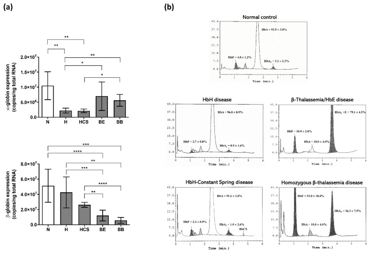
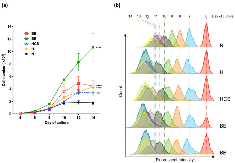
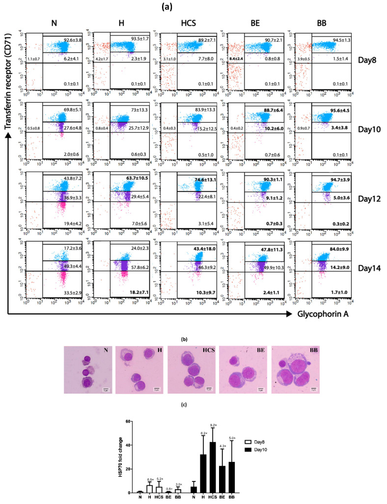
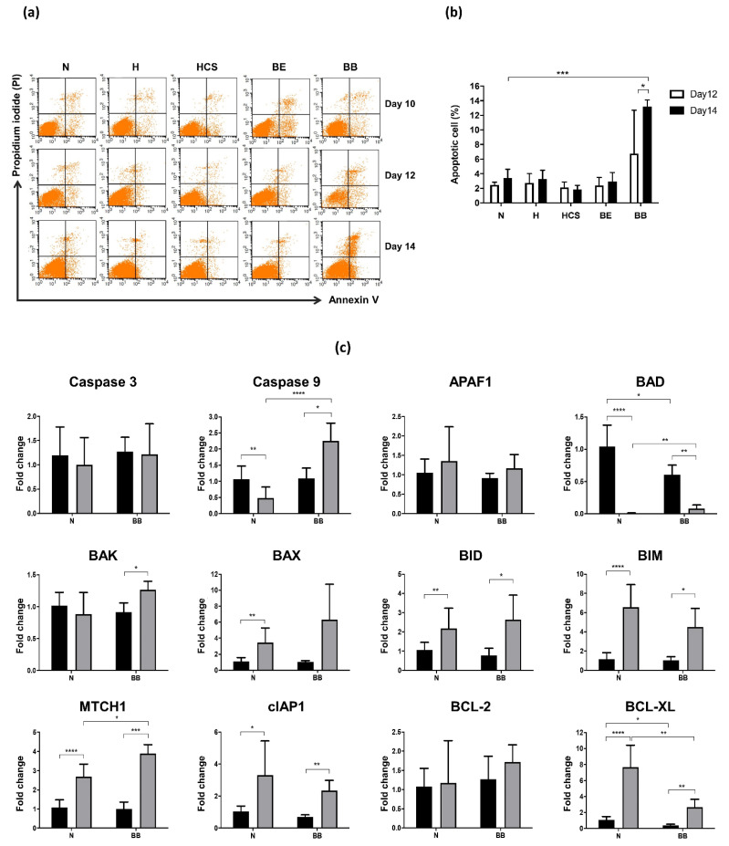
Defective hemoglobin production and ineffective erythropoiesis contribute to the pathophysiology of thalassemia syndromes. Previous studies in the field of erythropoiesis mainly focused on the severe forms of thalassemia, such as β-thalassemia major, while mechanisms underlying the pathogenesis of other thalassemia syndromes remain largely unexplored. The current study aimed to investigate the intrinsic pathophysiological properties of erythroid cells derived from the most common forms of thalassemia diseases, including α-thalassemia (hemoglobin H and hemoglobin H-Constant Spring diseases) and β-thalassemia (homozygous β0-thalassemia and β0-thalassemia/hemoglobin E diseases), under an identical in vitro erythroid culture system. Cell proliferation capacity, differentiation velocity, cell death, as well as globin synthesis and the expression levels of erythropoiesis modifying factors were determined. Accelerated expansion was found in erythroblast cells derived from all types of thalassemia, with the highest degree in β0-thalassemia/hemoglobin E. Likewise, all types of thalassemia showed limited erythroid cell differentiation, but each of them manifested varying degrees of erythroid maturation arrest corresponding with the clinical severity. Robust induction of HSP70 transcripts, an erythroid maturation-related factor, was found in both α- and β-thalassemia erythroid cells. Increased cell death was distinctly present only in homozygous β0-thalassemia erythroblasts and associated with the up-regulation of pro-apoptotic (Caspase 9, BAD, and MTCH1) genes and down-regulation of the anti-apoptotic BCL-XL gene.
Keywords: apoptosis; cell death; erythropoiesis; globin; hemoglobin; thalassemia.
Conflict of interest statement
The authors declare no conflict of interest.
Figures
Similar articles
-
Association of the Degree of Erythroid Expansion and Maturation Arrest with the Clinical Severity of β0-Thalassemia/Hemoglobin E Patients.Acta Haematol. 2021;144(6):660-671. doi: 10.1159/000518310. Epub 2021 Sep 14. Acta Haematol. 2021. PMID: 34535581
-
Elevated CDKN1A (P21) mediates β-thalassemia erythroid apoptosis, but its loss does not improve β-thalassemic erythropoiesis.Blood Adv. 2023 Nov 28;7(22):6873-6885. doi: 10.1182/bloodadvances.2022007655. Blood Adv. 2023. PMID: 37672319 Free PMC article.
-
A correlation of erythrokinetics, ineffective erythropoiesis, and erythroid precursor apoptosis in thai patients with thalassemia.Blood. 2000 Oct 1;96(7):2606-12. Blood. 2000. PMID: 11001918
-
Pathophysiology of thalassemia.Curr Opin Hematol. 2002 Mar;9(2):123-6. doi: 10.1097/00062752-200203000-00007. Curr Opin Hematol. 2002. PMID: 11844995 Review.
-
Ineffective erythropoiesis in β -thalassemia.ScientificWorldJournal. 2013 Mar 28;2013:394295. doi: 10.1155/2013/394295. Print 2013. ScientificWorldJournal. 2013. PMID: 23606813 Free PMC article. Review.
Cited by
-
Erythropoiesis and Gene Expression Analysis in Erythroid Progenitor Cells Derived from Patients with Hemoglobin H/Constant Spring Disease.Int J Mol Sci. 2024 Oct 19;25(20):11246. doi: 10.3390/ijms252011246. Int J Mol Sci. 2024. PMID: 39457028 Free PMC article.
-
Reticulocyte Antioxidant Enzymes mRNA Levels versus Reticulocyte Maturity Indices in Hereditary Spherocytosis, β-Thalassemia and Sickle Cell Disease.Int J Mol Sci. 2024 Feb 10;25(4):2159. doi: 10.3390/ijms25042159. Int J Mol Sci. 2024. PMID: 38396832 Free PMC article.
-
Activating transcription factor 4 in erythroid development and -thalassemia: a powerful regulator with therapeutic potential.Ann Hematol. 2024 Aug;103(8):2659-2670. doi: 10.1007/s00277-023-05508-8. Epub 2023 Oct 31. Ann Hematol. 2024. PMID: 37906269 Review.
-
MiR-223-3p regulates erythropoiesis by targeting TGFBR3/Smad signaling pathway in hemoglobin H-Constant Spring disease.Ann Med. 2025 Dec;57(1):2530690. doi: 10.1080/07853890.2025.2530690. Epub 2025 Jul 11. Ann Med. 2025. PMID: 40646708 Free PMC article.
-
Exosomes from Tregs mitigate lung damage caused by smoking via inhibiting inflammation and altering T lymphocyte subsets in COPD rats.BMC Pulm Med. 2025 Apr 14;25(1):181. doi: 10.1186/s12890-025-03632-x. BMC Pulm Med. 2025. PMID: 40229730 Free PMC article.
References
LinkOut - more resources
Full Text Sources
Research Materials
