

Management of Inadvertent Arterial Catheterization during Central Venous Catheter Placement: A Case Series
- PMID: 36143321
- PMCID: PMC9503793
- DOI: 10.3390/jpm12091537
Management of Inadvertent Arterial Catheterization during Central Venous Catheter Placement: A Case Series
Abstract
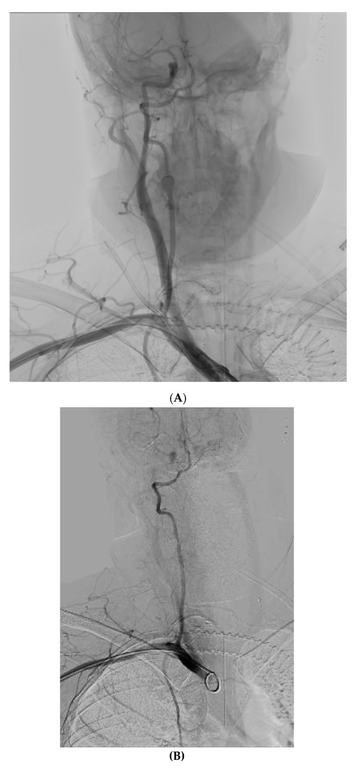
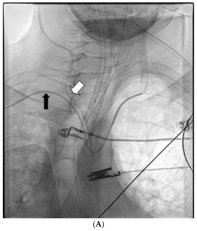
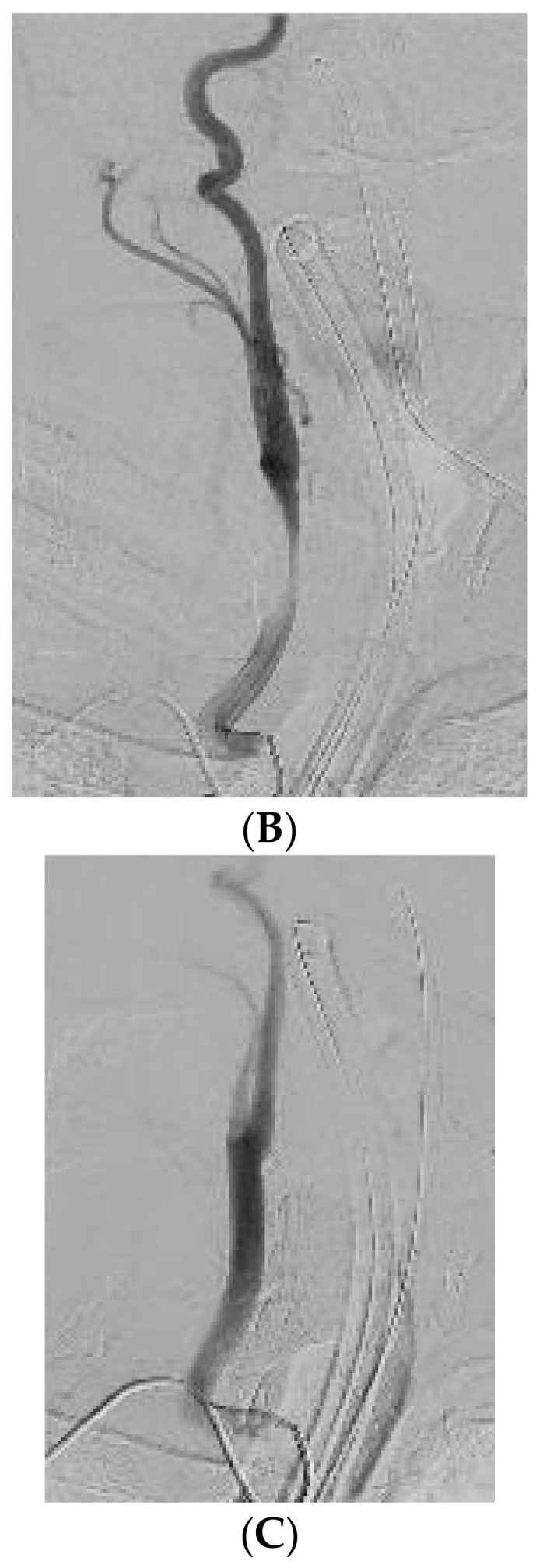
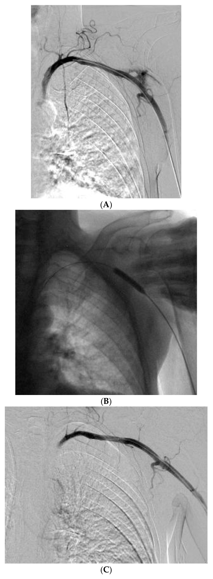
Percutaneous central venous catheterization, although a widely used technique in ICU patients worldwide, is not devoid of complications even under real-time ultrasound guidance. Arterial puncture is a well-recognized complication, while unintended subclavian or carotid artery cannulations during attempted central venous catheterization are infrequent, but documented complications with potentially deleterious consequences. Recently, endovascular balloon tamponade has emerged as the preferred initial approach to repair inadvertent arterial cannulations. Herein, we present a case series of inadvertent arterial catheterization during an attempted ultrasound-guided access of the right internal jugular and the left subclavian vein that were successfully managed with endovascular balloon tamponade.
Keywords: central venous catheter; inadvertent arterial catheterization; ultrasound guidance.
Conflict of interest statement
The authors declare no conflict of interest.
Figures
Similar articles
-
Endovascular management of inadvertent brachiocephalic arterial catheterization.J Neurosurg. 2011 Jan;114(1):146-52. doi: 10.3171/2009.10.JNS09940. Epub 2009 Nov 20. J Neurosurg. 2011. PMID: 19929193
-
Inadvertent arterial puncture involving the subclavian artery and the aorta during central venous catheterization: a case report.J Med Case Rep. 2021 May 28;15(1):303. doi: 10.1186/s13256-021-02871-w. J Med Case Rep. 2021. PMID: 34044882 Free PMC article.
-
Inadvertent catheter misplacement into the subclavian artery during ultrasound-guided internal jugular venous catheterization: a case report.JA Clin Rep. 2023 Sep 6;9(1):58. doi: 10.1186/s40981-023-00649-1. JA Clin Rep. 2023. PMID: 37672125 Free PMC article.
-
Arterial trauma during central venous catheter insertion: Case series, review and proposed algorithm.J Vasc Surg. 2008 Oct;48(4):918-25; discussion 925. doi: 10.1016/j.jvs.2008.04.046. Epub 2008 Aug 13. J Vasc Surg. 2008. PMID: 18703308 Review.
-
Iatrogenic intramural hematoma of the ascending aorta complicating inadvertent arterial cannulation during central venous catheter placement. A case report and review of the literature.Ann Card Anaesth. 2020 Oct-Dec;23(4):502-504. doi: 10.4103/aca.ACA_169_19. Ann Card Anaesth. 2020. PMID: 33109812 Free PMC article. Review.
Cited by
-
"It Looks Arterial": The Chest X-Ray of Nightmares.Cureus. 2025 Feb 23;17(2):e79532. doi: 10.7759/cureus.79532. eCollection 2025 Feb. Cureus. 2025. PMID: 40144411 Free PMC article.
-
Use of suture-mediated closure device system after inadvertent medport placement in the subclavian artery leading to multi-focal ischaemic infarct: a case report.Eur Heart J Case Rep. 2024 Oct 22;8(11):ytae565. doi: 10.1093/ehjcr/ytae565. eCollection 2024 Nov. Eur Heart J Case Rep. 2024. PMID: 39502261 Free PMC article.
-
Observational, retrospective real-world evidence study demonstrates safety, performance and usefulness of antimicrobial central venous catheters.J Int Med Res. 2024 Sep;52(9):3000605241279236. doi: 10.1177/03000605241279236. J Int Med Res. 2024. PMID: 39308254 Free PMC article.
-
Minimally Invasive Management of Subclavian Artery Catheter Misplacement: The New Standard?J Clin Med. 2025 Apr 12;14(8):2650. doi: 10.3390/jcm14082650. J Clin Med. 2025. PMID: 40283480 Free PMC article.
-
Two Cases of Malpositioning of Internal Jugular Central Venous Catheters: A Review of Literature and Current Treatment Recommendations.Cureus. 2024 May 7;16(5):e59814. doi: 10.7759/cureus.59814. eCollection 2024 May. Cureus. 2024. PMID: 38846204 Free PMC article.
References
-
- Marie P. Accidental Carotid Artery Catheterization during Attempted Central Venous Catheter Placement: A Case Report. [(accessed on 8 May 2022)];AANA J. 2012 80:251–255. Available online: www.aana.com/aanajournalonline. - PubMed
LinkOut - more resources
Full Text Sources
