Can Dynamic Whole-Body FDG PET Imaging Differentiate between Malignant and Inflammatory Lesions?
- PMID: 36143386
- PMCID: PMC9501027
- DOI: 10.3390/life12091350
Can Dynamic Whole-Body FDG PET Imaging Differentiate between Malignant and Inflammatory Lesions?
Abstract
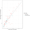
Background: Investigation of the clinical feasibility of dynamic whole-body (WB) [18F]FDG PET, including standardized uptake value (SUV), rate of irreversible uptake (Ki), and apparent distribution volume (Vd) in physiologic tissues, and comparison between inflammatory/infectious and cancer lesions. Methods: Twenty-four patients were prospectively included to undergo dynamic WB [18F]FDG PET/CT for clinically indicated re-/staging of oncological diseases. Parametric maps of Ki and Vd were generated using Patlak analysis alongside SUV images. Maximum parameter values (SUVmax, Kimax, and Vdmax) were measured in liver parenchyma and in malignant or inflammatory/infectious lesions. Lesion-to-background ratios (LBRs) were calculated by dividing the measurements by their respective mean in the liver tissue. Results: Seventy-seven clinical target lesions were identified, 60 malignant and 17 inflammatory/infectious. Kimax was significantly higher in cancer than in inflammatory/infections lesions (3.0 vs. 2.0, p = 0.002) while LBRs of SUVmax, Kimax, and Vdmax did not differ significantly between the etiologies: LBR (SUVmax) 3.3 vs. 2.9, p = 0.06; LBR (Kimax) 5.0 vs. 4.4, p = 0.05, LBR (Vdmax) 1.1 vs. 1.0, p = 0.18). LBR of inflammatory/infectious and cancer lesions was higher in Kimax than in SUVmax (4.5 vs. 3.2, p < 0.001). LBRs of Kimax and SUVmax showed a strong correlation (Spearman’s rho = 0.83, p < 0.001). Conclusions: Dynamic WB [18F]FDG PET/CT is feasible in a clinical setting. LBRs of Kimax were higher than SUVmax. Kimax was higher in malignant than in inflammatory/infectious lesions but demonstrated a large overlap between the etiologies.
Keywords: FDG PET/CT; Patlak; dynamic whole-body positron emission tomography; fluorodeoxyglucose; infection; molecular imaging; oncologic imaging.
Conflict of interest statement
Irene A. Burger and Martin W. Huellner received grants from GE Healthcare. The University Hospital of Zurich holds a research agreement with GE Healthcare. Fotis Kotasidis is an employee of GE Healthcare. Other than that, the authors declare that they have no competing interests.
Figures


Similar articles
-
Does whole-body Patlak 18F-FDG PET imaging improve lesion detectability in clinical oncology?Eur Radiol. 2019 Sep;29(9):4812-4821. doi: 10.1007/s00330-018-5966-1. Epub 2019 Jan 28. Eur Radiol. 2019. PMID: 30689031
-
Prognostic value of whole-body dynamic 18F-FDG PET/CT Patlak in diffuse large B-cell lymphoma.Heliyon. 2023 Sep 1;9(9):e19749. doi: 10.1016/j.heliyon.2023.e19749. eCollection 2023 Sep. Heliyon. 2023. PMID: 37809527 Free PMC article.
-
Assessment of Lesion Detectability in Dynamic Whole-Body PET Imaging Using Compartmental and Patlak Parametric Mapping.Clin Nucl Med. 2020 May;45(5):e221-e231. doi: 10.1097/RLU.0000000000002954. Clin Nucl Med. 2020. PMID: 32108696
-
Dual-time point 18F-FDG-PET and PET/CT for Differentiating Benign From Malignant Musculoskeletal Lesions: Opportunities and Limitations.Semin Nucl Med. 2017 Jul;47(4):373-391. doi: 10.1053/j.semnuclmed.2017.02.009. Epub 2017 Apr 19. Semin Nucl Med. 2017. PMID: 28583277 Review.
-
Hyperaccumulation of (18)F-FDG in order to differentiate solid pseudopapillary tumors from adenocarcinomas and from neuroendocrine pancreatic tumors and review of the literature.Hell J Nucl Med. 2013 May-Aug;16(2):97-102. doi: 10.1967/s002449910084. Epub 2013 May 20. Hell J Nucl Med. 2013. PMID: 23687644 Review.
Cited by
-
Incidental Uptake in Gastrointestinal Tract on 18F-FDG-PET/CT: Is It Worth to Investigate? A Study with 371 Patients.GE Port J Gastroenterol. 2024 Oct 9;32(3):174-184. doi: 10.1159/000541209. eCollection 2025 Jun. GE Port J Gastroenterol. 2024. PMID: 40469205 Free PMC article.
-
Fluorodeoxyglucose Positron Emission Tomography Evaluation of Chronic Recurrent Multifocal Osteomyelitis.Cureus. 2024 Sep 19;16(9):e69735. doi: 10.7759/cureus.69735. eCollection 2024 Sep. Cureus. 2024. PMID: 39429331 Free PMC article.
-
Long-Axial Field-of-View PET Imaging in Patients with Lymphoma: Challenges and Opportunities.PET Clin. 2024 Oct;19(4):495-504. doi: 10.1016/j.cpet.2024.05.005. Epub 2024 Jul 4. PET Clin. 2024. PMID: 38969563 Review.
-
Assessment of image-derived input functions from small vessels for patlak parametric imaging using total-body PET/CT.Eur J Nucl Med Mol Imaging. 2025 Jan;52(2):648-659. doi: 10.1007/s00259-024-06926-0. Epub 2024 Sep 26. Eur J Nucl Med Mol Imaging. 2025. PMID: 39325156 Free PMC article.
-
Case report: When infection lurks behind malignancy: a unique case of primary bone lymphoma mimicking infectious process in the spine.Front Nucl Med. 2024 Jul 15;4:1402552. doi: 10.3389/fnume.2024.1402552. eCollection 2024. Front Nucl Med. 2024. PMID: 39355207 Free PMC article.
References
-
- Boellaard R., Delgado-Bolton R., Oyen W.J., Giammarile F., Tatsch K., Eschner W., Verzijlbergen F.J., Barrington S.F., Pike L.C., Weber W.A., et al. FDG PET/CT: EANM procedure guidelines for tumour imaging: Version 2.0. Eur. J. Nucl. Med. Mol. Imaging. 2015;42:328–354. doi: 10.1007/s00259-014-2961-x. - DOI - PMC - PubMed
Grants and funding
LinkOut - more resources
Full Text Sources
Medical

