Review
. 2022 Sep 14;10(9):1835.
doi: 10.3390/microorganisms10091835.

Past, Present, and Future of Genome Modification in Escherichia coli

Affiliations
Review

Past, Present, and Future of Genome Modification in Escherichia coli

Hirotada Mori et al. Microorganisms. .

Abstract

Escherichia coli K-12 is one of the most well-studied species of bacteria. This species, however, is much more difficult to modify by homologous recombination (HR) than other model microorganisms. Research on HR in E. coli has led to a better understanding of the molecular mechanisms of HR, resulting in technical improvements and rapid progress in genome research, and allowing whole-genome mutagenesis and large-scale genome modifications. Developments using λ Red (exo, bet, and gam) and CRISPR-Cas have made E. coli as amenable to genome modification as other model microorganisms, such as Saccharomyces cerevisiae and Bacillus subtilis. This review describes the history of recombination research in E. coli, as well as improvements in techniques for genome modification by HR. This review also describes the results of large-scale genome modification of E. coli using these technologies, including DNA synthesis and assembly. In addition, this article reviews recent advances in genome modification, considers future directions, and describes problems associated with the creation of cells by design.

Keywords: Escherichia coli K-12; HR; P1 transduction; genome modification; homologous recombination; mutation; recombineering; site-specific recombination; λ Red.


Conflict of interest statement

The authors declare no conflict of interest.

Figures

Figure 1
Figure 1
Schematic illustration of genome-scale modification methods. (A) Two methods have been used to block RecD exonuclease: (1) using recBC mutations and (2) λ Red Gam synthesis. Cells are transformed with linear double-stranded (ds) DNA encoding an antibiotic resistance (AbR) cassette and ends at homology regions (hr) of upstream (up) and downstream (down) regions of the target. (B1) Scarless deletion using I-SceI nuclease. The drug-resistant fragment flanked by I-SceI restriction enzyme sites is amplified with up and next to the downside homology regions (u and hr) and introduced into the genome by λ Red homologous recombination [16]. The I-SceI-flanked segment is eliminated by expressing the meganuclease I-SceI, resulting in a double-strand break (DSB), DSB-stimulated DNA repair, and RecA-dependent recombination between the d direct repeats. This figure is modified from Kolisnychenko et al. [16] (B2) The Ab (antibiotic resistance gene) with the killing gene, such as sacB, ccdB, parE, or phage T7 0.7, under the control of the tightly regulated promoter, such as rhaBp [51], is amplified with 36 to 40 nt homology region (hr) to target. The amplified fragment is then transformed into a strain expressing λ Red to insert into the genome. Double-stranded “Substitution Sequence (SS)” with flanking hrs is transformed into the Ab-resistant fragment integrated strain expressing λ Red. The transformants are selected in the presence of L-rhamnose, preferably with L-rhamnose as the sole carbon source. (C) Random insertion mutagenesis by Tn. (C1) Mutant Tns with less sequence specificity for insertion sites on chromosomes have been developed, with transposons such as Tn5, Tn10, and Mariner often used. Two different Tns of two different drug resistance genes were randomly mutated, with the location of insertion on the genome determined by PCR and sequencing. E. coli strains with insertions at appropriate positions were selected and combined into a single E. coli strain using the P1 transduction method. This method used site-specific recombination at each Tn and induced recombination by increasing the production of site-specific recombinase and deleting the region between Tns. (C2) A complex with Tnp that recognizes IE is introduced into the cell to obtain the first random insertion mutation. Synthetic induction of Tnp recognizing the internal ME is then performed to obtain a transition mutation; the direction of the second transition results in a deletion between two different Tn insertion sites. This figure is modified from Goryshin et al. [52] (C3) Insertion of a Tn fragment into the genome, followed by CRISPR-Cas cutting of the inside of the Tn fragment. This yielded a strain in which nuclease activity deleted the periphery. (D1) DSB was induced by CRISPR-Cas, with DNA fragments transformed by bridging homologous regions at both ends of each double-strand break, resulting in genome repair and yielding to the circular genome. (D2) Genome editing by fusion protein with a function different from that of Cas protein. Left panel: fusion of cytosine deaminase to a Cas protein with mutation-inactivated DNase activities [53]. Right panel: fusion of reverse transcriptase to a Cas, which inactivates only the nick on the other strand, providing a template for repairing the nick site and introducing the mutation by a reverse-transcribed sequence 77. (E) Introduction of synthetic DNA fragments into cells, generally yeast cells, resulting in assembly of the fragments by in vivo homologous recombination. After the assembly, the assembled fragment is collected and transformed into E. coli cell and replacement of the target region in the λ Red-induced strain by homologous recombination. (F) Assembly of the synthetic DNA fragments in the cell, followed by circularization to reconstruct the genome. The synthetic genome was subsequently transferred to bacterial cells by cell fusion [17,18]. This figure is summarized from Gibson et al. [17] (G) Introduction of an ssDNA about 90 bp in length to be mutated in the cell via the induction of λ Red β protein, which promoted the introduction of mutations on the lagging strand during DNA replication and accelerated the introduction of mutations throughout the genome. This figure is modified from Wang et al. [54] and Costantino and Court [55].
Figure 1
Figure 1
Schematic illustration of genome-scale modification methods. (A) Two methods have been used to block RecD exonuclease: (1) using recBC mutations and (2) λ Red Gam synthesis. Cells are transformed with linear double-stranded (ds) DNA encoding an antibiotic resistance (AbR) cassette and ends at homology regions (hr) of upstream (up) and downstream (down) regions of the target. (B1) Scarless deletion using I-SceI nuclease. The drug-resistant fragment flanked by I-SceI restriction enzyme sites is amplified with up and next to the downside homology regions (u and hr) and introduced into the genome by λ Red homologous recombination [16]. The I-SceI-flanked segment is eliminated by expressing the meganuclease I-SceI, resulting in a double-strand break (DSB), DSB-stimulated DNA repair, and RecA-dependent recombination between the d direct repeats. This figure is modified from Kolisnychenko et al. [16] (B2) The Ab (antibiotic resistance gene) with the killing gene, such as sacB, ccdB, parE, or phage T7 0.7, under the control of the tightly regulated promoter, such as rhaBp [51], is amplified with 36 to 40 nt homology region (hr) to target. The amplified fragment is then transformed into a strain expressing λ Red to insert into the genome. Double-stranded “Substitution Sequence (SS)” with flanking hrs is transformed into the Ab-resistant fragment integrated strain expressing λ Red. The transformants are selected in the presence of L-rhamnose, preferably with L-rhamnose as the sole carbon source. (C) Random insertion mutagenesis by Tn. (C1) Mutant Tns with less sequence specificity for insertion sites on chromosomes have been developed, with transposons such as Tn5, Tn10, and Mariner often used. Two different Tns of two different drug resistance genes were randomly mutated, with the location of insertion on the genome determined by PCR and sequencing. E. coli strains with insertions at appropriate positions were selected and combined into a single E. coli strain using the P1 transduction method. This method used site-specific recombination at each Tn and induced recombination by increasing the production of site-specific recombinase and deleting the region between Tns. (C2) A complex with Tnp that recognizes IE is introduced into the cell to obtain the first random insertion mutation. Synthetic induction of Tnp recognizing the internal ME is then performed to obtain a transition mutation; the direction of the second transition results in a deletion between two different Tn insertion sites. This figure is modified from Goryshin et al. [52] (C3) Insertion of a Tn fragment into the genome, followed by CRISPR-Cas cutting of the inside of the Tn fragment. This yielded a strain in which nuclease activity deleted the periphery. (D1) DSB was induced by CRISPR-Cas, with DNA fragments transformed by bridging homologous regions at both ends of each double-strand break, resulting in genome repair and yielding to the circular genome. (D2) Genome editing by fusion protein with a function different from that of Cas protein. Left panel: fusion of cytosine deaminase to a Cas protein with mutation-inactivated DNase activities [53]. Right panel: fusion of reverse transcriptase to a Cas, which inactivates only the nick on the other strand, providing a template for repairing the nick site and introducing the mutation by a reverse-transcribed sequence 77. (E) Introduction of synthetic DNA fragments into cells, generally yeast cells, resulting in assembly of the fragments by in vivo homologous recombination. After the assembly, the assembled fragment is collected and transformed into E. coli cell and replacement of the target region in the λ Red-induced strain by homologous recombination. (F) Assembly of the synthetic DNA fragments in the cell, followed by circularization to reconstruct the genome. The synthetic genome was subsequently transferred to bacterial cells by cell fusion [17,18]. This figure is summarized from Gibson et al. [17] (G) Introduction of an ssDNA about 90 bp in length to be mutated in the cell via the induction of λ Red β protein, which promoted the introduction of mutations on the lagging strand during DNA replication and accelerated the introduction of mutations throughout the genome. This figure is modified from Wang et al. [54] and Costantino and Court [55].
Figure 2
Figure 2
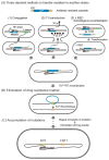
Common methods to transfer mutations to another strain using Keio collection mutants. (A) Methods of transferring mutations into the target host strain. (1) Conjugation, consisting of the recombination of an oriT onto the chromosome and use of an F plasmid to provide conjugative transfer factors in trans. (2) P1 transduction, using phage P1 lysate prepared on the mutant to infect new strains. (3) λ Red homologous recombination. (B) Elimination of drug-resistant marker by Flp-FRT site-specific recombination. After elimination, one copy of a 34 bp FRT scar remained. (C) Repetition of steps (A,B), resulting in the accumulation of mutations with FRT copies as a scar.
Figure 3
Figure 3
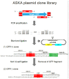
Construction of the ASKA plasmid clone library. The sequence corresponding to all of the amino acids in the coding region, except for the first Met codon, were PCR amplified, with additional GCC and CC nucleotides at the N- and C-termini, respectively. Translation from termination codon is shown by ***. The amplified fragments were subsequently cloned into the StuI site of pCA24N. Only clones with predicted orientation could generate fluorescence from the eGFP peptide. After the structures of the cloned plasmids were validated, the plasmids were cut with NotI and self-ligated to eliminate eGFP. The structures are of (1) a fusion type with eGFP and (2) a non-fusion type plasmid clone.
Figure 4
Figure 4
Structure of Keio single-gene deletion and ASKA barcode deletion collections. The coding regions of both deletion strains, except for the initiation codon and the codons encoding the six amino acids at the C-terminal, were replaced by drug resistance fragments. Site-directed recombination of FLP-FRT removed the drug resistance region. After removal from the Keio collection, the initiation codon and the codons encoding the C-terminal six-amino-acid region of the target gene were fused in frame with codons from the FRT site to suppress the polar effect of the downstream gene 35. The shared-primers contained the initiation codon (blue) and 50 bases upstream and downstream (gray), including the six C-terminal codons and the terminal codon (red) as chromosomal homologous regions. The black and blue sequences represent primers amplifying the template plasmid with the drug resistance gene and FRT sites both of Keio and ASKA barcode collections, respectively. For introducing 20 nt length random sequence as a barcode, PS for amplifying the resistant fragment (green), 20 nt random sequence (yellow), and PS of shared-primer (blue) was synthesized and amplified with PS (black) to prepare the template fragment. The resistant fragment with barcode was then amplified by shared-primers. The amplified drug resistance fragments for the Keio collection and ASKA barcode collection were used to transform λ Red-induced strains to generate deletion strains by homologous recombination. The barcode deletion strains are available for about 3000 genes.

Similar articles

Cited by

References

    1. Lederberg J., Tatum E.L. Gene Recombination in Escherichia Coli. Nature. 1946;158:558. doi: 10.1038/158558a0. - DOI - PubMed
    1. Lennox E. Transduction of linked genetic characters of the host by bacteriophage P1. Virology. 1955;1:190–206. doi: 10.1016/0042-6822(55)90016-7. - DOI - PubMed
    1. Griffith F. The Significance of Pneumococcal Types. J. Hyg. 1928;27:113–159. doi: 10.1017/S0022172400031879. - DOI - PMC - PubMed
    1. Alexander E.H., Leidy G. Transformation of type specificity of Hemophilus influenzae. A.M.A. Am. J. Dis. Child. 1950;80:877–878. - PubMed
    1. Spizizen J. Transformation of biochemically deficient strains of bacillus subtilis by deoxyribonucleate. Proc. Natl. Acad. Sci. USA. 1958;44:1072–1078. doi: 10.1073/pnas.44.10.1072. - DOI - PMC - PubMed

LinkOut - more resources