. 2022 Sep 19;10(9):1874.
doi: 10.3390/microorganisms10091874.

Antibacterial and Anti-Inflammatory Activities of Thymus vulgaris Essential Oil Nanoemulsion on Acne Vulgaris

Affiliations

Antibacterial and Anti-Inflammatory Activities of Thymus vulgaris Essential Oil Nanoemulsion on Acne Vulgaris

Farah M Abdelhamed et al. Microorganisms. .

Abstract

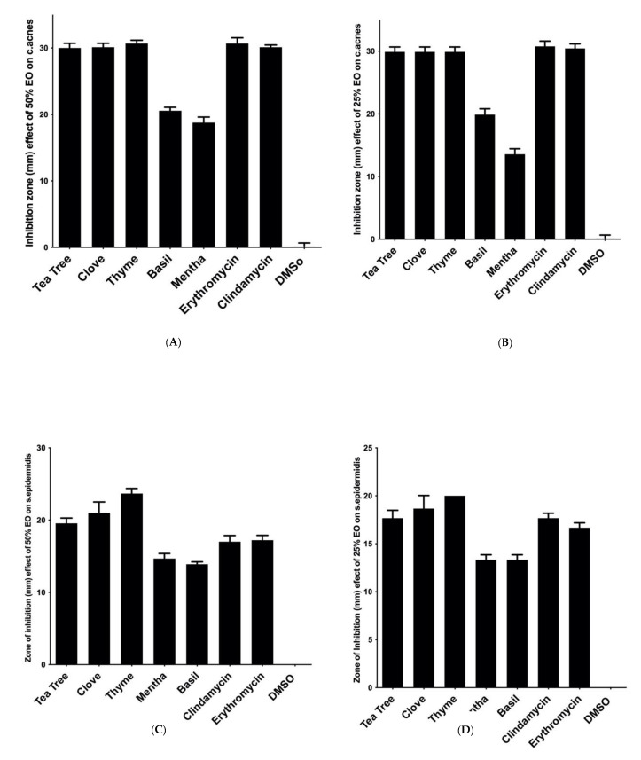
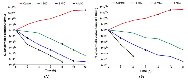
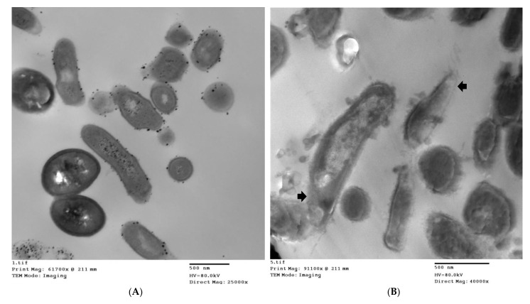
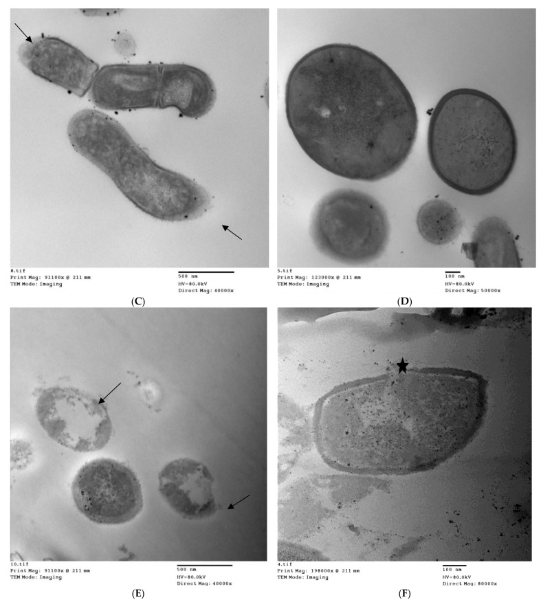
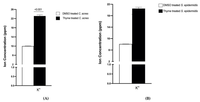
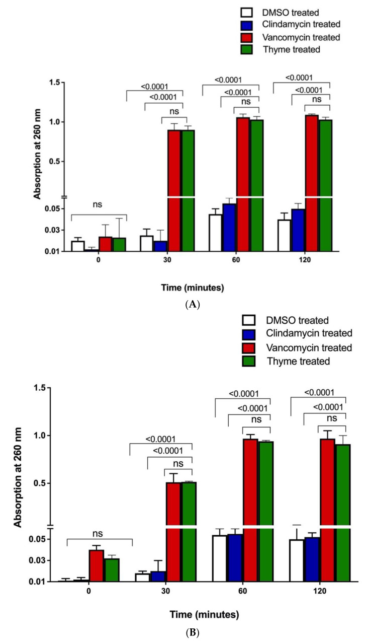
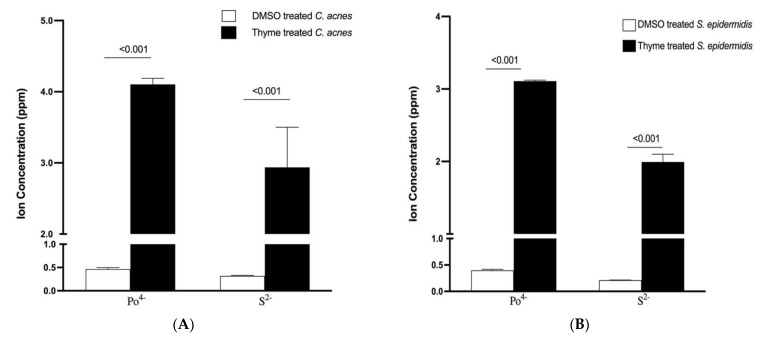
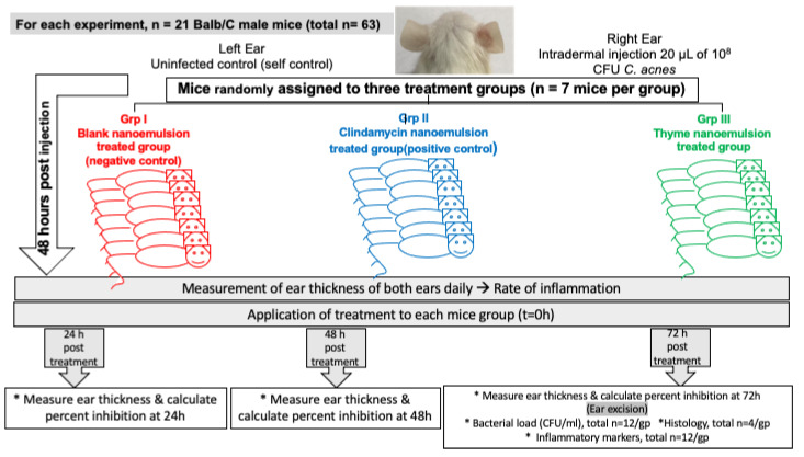
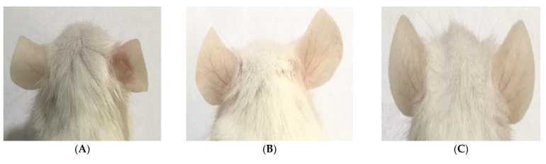
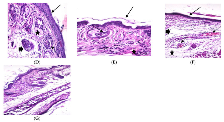
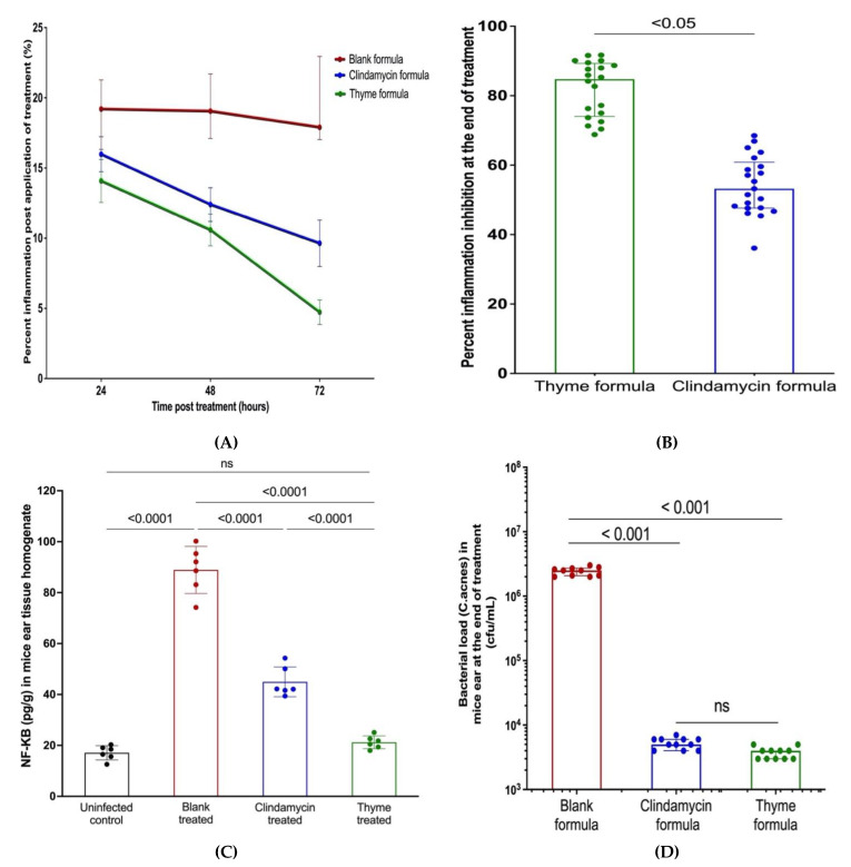
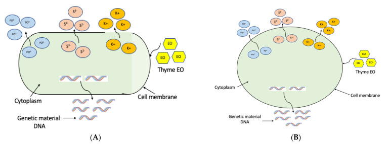
Antibiotics are frequently used in acne treatment and their prolonged use has led to an emergence of resistance. This study aimed to investigate the use of natural antimicrobials as an alternative therapy. The antimicrobial and anti-inflammatory activities of five commonly used essential oils (EOs) (tea tree, clove, thyme, mentha and basil EOs), and their possible mechanisms of action against Cutibacterium acnes and Staphylococcus epidermidis, were explored. The effect of the most potent EO on membrane permeability was elucidated and its anti-inflammatory action, when formulated as nanoemulsion, was tested in an in vivo acne model. The in vitro studies showed that thyme EO had the most potent antimicrobial and antibiofilm activity, with phenolics and terpenoids as main antimicrobial constituents of EO. Thyme EO affected cell membrane permeability of both bacterial species, evident by the detection of the leakage of intracellular ions and membrane integrity by the leakage of nucleic acids. Morphological alteration in bacterial cells was confirmed by transmission electron microscopy. Thyme EO nanoemulsion led to the suppression of an inflammatory response in acne animal models along with a bacterial load decrease and positive histopathological changes. Collectively, thyme EO nanoemulsion showed potent antimicrobial and anti-inflammatory effects compared to the reference antibiotics, suggesting its effectiveness as a natural alternative in acne treatment.

Keywords: Cutibacterium acnes; Staphylococcus epidermidis; acne vulgaris; antibiotic resistance; biofilms; inflammation; mode of action; nanoparticles; natural antimicrobials; plant extracts.


Conflict of interest statement

The authors declare no conflicts of interest.

Figures

Figure 1
Figure 1
Antibacterial activity of the screened essential oils (EOs) using the agar disc-diffusion method against (A) Cutibacterium acnes with EOs at a concentration of 50% v/v; (B) C. acnes with EOs at a concentration of 25% v/v; (C) Staphylococcus epidermidis with EOs at a concentration of 50% v/v; (D) S. epidermidis with EOs at a concentration of 25% v/v. Data are represented as means of inhibition zones (mm) ± standard deviation (SD). Controls used were clindamycin and erythromycin as positive controls, while dimethyl sulphoxide (DMSO) was used as a negative control.
Figure 2
Figure 2
Time–kill assay curves of thyme essential at 1, 2 and 4 MIC against (A) C. acnes, where it exerted its bactericidal activity after 10 h and (B) S. epidermidis, where thyme EO exerted its bactericidal effect after 6 h. All data are represented as superimposed dots. The experiment was performed in triplicate and the assay was repeated two independent times.
Figure 3
Figure 3
Transmission electron microscope (TEM) photographs of (A): C. acnes control bacteria with an intact cell wall and intracellular content, (B,C): C. acnes after treatment with MIC of thyme EO, (D): S. epidermidis control bacteria, (E,F): S. epidermidis after treatment with thyme EO. The arrows show the leakage in the cytoplasm while the thick arrowheads indicate the cellular damage, and the star shows pore formation in the cell membrane. Notice the amorphous vacuolated cells.
Figure 3
Figure 3
Transmission electron microscope (TEM) photographs of (A): C. acnes control bacteria with an intact cell wall and intracellular content, (B,C): C. acnes after treatment with MIC of thyme EO, (D): S. epidermidis control bacteria, (E,F): S. epidermidis after treatment with thyme EO. The arrows show the leakage in the cytoplasm while the thick arrowheads indicate the cellular damage, and the star shows pore formation in the cell membrane. Notice the amorphous vacuolated cells.
Figure 4
Figure 4
Effect of thyme EO on membrane integrity through the leakage of K+ ion from (A) C. acnes and (B) S. epidermidis. Thyme EO at its MBC resulted in a significant release of K+ ion compared to DMSO negative control. The means ± SDs for three replicates are illustrated; an untreated t-test was applied with p-value < 0.001.
Figure 5
Figure 5
Loss of nucleic acids. The appearance of 260 nm of absorbing material in the filtrates of (A) C. acnes and (B) S. epidermidis control suspensions (white bars) and after treatment with clindamycin (silver bars), vancomycin (grey bars) and the MICs of thyme EO (black bars) confirms the effect of thyme EO on bacterial membrane integrity. The means ± SD for at least three replicates are illustrated. A two-way ANOVA test was performed, p-value < 0.0001. ns means not statistically significant.
Figure 6
Figure 6
Extracellular concentration of phosphate (PO4−) and sulfur ions (S2−) in aliquots of C. acnes (A) and S. epidermidis (B) after treatment with DMSO (control) and thyme EO at its MBC for 6 h. Data are represented as mean ± SD of three independent experiments. A multiple unpaired t-test test was performed, p-value < 0.001.
Figure 7
Figure 7
Diagrammatic representation for the in vivo experiment.
Figure 8
Figure 8
Morphological and histopathological changes in BALB/c mice ear skins at the end of the experiment. (A) Appearance of microcomedones and inflammation in the right mouse ear in blank formula-treated mice (negative control); (B) absence of inflammatory signs in the right mouse ear treated with thyme formula; (C) absence of inflammation in the right mouse ear treated with clindamycin (positive control); (D) normal mouse ear tissue with basal layer and epidermal cell maturation was preserved; squamous epithelium (thick arrow), hair follicles (thin arrow), sebaceous glands (*), and subcutaneous tissue (arrow head) were normal, (H&E ×200); (E) mouse ear tissue treated with thyme EO nanoemulsion; normal epidermal thickening (thick arrow) with markedly reduced inflammatory cells infiltration in the dermis (*), hair follicles and sebaceous glands were normal (thin arrow), (H&E ×400); (F) mouse ear tissue treated with blank formula; epidermal hypoplasia (thick arrow) and dermal thickening with edema (arrow head), note the congested blood vessel (thin arrow), and mononuclear cells infiltrations (*), (H&E ×200) and (G) mouse ear tissue treated with 1% clindamycin formula showing skin squamous epithelial cells show proliferous thickening (arrow) and the subcutaneous tissue; thickening with edema (arrow head) and small amount of (inflammatory cells invasion (*), (H&E ×100).
Figure 8
Figure 8
Morphological and histopathological changes in BALB/c mice ear skins at the end of the experiment. (A) Appearance of microcomedones and inflammation in the right mouse ear in blank formula-treated mice (negative control); (B) absence of inflammatory signs in the right mouse ear treated with thyme formula; (C) absence of inflammation in the right mouse ear treated with clindamycin (positive control); (D) normal mouse ear tissue with basal layer and epidermal cell maturation was preserved; squamous epithelium (thick arrow), hair follicles (thin arrow), sebaceous glands (*), and subcutaneous tissue (arrow head) were normal, (H&E ×200); (E) mouse ear tissue treated with thyme EO nanoemulsion; normal epidermal thickening (thick arrow) with markedly reduced inflammatory cells infiltration in the dermis (*), hair follicles and sebaceous glands were normal (thin arrow), (H&E ×400); (F) mouse ear tissue treated with blank formula; epidermal hypoplasia (thick arrow) and dermal thickening with edema (arrow head), note the congested blood vessel (thin arrow), and mononuclear cells infiltrations (*), (H&E ×200) and (G) mouse ear tissue treated with 1% clindamycin formula showing skin squamous epithelial cells show proliferous thickening (arrow) and the subcutaneous tissue; thickening with edema (arrow head) and small amount of (inflammatory cells invasion (*), (H&E ×100).
Figure 9
Figure 9
(A) Rate of inhibition of inflammation by thyme EO nanoemulsion and controls in acne mouse model showing that the reduction in mice ear thickness post-treatment with thyme EO was superior to clindamycin as a positive control; (B) Percent of inflammation inhibition at the end of treatment showing thyme EO nanoemulsion resulting in more than 80% inhibition of inflammation at the end of the treatment period, while clindamycin resulted in a reduction in inflammation by 55%. Mann–Whitney test was performed, p-value < 0.05; (C) Thyme EO resulted in a 5-fold reduction in NF-κB levels while clindamycin caused only 2.5-fold reduction in the transcription protein. Ordinary one-way ANOVA—Tukey’s multiple comparison test was performed, p-value < 0.001; (D) In vivo antimicrobial activity of thyme EO nanoemulsion and clindamycin control exerting potent antimicrobial activity against C. acnes as the bacterial load was reduced to ~103 cfu/mL in both groups, while the bacterial load in the blank formula-treated mice group (negative control) was kept at 105 cfu/mL. Ordinary one-way ANOVA—Tukey’s multiple comparison test was performed, p-value < 0.0001. Data are represented as mean ± SD of three independent experiments.
Figure 10
Figure 10
Schematic diagram of mechanism of action of thyme EO against (A) C. acnes and (B) S. epidermidis, showing that the drug affects bacterial cell membrane, leading to a leakage of cytoplasmic components, including genetic material and intracellular ions.

References

    1. Tuchayi S., Makrantonaki E., Ganceviciene R., Feldman S.R., Zouboulis C.C. Acne vulgaris. Nat. Rev. Dis. Primers. 2015;1:15029. doi: 10.1038/nrdp.2015.29. - DOI - PubMed
    1. Dunn L.K., O’Neill J.L., Feldman S.R. Acne in adolescents: Quality of life, self-esteem, mood and psychological disorders. Dermatol. Online J. 2011;17:1. doi: 10.5070/D34HP8N68P. - DOI - PubMed
    1. Yan H.M., Zhao H.J., Guo D.Y., Zhu P.Q., Zhang C.L., Jiang W. Gut microbiota alterations in moderate to severe acne vulgaris patients. J. Dermatol. 2018;45:1166–1171. doi: 10.1111/1346-8138.14586. - DOI - PubMed
    1. Nagy I., Pivarcsi A., Kis K., Koreck A., Bodai L., McDowell A., Kemény L. Propionibacterium acnes and lipopolysaccharide induce the expression of antimicrobial peptides and proinflammatory cytokines/chemokines in human sebocytes. Microbes Infect. 2006;8:2195–2205. doi: 10.1016/j.micinf.2006.04.001. - DOI - PubMed
    1. Fournière M., Latire T., Souak D., Feuilloley M.G.J., Bedoux G. Staphylococcus epidermidis and Cutibacterium acnes: Two major sentinels of skin microbiota and the influence of cosmetics. Microorganisms. 2020;8:1752. doi: 10.3390/microorganisms8111752. - DOI - PMC - PubMed

LinkOut - more resources