

Evaluation of Novel Inhibitors of Tryptophan Dioxygenases for Enzyme and Species Selectivity Using Engineered Tumour Cell Lines Expressing Either Murine or Human IDO1 or TDO2
- PMID: 36145311
- PMCID: PMC9501369
- DOI: 10.3390/ph15091090
Evaluation of Novel Inhibitors of Tryptophan Dioxygenases for Enzyme and Species Selectivity Using Engineered Tumour Cell Lines Expressing Either Murine or Human IDO1 or TDO2
Abstract
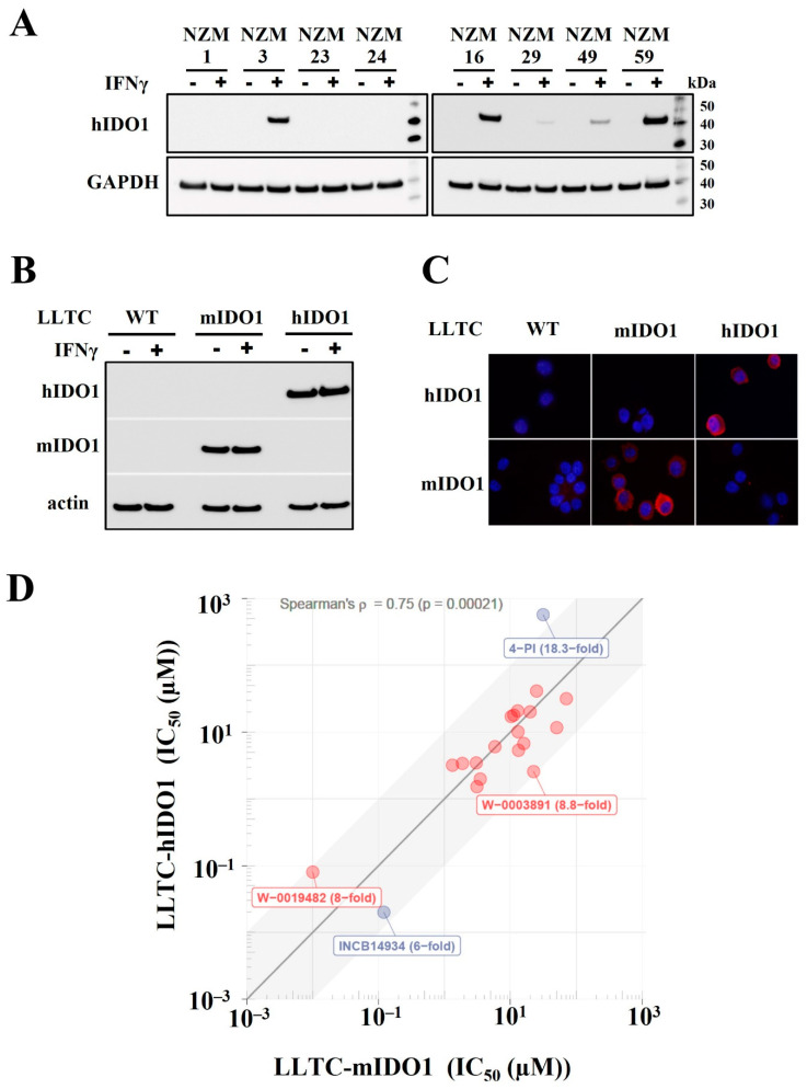
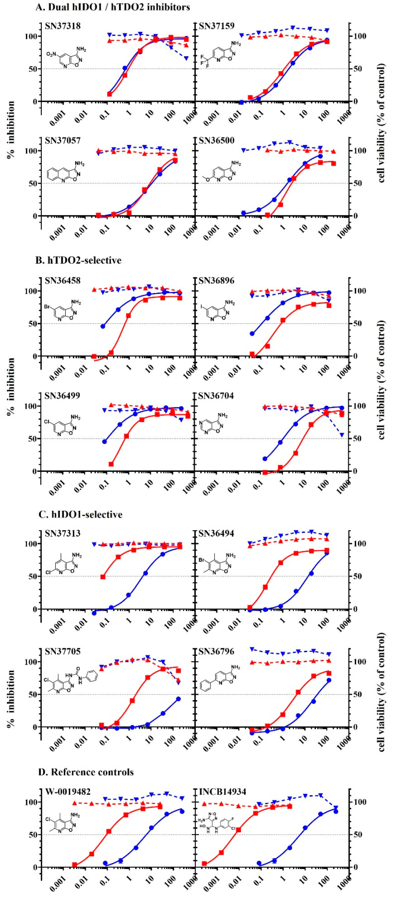
Indoleamine 2, 3-dioxygenase 1 (IDO1) is commonly expressed by cancers as a mechanism for evading the immune system. Preclinical and clinical studies have indicated the potential of combining IDO1 inhibitors with immune therapies for the treatment of cancer, strengthening an interest in the discovery of novel dioxygenase inhibitors for reversing tumour-mediated immune suppression. To facilitate the discovery, development and investigation of novel small molecule inhibitors of IDO1 and its hepatic isozyme tryptophan dioxygenase (TDO2), murine tumour cells were engineered to selectively express either murine or human IDO1 and TDO2 for use as tools to dissect both the species specificity and isoenzyme selectivity of newly discovered inhibitors. Lewis lung carcinoma (LLTC) lines were engineered to express either murine or human IDO1 for use to test species selectivity of the novel inhibitors; in addition, GL261 glioma lines were engineered to express either human IDO1 or human TDO2 and used to test the isoenzyme selectivity of individual inhibitors in cell-based assays. The 20 most potent inhibitors against recombinant human IDO1 enzyme, discovered from a commissioned screening of 40,000 compounds in the Australian WEHI compound library, returned comparable IC50 values against murine or human IDO1 in cell-based assays using the LLTC-mIDO1 and LLTC-hIDO1 line, respectively. To test the in vivo activity of the hits, transfected lines were inoculated into syngeneic C57Bl/6 mice. Individual LLTC-hIDO1 tumours showed variable expression of human IDO1 in contrast to GL261-hIDO1 tumours which were homogenous in their IDO1 expression and were subsequently used for in vivo studies. W-0019482, the most potent IDO1 inhibitor identified from cell-based assays, reduced plasma and intratumoural ratios of kynurenine to tryptophan (K:T) and delayed the growth of subcutaneous GL261-hIDO1 tumours in mice. Synthetic modification of W-0019482 generated analogues with dual IDO1/TDO2 inhibitory activity, as well as inhibitors that were selective for either TDO2 or IDO1. These results demonstrate the versatility of W-0019482 as a lead in generating all three subclasses of tryptophan dioxygenase inhibitors which can be applied for investigating the individual roles and interactions between IDO1 and TDO2 in driving cancer-mediated immune suppression.
Keywords: cancer models; immune modulation; tryptophan dioxygenase inhibitors.
Conflict of interest statement
The authors declare no conflict of interest.
Figures


Similar articles
-
Discovery and Characterisation of Dual Inhibitors of Tryptophan 2,3-Dioxygenase (TDO2) and Indoleamine 2,3-Dioxygenase 1 (IDO1) Using Virtual Screening.Molecules. 2019 Nov 28;24(23):4346. doi: 10.3390/molecules24234346. Molecules. 2019. PMID: 31795096 Free PMC article.
-
Parallel discovery of selective and dual inhibitors of tryptophan dioxygenases IDO1 and TDO2 with a newly-modified enzymatic assay.Bioorg Med Chem. 2021 Jun 1;39:116160. doi: 10.1016/j.bmc.2021.116160. Epub 2021 Apr 20. Bioorg Med Chem. 2021. PMID: 33901770
-
Preparation and evaluation of L- and D-5-[18F]fluorotryptophan as PET imaging probes for indoleamine and tryptophan 2,3-dioxygenases.Nucl Med Biol. 2017 Aug;51:10-17. doi: 10.1016/j.nucmedbio.2017.05.001. Epub 2017 May 5. Nucl Med Biol. 2017. PMID: 28511073
-
Targeting the IDO1/TDO2-KYN-AhR Pathway for Cancer Immunotherapy - Challenges and Opportunities.Trends Pharmacol Sci. 2018 Mar;39(3):307-325. doi: 10.1016/j.tips.2017.11.007. Epub 2017 Dec 15. Trends Pharmacol Sci. 2018. PMID: 29254698 Review.
-
Tryptophan: A Rheostat of Cancer Immune Escape Mediated by Immunosuppressive Enzymes IDO1 and TDO.Front Immunol. 2021 Feb 23;12:636081. doi: 10.3389/fimmu.2021.636081. eCollection 2021. Front Immunol. 2021. PMID: 33708223 Free PMC article. Review.
Cited by
-
The kynurenine pathway presents multi-faceted metabolic vulnerabilities in cancer.Front Oncol. 2023 Oct 9;13:1256769. doi: 10.3389/fonc.2023.1256769. eCollection 2023. Front Oncol. 2023. PMID: 37876966 Free PMC article. Review.
-
From Microbial Switches to Metabolic Sensors: Rewiring the Gut-Brain Kynurenine Circuit.Biomedicines. 2025 Aug 19;13(8):2020. doi: 10.3390/biomedicines13082020. Biomedicines. 2025. PMID: 40868271 Free PMC article. Review.
References
-
- Takikawa O., Kuroiwa T., Yamazaki F., Kido R. Mechanism of interferon-gamma action-characterizarion of indoleamine 2,3-dioxygenase in cultured human cells induced by interferon-gamma and evaluation of the enzyme-mediated tryptophan degradation in its anticellular activity. J. Biol. Chem. 1998;189:461–466. - PubMed
LinkOut - more resources
Full Text Sources
Other Literature Sources
Research Materials
