. 2022 Aug 26;11(9):972.
doi: 10.3390/pathogens11090972.

Microbiota Transplantation in Day-Old Broiler Chickens Ameliorates Necrotic Enteritis via Modulation of the Intestinal Microbiota and Host Immune Responses

Affiliations

Microbiota Transplantation in Day-Old Broiler Chickens Ameliorates Necrotic Enteritis via Modulation of the Intestinal Microbiota and Host Immune Responses

Sarah J M Zaytsoff et al. Pathogens. .

Abstract

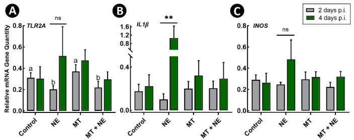
A microbiota transplant (MT) originating from mature adult chicken ceca and propagated in bioreactors was administered to day-old broiler chicks to ascertain the degree to which, and how, the MT affects Clostridium perfringens (Cp)-incited necrotic enteritis (NE). Using a stress predisposition model of NE, birds administered the MT and challenged with Cp showed fewer necrotic lesions, and exhibited a substantially higher α- and β-diversity of bacteria in their jejunum and ceca. Birds challenged with Cp and not administered the MT showed decreased Lactobacillus and increased Clostridium sensu strico 1 in the jejunum. In ceca, Megamonas, a genus containing butyrate-producing bacteria, was only present in birds administered the MT, and densities of this genus were increased in birds challenged with Cp. Metabolite profiles in cecal digesta were altered in birds administered the MT and challenged with the pathogen; 59 metabolites were differentially abundant following MT treatment, and the relative levels of short chain fatty acids, butyrate, valerate, and propionate, were decreased in birds with NE. Birds administered the MT and challenged with Cp showed evidence of enhanced restoration of intestinal barrier functions, including elevated mRNA of MUC2B, MUC13, and TJP1. Likewise, birds administered the MT exhibited higher mRNA of IL2, IL17A, and IL22 at 2-days post-inoculation with Cp, indicating that these birds were better immunologically equipped to respond to pathogen challenge. Collectively, study findings demonstrated that administering a MT containing a diverse mixture of microorganisms to day-old birds ameliorated NE in broilers by increasing bacterial diversity and promoting positive immune responses.

Keywords: Clostridium perfringens; immune responses; metabolomics; microbiota transplantation; necrotic enteritis.


Conflict of interest statement

The authors declare no conflict of interest.

Figures

Figure 1
Figure 1
Gross pathologies, histopathologic changes, and quantitative PCR for Clostridium perfringens in the jejunum. At 1-day post-hatch, birds were orally administered a microbiota transplant (MT) originating from adult broiler breeder birds and propagated within bioreactors. On days 12 and 13 post-hatch, birds were orally administered 1–2 × 10⁸ colony forming units of C. perfringens, the incitant of necrotic enteritis (NE) (i.e., NE and MT + NE treatments) or buffer alone (i.e., Control and MT treatments). (A) Longitudinally incised jejunum showing fibrin development in NE treatment (arrows) relative to a healthy intestine (i.e., Control treatment). (B) Necrotic lesion scoring. (C) Densities of the C. perfringens 16S rRNA gene in jejunum digesta as measured by qPCR. (D) Micrographs showing bacteria in association with jejunal mucosa (arrows). (E) Stacked bar plot showing total histopathologic scores by metric. Six replicate birds were analyzed per treatment. * p ≤ 0.050. ** p ≤ 0.010.
Figure 2
Figure 2
Bacterial diversity within jejunal and cecal digesta. At 1-day post-hatch, birds were orally administered a microbiota transplant (MT) originating from adult broiler breeder birds and propagated within bioreactors. On days 12 and 13 post-hatch, birds were orally administered 1–2 × 10⁸ colony forming units of Clostridium perfringens, the incitant of necrotic enteritis (NE) (i.e., NE and MT + NE treatments) or buffer alone (i.e., Control and MT treatments). (A,B) Faith’s phylogenetic diversity within the jejunum (A) and ceca (B). (C,D) Unweighted UniFrac principal coordinate analysis plot of β-diversity in the jejunum (C) and ceca (D). Bacterial community structure between birds ± MT treatment in jejunum and ceca differed (p = 0.001). Six replicate birds were analyzed per treatment.* p < 0.050 and *** p < 0.001.
Figure 3
Figure 3
Composition of the microbiota within jejunal and cecal digesta. At 1-day post-hatch, birds were orally administered a microbiota transplant (MT) originating from adult broiler breeder birds and propagated within bioreactors. On days 12 and 13 post-hatch, birds were orally administered 1–2 × 10⁸ colony forming units of Clostridium perfringens, the incitant of necrotic enteritis (NE) (i.e., NE and MT + NE treatments) or buffer alone (i.e., Control and MT treatments). (A) Percent abundance of total taxa in jejunal digesta, with taxa that comprised >1% of total abundance shown. (B) Percent abundance of total taxa in the cecal digesta, with taxa that comprised >3% of total abundance shown. (C) Percent abundance of Lactobacillus spp. and Clostridium sensu stricto 1 (including C. perfringens) in jejunal digesta. (D) Percent abundance of Bacteroides spp. and Megamonas spp. in cecal digesta. # denotes taxa delineated at the family level. Six replicate birds were analyzed per treatment. * p < 0.050, ** p < 0.010, and *** p < 0.001.
Figure 4
Figure 4
Orthogonal Projections to Latent Structures Discriminant Analysis (OPLS-DA) scores plot of metabolites within cecal digesta. At 1-day post-hatch, birds were orally administered a microbiota transplant (MT) originating from adult broiler breeder birds and propagated within bioreactors. On days 12 and 13 post-hatch, birds were orally administered 1–2 × 10⁸ colony forming units of Clostridium perfringens, the incitant of necrotic enteritis (NE) (i.e., NE and MT + NE treatments). Each triangle or square represents one bird, and data were plotted using metabolites identified to be significant by a Mann–Whitney U test and/or Variable Importance Analysis based on Variable Combination (VIAVC) machine learning. Cross-validation of the OPLS-DA model for the cecal digesta provided an excellent model fit (Q2 = 0.873, p = 0.002) and explained most of the variance (R2 = 0.994, p = 0.007), indicating that the multivariate difference observed between the NE and MT + NE treatments was real. Six replicate birds were analyzed per treatment.
Figure 5
Figure 5
Metabolite profiles and associated functional pathways within cecal digesta. At 1-day post-hatch, birds were orally administered a microbiota transplant (MT) originating from adult broiler breeder birds and propagated within bioreactors. On days 12 and 13 post-hatch, birds were orally administered 1–2 × 10⁸ colony forming units of Clostridium perfringens, the incitant of necrotic enteritis (NE) (i.e., NE and MT + NE treatments). Metabolites that were differentially abundant between NE treatment and MT + NE treatment birds are shown. Six replicate birds were analyzed per treatment.
Figure 6
Figure 6
Relative mRNA gene quantities in jejunal tissue 2- and 4-days post-inoculation of birds with Clostridium perfringens, the incitant of necrotic enteritis (NE). At 1-day post-hatch, birds were orally administered a microbiota transplant (MT) originating from adult broiler breeder birds and propagated within bioreactors. On days 12 and 13 post-hatch, birds were orally administered 1–2 × 10⁸ colony forming units of C. perfringens (i.e., NE and MT + NE treatments) or buffer alone (i.e., Control and MT treatments). (A) MUC2B. (B) MUC13. (C) CLD3. (D) TJP1. (E) CATH1. (F) AvBD6. Three replicate birds were analyzed per treatment and time. Histogram bars denoted with different letters differ (p < 0.050) among treatments for the 2-day post-inoculation (p.i.) time point. The asterisk indicates a difference between NE and non-NE treatments (* p < 0.001). # denotes a difference between the 2- and 4-day p.i. time points within the same treatment at p < 0.050, and ## denotes a difference at p < 0.010.
Figure 7
Figure 7
Relative mRNA gene quantities in jejunal tissue 2- and 4-days post-inoculation (p.i.) of birds with Clostridium perfringens, the incitant of necrotic enteritis (NE). At 1-day post-hatch, birds were orally administered a microbiota transplant (MT) originating from adult broiler breeder birds and propagated within bioreactors. On days 12 and 13 post-hatch, birds were orally administered 1–2 × 10⁸ colony forming units of C. perfringens (i.e., NE and MT + NE treatments) or buffer alone (i.e., Control and MT treatments). (A) TLR2A; (B) IL1β; and (C) INOS. Three replicate birds were analyzed per treatment and time. Histogram bars denoted with different letters differ (p < 0.050) among treatments for the 2-day p.i. time point. Double asterisks denote a difference between the 2- and 4-day p.i. time points within the same treatment (p < 0.010). ns denotes “not significant”.
Figure 8
Figure 8
Relative mRNA gene quantities in jejunal tissue 2- and 4-days post-inoculation of birds with Clostridium perfringens, the incitant of necrotic enteritis (NE). At 1-day post-hatch, birds were orally administered a microbiota transplant (MT) originating from adult broiler breeder birds and propagated within bioreactors. On days 12 and 13 post-hatch, birds were orally administered 1–2 × 10⁸ colony forming units of C. perfringens (i.e., NE and MT + NE treatments) or buffer alone (i.e., Control and MT treatments). (A) IL2. (B) IL17A. (C) IL22. Three replicate birds were analyzed per treatment and time. Histogram bars denoted with different letters differ (p < 0.050) between the two time points among the same treatment. Asterisks indicate treatments that differ within individual time points, where * is p < 0.050, and ** is p < 0.010.

Similar articles

Cited by

References

    1. Bojanova D.P., Bordenstein S.R. Fecal transplants: What is being transferred? PLoS Biol. 2016;14:e1002503. doi: 10.1371/journal.pbio.1002503. - DOI - PMC - PubMed
    1. Quraishi M.N., Shaheen W., Oo Y.H., Iqbal T.H. Immunological mechanisms underpinning faecal microbiota transplantation for the treatment of inflammatory bowel disease. Clin. Exp. Immunol. 2020;199:24–38. doi: 10.1111/cei.13397. - DOI - PMC - PubMed
    1. Niederwerder M.C. Fecal microbiota transplantation as a tool to treat and reduce susceptibility to disease in animals. Vet. Immunol. Immunopathol. 2018;206:65–72. doi: 10.1016/j.vetimm.2018.11.002. - DOI - PMC - PubMed
    1. Moore R.J. Necrotic enteritis predisposing factors in broiler chickens. Avian Pathol. 2016;45:275–281. doi: 10.1080/03079457.2016.1150587. - DOI - PubMed
    1. Dahiya J.P., Wilkie D.C., Van Kessel A.G., Drew M.D. Potential strategies for controlling necrotic enteritis in broiler chickens in post-antibiotic era. Anim. Feed Sci. Technol. 2006;129:60–88. doi: 10.1016/j.anifeedsci.2005.12.003. - DOI

LinkOut - more resources