

Nc GRA7 and Nc ROP40 Play a Role in the Virulence of Neospora caninum in a Pregnant Mouse Model
- PMID: 36145430
- PMCID: PMC9506596
- DOI: 10.3390/pathogens11090998
Nc GRA7 and Nc ROP40 Play a Role in the Virulence of Neospora caninum in a Pregnant Mouse Model
Abstract
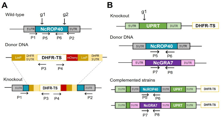
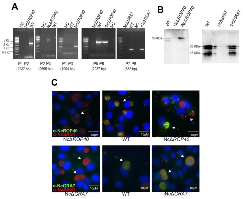
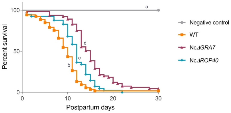
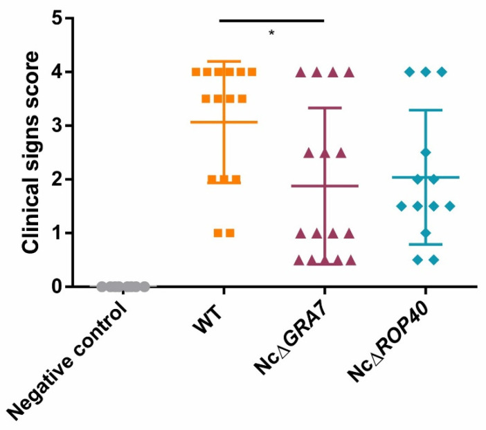
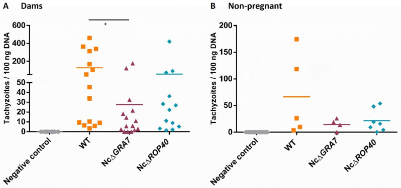
The intraspecific variability among Neospora caninum isolates in their in vitro behaviour and in vivo virulence has been widely studied. In particular, transcriptomic and proteomic analyses have shown a higher expression/abundance of specific genes/proteins in high-virulence isolates. Consequently, the dense granule protein NcGRA7 and the rhoptry protein NcROP40 were proposed as potential virulence factors. The objective of this study was to characterize the role of these proteins using CRISPR/Cas9 knockout (KO) parasites in a well-established pregnant BALB/c mouse model of N. caninum infection at midgestation. The deletion of NcGRA7 and NcROP40 was associated with a reduction of virulence, as infected dams displayed milder clinical signs, lower parasite burdens in the brain, and reduced mortality rates compared to those infected with the wild-type parasite (Nc-Spain7). Specifically, those infected with the NcGRA7 KO parasites displayed significantly milder clinical signs and a lower brain parasite burden. The median survival time of the pups from dams infected with the two KO parasites was significantly increased, but differences in neonatal mortality rates were not detected. Overall, the present study indicates that the disruption of NcGRA7 considerably impairs virulence in mice, while the impact of NcROP40 deletion was more modest. Further research is needed to understand the role of these virulence factors during N. caninum infection.
Keywords: BALB/c; CRISPR/Cas9; NcGRA7 protein; NcROP40 protein; Neospora caninum; pregnant mouse model; virulence factor.
Conflict of interest statement
The funders had no role in study design, data collection and analysis, decision to publish, or preparation of the manuscript. The authors declare that they have no conflict of interests.
Figures
Similar articles
-
Characterization of Neospora caninum virulence factors NcGRA7 and NcROP40 in bovine target cells.Vet Parasitol. 2023 Aug;320:109973. doi: 10.1016/j.vetpar.2023.109973. Epub 2023 Jun 14. Vet Parasitol. 2023. PMID: 37356132
-
Neospora caninum Dense Granule Protein 7 Regulates the Pathogenesis of Neosporosis by Modulating Host Immune Response.Appl Environ Microbiol. 2018 Aug 31;84(18):e01350-18. doi: 10.1128/AEM.01350-18. Print 2018 Sep 15. Appl Environ Microbiol. 2018. PMID: 30006392 Free PMC article.
-
A vaccine formulation combining rhoptry proteins NcROP40 and NcROP2 improves pup survival in a pregnant mouse model of neosporosis.Vet Parasitol. 2015 Jan 30;207(3-4):203-15. doi: 10.1016/j.vetpar.2014.12.009. Epub 2014 Dec 25. Vet Parasitol. 2015. PMID: 25579396
-
Role of dense granule antigen 7 in vertical transmission of Neospora caninum in C57BL/6 mice infected during early pregnancy.Parasitol Int. 2022 Aug;89:102576. doi: 10.1016/j.parint.2022.102576. Epub 2022 Mar 15. Parasitol Int. 2022. PMID: 35301119
-
Function of Neospora caninum dense granule protein 7 in innate immunity in mice.Parasitol Res. 2021 Jan;120(1):197-207. doi: 10.1007/s00436-020-06961-4. Epub 2020 Nov 8. Parasitol Res. 2021. PMID: 33164154
Cited by
-
Neospora caninum surface antigen 1 is a major determinant of the pathogenesis of neosporosis in nonpregnant and pregnant mice.Front Microbiol. 2024 Jan 8;14:1334447. doi: 10.3389/fmicb.2023.1334447. eCollection 2023. Front Microbiol. 2024. PMID: 38260884 Free PMC article.
-
Immunization with the NcMYR1 gene knockout strain effectively protected C57BL/6 mice and their pups against the Neospora caninum challenge.Virulence. 2024 Dec;15(1):2427844. doi: 10.1080/21505594.2024.2427844. Epub 2024 Nov 28. Virulence. 2024. PMID: 39607301 Free PMC article.
-
Protective efficacy of the NcGRA7-deficient parasite as a live attenuated vaccine against Neospora caninum infection in mice.J Vet Med Sci. 2025 May 1;87(5):472-480. doi: 10.1292/jvms.24-0460. Epub 2025 Mar 20. J Vet Med Sci. 2025. PMID: 40128980 Free PMC article.
-
TaqMan-quantitative PCR assays applied in Neospora caninum knock-outs generated through CRISPR-Cas9 allow to determine the copy numbers of integrated dihydrofolate reductase-thymidylate synthase drug selectable markers.Front Cell Infect Microbiol. 2024 Jun 21;14:1419209. doi: 10.3389/fcimb.2024.1419209. eCollection 2024. Front Cell Infect Microbiol. 2024. PMID: 38975328 Free PMC article.
-
Evaluation of Protective Immune Responses Induced in BALB/c Mice and Goats by the Neospora caninum Surface SRS Proteins and Interleukin-18.Animals (Basel). 2022 Oct 27;12(21):2952. doi: 10.3390/ani12212952. Animals (Basel). 2022. PMID: 36359077 Free PMC article.
References
-
- Rojo-Montejo S., Collantes-Fernández E., Regidor-Cerrillo J., Álvarez-García G., Marugan-Hernández V., Pedraza-Díaz S., Blanco-Murcia J., Prenafeta A., Ortega-Mora L.M. Isolation and characterization of a bovine isolate of Neospora caninum with low virulence. Vet. Parasitol. 2009;159:7–16. doi: 10.1016/j.vetpar.2008.10.009. - DOI - PubMed
-
- Regidor-Cerrillo J., Gámez-Bautista M., Sodupe I., Aduriz G., Álvarez-García G., Del Pozo I., Ortega-Mora L. In vitro invasion efficiency and intracellular proliferation rate comprise virulence-related phenotypic traits of Neospora caninum. Vet. Res. 2011;42:41. doi: 10.1186/1297-9716-42-41. - DOI - PMC - PubMed
-
- Dellarupe A., Regidor-Cerrillo J., Jiménez-Ruiz E., Schares G., Unzaga J.M., Venturini M.C., Ortega-Mora L.M. Comparison of host cell invasion and proliferation among Neospora caninum isolates obtained from oocysts and from clinical cases of naturally infected dogs. Exp. Parasitol. 2014;145:22–28. doi: 10.1016/j.exppara.2014.07.003. - DOI - PubMed
