. 2022 Sep 8;14(18):3754.
doi: 10.3390/polym14183754.

Matrix Regeneration Ability In Situ Induced by a Silk Fibroin Small-Caliber Artificial Blood Vessel In Vivo

Affiliations

Matrix Regeneration Ability In Situ Induced by a Silk Fibroin Small-Caliber Artificial Blood Vessel In Vivo

Helei Li et al. Polymers (Basel). .

Abstract

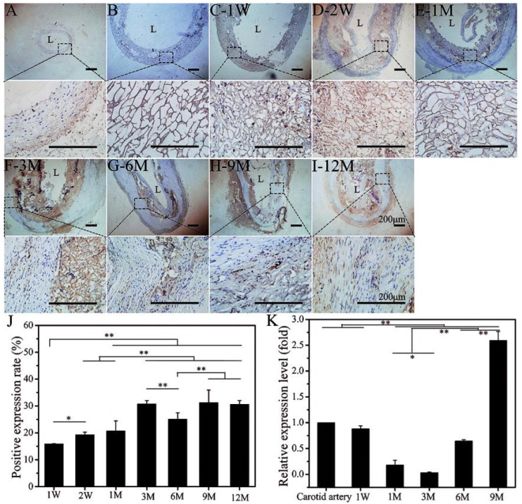
The success of a small-caliber artificial vascular graft in the host in order to obtain functional tissue regeneration and remodeling remains a great challenge in clinical application. In our previous work, a silk-based, small-caliber tubular scaffold (SFTS) showed excellent mechanical properties, long-term patency and rapid endothelialization capabilities. On this basis, the aim of the present study was to evaluate the vascular reconstruction process after implantation to replace the common carotid artery in rabbits. The new tissue on both sides of the SFTSs at 1 month was clearly observed. Inside the SFTSs, the extracellular matrix (ECM) was deposited on the pore wall at 1 month and continued to increase during the follow-up period. The self-assembled collagen fibers and elastic fibers were clearly visible in a circumferential arrangement at 6 months and were similar to autologous blood vessels. The positive expression rate of Lysyl oxidase-1 (LOXL-1) was positively correlated with the formation and maturity of collagen fibers and elastic fibers. In summary, the findings of the tissue regeneration processes indicated that the bionic SFTSs induced in situ angiogenesis in defects.

Keywords: ECM; matrix fiber self-assembly; silk fibroin; small-caliber artificial vessels.


Conflict of interest statement

The authors declare no conflict of interest.

Figures

Figure 1
Figure 1
SEM images (A) and HE staining (B) of SFTSs after implantation. (a) Autogenous carotid artery; (be) represent the different time points after implantation; (f) non-implanted SFTS. The scale bar of magnified views is 50 μm, and others are 200 μm.
Figure 2
Figure 2
Immunoenzyme staining of elastin. (A): Autogenous carotid artery; (B): non-implanted SFTS; (C–I): different time points after implantation of SFTSs; (J): semi-quantitative results of positive expression. “L” represents the lumen. The scale bar is 200 μm. “*” represents p < 0.05; “**” represents p < 0.01.
Figure 3
Figure 3
Immunoenzyme staining of COL-I. (A): Autogenous carotid artery; (B): non-implanted SFTS; (C–I): different time points after implantation of SFTSs; (J): semi-quantitative results of positive expression. “L” represents the lumen. The scale bar is 200 μm. “**” represents p < 0.01.
Figure 4
Figure 4
Immunoenzyme staining of COL-III. (A): Autogenous carotid artery; (B): non-implanted SFTS; (C–I): different time points after implantation of SFTSs; (J): semi-quantitative results of positive expression; “L” represents the lumen. The scale bar is 200 μm. “*” represents p < 0.05; “**” represents p < 0.01.
Figure 5
Figure 5
Quantitative analysis of mRNA expression level for elastin (A), COL-I (B), and COL-III (C). Data are presented as mean ± SD, n = 3. “*” represents p < 0.05; “**” represents p < 0.01.
Figure 6
Figure 6
SEM images of matrix fibers in different structural layers of SFTSs after implantation. (AD) Represent the different time points after implantation; (ac) represent the different layers of the wall. The scale bar is 30 μm.
Figure 7
Figure 7
Masson (A) and WVG staining (B) of SFTSs after implantation. (a) Autogenous carotid artery; (b) non-implanted SFTS; (ci) represent the different time points after implantation. The scale bar is 200 μm.
Figure 8
Figure 8
Immunoenzyme staining of LOXL-1. (A): Autogenous carotid artery; (B): non-implanted SFTS; (CI): different time points after implantation; (J): semi-quantitative results of positive expression rates; (K): mRNA expression level of LOXL-1. Data are presented as mean ± SD, n = 3. The scale bar is 200 μm. “*” represents p < 0.05; “**” represents p < 0.01.

References

    1. Mathers C.D., Loncar D. Projections of global mortality and burden of disease from 2002 to 2030. PLoS Med. 2006;3:e442. doi: 10.1371/journal.pmed.0030442. - DOI - PMC - PubMed
    1. Catto V., Fare S., Cattaneo I., Figliuzzi M., Alessandrino A., Freddi G., Remuzzi A., Tanzi M.C. Small diameter electrospun silk fibroin vascular grafts: Mechanical properties, in vitro biodegradability, and in vivo biocompatibility. Mater. Sci. Eng. C. 2015;54:101–111. doi: 10.1016/j.msec.2015.05.003. - DOI - PubMed
    1. Pashneh-Tala S., MacNeil S., Claeyssens F. The tissue-engineered vascular graft-past, present, and future. Tissue Eng. Part B Rev. 2015;22:68–100. doi: 10.1089/ten.teb.2015.0100. - DOI - PMC - PubMed
    1. Cabrera P.O. Esthetic root coverage in periodontics: A review. CDS Rev. 1995;88:30–34. - PubMed
    1. Cleary M.A., Geiger E., Grady C., Best C., Naito Y., Breuer C. Vascular tissue engineering: The next generation. Trends Mol. Med. 2012;18:394–404. doi: 10.1016/j.molmed.2012.04.013. - DOI - PubMed

LinkOut - more resources