. 2022 Aug 30;14(9):1919.
doi: 10.3390/v14091919.

IFIT3 and IFIT5 Play Potential Roles in Innate Immune Response of Porcine Pulmonary Microvascular Endothelial Cells to Highly Pathogenic Porcine Reproductive and Respiratory Syndrome Virus

Affiliations

IFIT3 and IFIT5 Play Potential Roles in Innate Immune Response of Porcine Pulmonary Microvascular Endothelial Cells to Highly Pathogenic Porcine Reproductive and Respiratory Syndrome Virus

Yanmei Wu et al. Viruses. .

Abstract

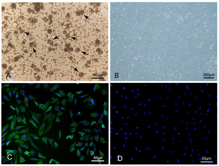
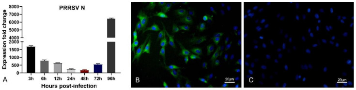
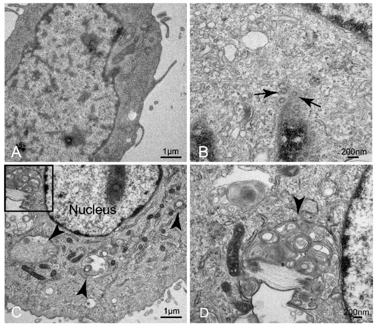
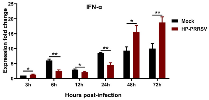
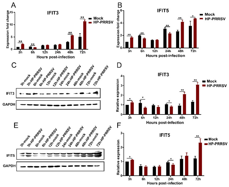
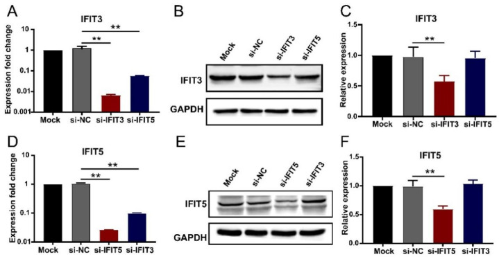
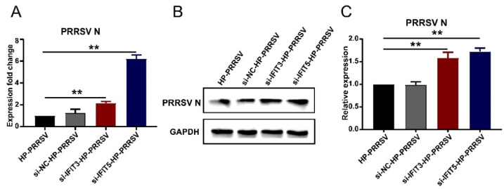
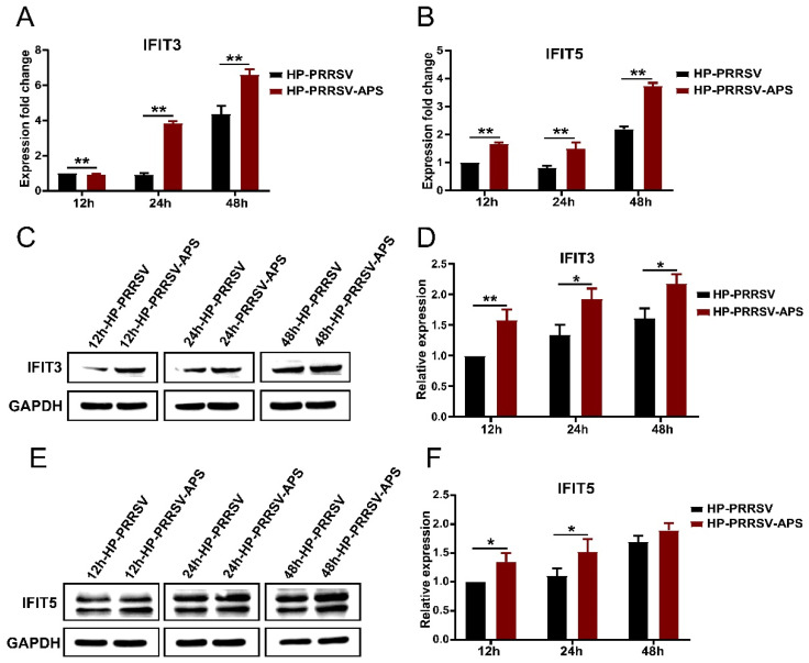
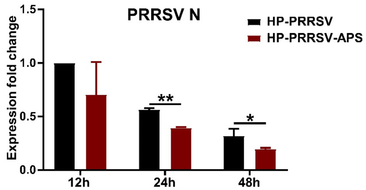
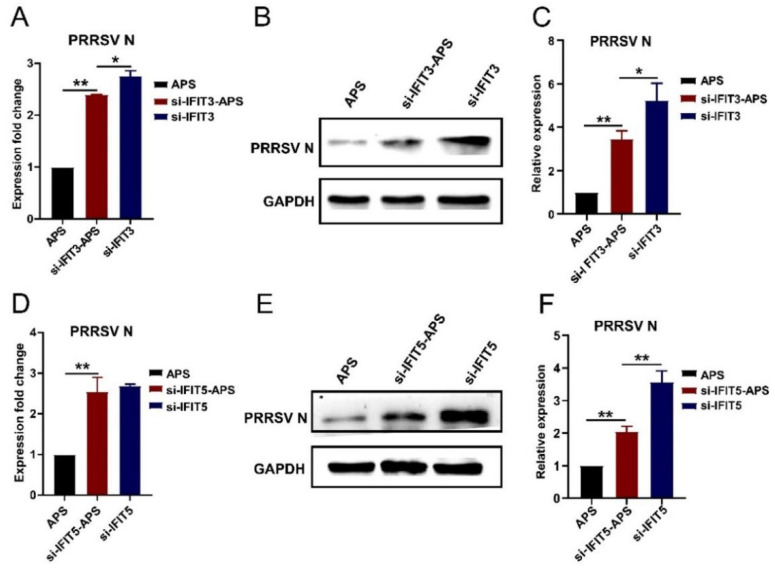
Our previous study has demonstrated that porcine pulmonary microvascular endothelial cells (MVECs) are susceptible to highly pathogenic porcine reproductive and respiratory syndrome virus (HP-PRRSV). The innate immune response of MVECs infected with HP-PRRSV would play important roles in controlling virus proliferation, resisting cellular injury, and preventing the virus from spreading to other tissues and organs. Type I interferon is one of the most effective antiviral cytokines in the innate immune response, and interferon-induced proteins with tetratricopeptide repeats (IFITs) are members of interferon-stimulated genes induced by viruses and other pathogens, which are crucial in inhibiting virus proliferation and regulating the innate immune response. However, their effects on HP-PRRSV-induced innate immunity in porcine pulmonary MVECs remain unclear. Here, the roles of IFITs in porcine pulmonary MVECs infected with the HP-PRRSV HN strain were investigated, and the effects of astragalus polysaccharides (APS), a widely used traditional Chinese herbal ingredient with the immunopotentiating effect, on them were studied. The results showed that more autophagosomes were observed in HP-PRRSV-infected MVECs, and the expression of IFN-α, IFIT3, and IFIT5 decreased or increased at different time points after infection. When silencing the genes of IFIT3 or IFIT5, the HP-PRRSV replication in MVECs was significantly increased. The expression of IFIT3 and IFIT5 could be upregulated by APS, whose inhibitory effects on the HP-PRRSV replication significantly declined when the genes of IFIT3 or IFIT5 were silenced. The results suggest that IFIT3 and IFIT5 play an important role in inhibiting the HP-PRRSV replication in porcine pulmonary MVECs, and APS suppress the multiplication of HP-PRRSV by upregulating their expression.

Keywords: HP-PRRSV; MVECs; astragalus polysaccharide; autophagosome; interferon; interferon-inducible protein.


Conflict of interest statement

The authors declare no competing interest.

Figures

Figure 1
Figure 1
Microscopic morphology and CD31 immunofluorescence staining of porcine pulmonary MVECs. (A) Porcine pulmonary MVECs conjugated with CD31 immunomagnetic beads (B) Sub-confluent MVECs. (C) Positive staining for CD31. (D) Negative control of CD31 immunofluorescence staining.
Figure 2
Figure 2
Determination of PRRSV N in porcine pulmonary MVECs. (A) The expression of PRRSV N mRNA was detected by quantitative RT-PCR from 3 hpi to 96 hpi. (B) Immunofluorescence staining of PRRSV N protein at 24 hpi. (C) Negative control to PRRSV N protein. Data are mean ± SD from three independent experiments. Statistical analysis was performed by Student’s t-test.
Figure 3
Figure 3
Ultrastructure analysis of HP-PRRSV-infected porcine pulmonary MVECs. (A) Normal control group. (BD) HP-PRRSV-infected group. (D) is the enlarged insert from (C) in the solid line rectangular frame. Black arrows indicate virus particles at 24 hpi. (B) Arrowheads indicate autophagosomes at 72 hpi.
Figure 4
Figure 4
Kinetics of the mRNA expression of IFN-α in HP-PRRSV-infected porcine pulmonary MVECs. MVECs were infected with the HP-PRRSV HN strain, and total mRNA was extracted at 3, 6, 12, 24, 48, and 72 hpi. Its gene expression was analyzed by quantitative RT-PCR. Data are mean ± SD from three independent experiments. Statistical analysis was performed by Student’s t-test. * p < 0.05; ** p < 0.01.
Figure 5
Figure 5
Effects of HP-PRRSV infection on the expression of IFIT3 and IFIT5 in porcine pulmonary MVECs. Porcine pulmonary MVECs were infected with the HP-PRRSV HN strain, and total cellular RNA or protein was extracted at 3, 6, 12, 24, 48, and 72 hpi. (A,B) The expression of IFIT3 and IFIT5 was detected by quantitative RT-PCR. (C,E) The expression of IFIT3 and IFIT5 protein was detected by Western blotting. (D,F) The gray values of the IFIT3 and IFIT5 protein bands were analyzed by ImageJ software and normalized to that of the control groups at 3 hpi. Data are mean ± SD from three independent experiments. Statistical analysis was performed by Student’s t-test. * p < 0.05; ** p < 0.01.
Figure 6
Figure 6
Effects of siRNA transfection on the expression of IFIT3 and IFIT5 in porcine pulmonary MVECs. Porcine pulmonary MVECs were transfected with siRNA of Negative Control (NC), IFIT3, or IFIT5 for 36 h. (A,D) The total cellular RNA was extracted for detection of the IFIT3 and IFIT5 expression by quantitative RT-PCR. (B,E) The total cellular protein was extracted for detection of IFIT3 and IFIT5 protein by Western blotting. (C,F) The gray values of the IFIT3 and IFIT5 protein bands were analyzed by ImageJ software and normalized to that of the negative control group. Data are mean ± SD from three independent experiments. Statistical analysis was performed by Student’s t-test. ** p < 0.01.
Figure 7
Figure 7
Effects of silencing IFI3 and IFIT5 expression on the HP-PRRSV multiplication. Porcine pulmonary MVECs were transfected with siRNA of IFIT3 and IFIT5 or Negative Control (NC) for 36 h and then infected with the HP-PRRSV HN strain for 24 h. (A) The expression of PRRSV N mRNA was detected by quantitative RT-PCR, (B)and its protein was detected by Western blotting. (C) The gray values of the PRRSV N protein bands were analyzed by ImageJ software and normalized to the control group. Data are mean ± SD from three independent experiments. Statistical analysis was performed by Student’s t-test. ** p < 0.01.
Figure 8
Figure 8
Effects of APS on the expression of IFIT3 and IFIT5 in HP-PRRSV-infected MVECs. Porcine pulmonary MVECs were infected with the HP-PRRSV HN strain for 1 h and then incubated in the maintenance medium containing 100 μg/mL APS. (A,B) The total cellular RNA was extracted at 12, 24, and 48 hpi for the detection of IFIT3 and IFIT5 by quantitative RT-PCR. (C,E) The total cellular protein was extracted at 12, 24, and 48 hpi for the detection of IFIT3 and IFIT5 by Western blotting. (D,F) The gray values of the IFIT3 and IFIT5 protein bands were analyzed by ImageJ software and normalized to the 12h virus control group. Data are mean ± SD from three independent experiments. Statistical analysis was performed by Student’s t-test. * p < 0.05; ** p < 0.01.
Figure 9
Figure 9
Effects of APS on HP-PRRSV multiplication in porcine pulmonary MVECs. Porcine pulmonary MVECs were infected with the HP-PRRSV HN strain and then incubated in the maintenance medium containing 100 μg/mL APS. The total cellular RNA was extracted at 12, 24, and 48 hpi for the detection of PRRSV N mRNA by quantitative RT-PCR. Data are mean ± SD from three independent experiments. Statistical analysis was performed by Student’s t-test. * p < 0.05; ** p < 0.01.
Figure 10
Figure 10
Effects of silencing IFIT3 and IFIT5 on the anti-HP-PRRSV action of APS. Porcine pulmonary MVECs were infected with the HP-PRRSV HN strain and incubated in the maintenance medium with 100 μg/mL APS for 24 h after silencing the IFIT3 or IFIT5 gene. (A,D) The total cellular RNA was extracted for the detection of PRRSV N mRNA by quantitative RT-PCR. (B,E) The total cellular protein was extracted for the detection of PRRSV N protein by Western blotting. (C,F) The gray values of the PRRSV N protein bands were analyzed by ImageJ software and normalized to the APS group. Data are mean ± SD from three independent experiments. Statistical analysis was performed by Student’s t-test. * p < 0.05; ** p < 0.01.

Similar articles

Cited by

References

    1. Han J., Zhou L., Ge X., Guo X., Yang H. Pathogenesis and control of the Chinese highly pathogenic porcine reproductive and respiratory syndrome virus. Vet. Microbiol. 2017;209:30–47. doi: 10.1016/j.vetmic.2017.02.020. - DOI - PubMed
    1. Li P., Yang Z., Ma S., Hu G., Dong H., Zhang T. Susceptibility of porcine pulmonary microvascular endothelial cells to porcine reproductive and respiratory syndrome virus. J. Vet. Med. Sci. 2020;82:1404–1409. doi: 10.1292/jvms.20-0324. - DOI - PMC - PubMed
    1. Shao Y., Saredy J., Yang W.Y., Sun Y., Lu Y., Saaoud F., Drummer C., IV, Johnson C., Xu K., Jiang X., et al. Vascular endothelial cells and innate immunity. Arterioscler. Thromb. Vasc. Biol. 2020;40:e138–e152. doi: 10.1161/ATVBAHA.120.314330. - DOI - PMC - PubMed
    1. Vladimer G.I., Górna M.W., Superti-Furga G. IFITs: Emerging roles as key anti-viral proteins. Front. Immunol. 2014;5:94. doi: 10.3389/fimmu.2014.00094. - DOI - PMC - PubMed
    1. Hui X., Zhang L., Cao L., Huang K., Zhao Y., Zhang Y., Chen X., Lin X., Chen M., Jin M. SARS-CoV-2 promote autophagy to suppress type I interferon response. Signal Transduct. Target. Ther. 2021;6:180. doi: 10.1038/s41392-021-00574-8. - DOI - PMC - PubMed

Publication types