

A Potential Role for Substance P in West Nile Virus Neuropathogenesis
- PMID: 36146768
- PMCID: PMC9503494
- DOI: 10.3390/v14091961
A Potential Role for Substance P in West Nile Virus Neuropathogenesis
Abstract
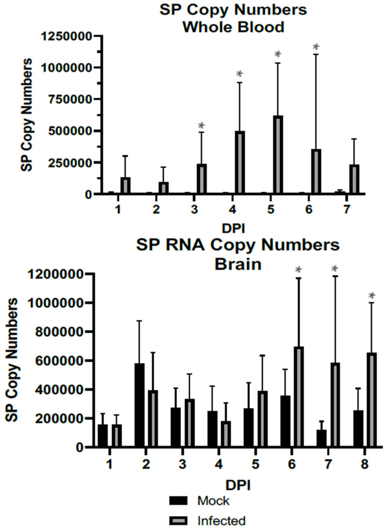
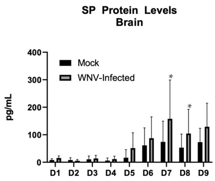
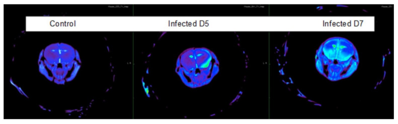
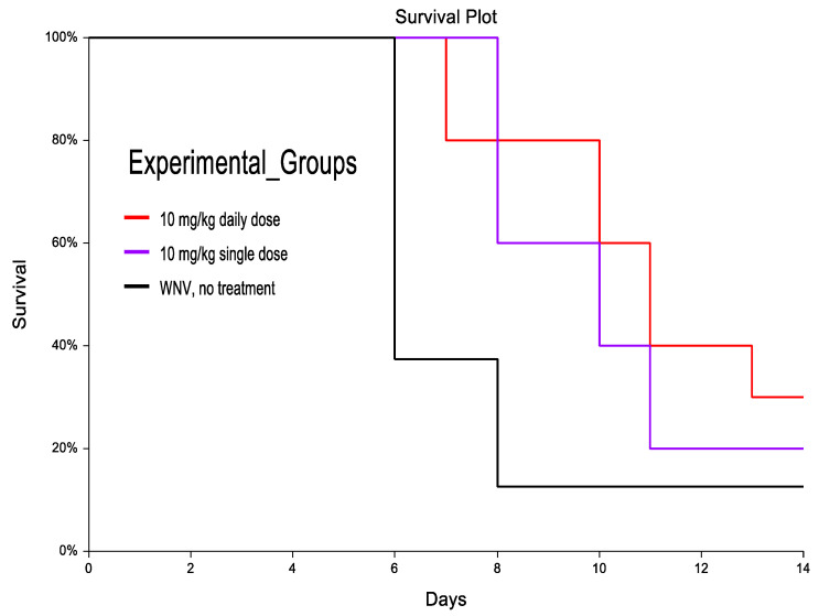
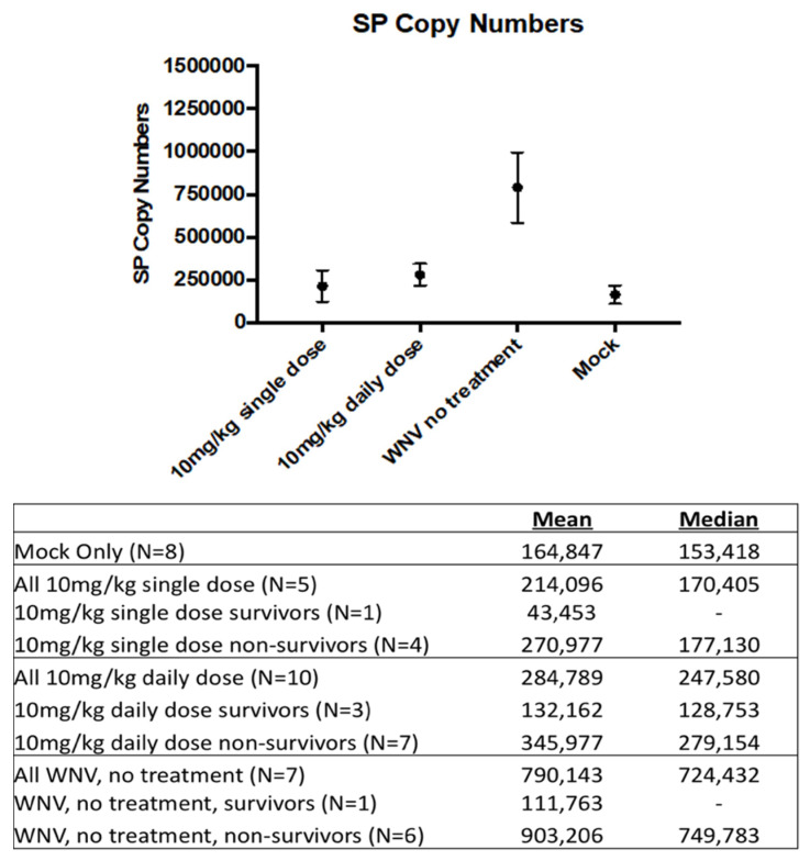
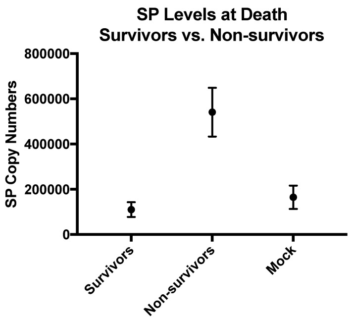
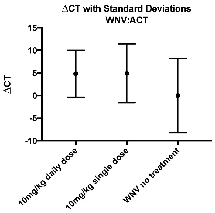
Of individuals who develop West Nile neuroinvasive disease (WNND), ~10% will die and >40% will develop long-term complications. Current treatment recommendations solely focus on supportive care; therefore, we urgently need to identify novel and effective therapeutic options. We observed a correlation between substance P (SP), a key player in neuroinflammation, and its receptor Neurokinin-1 (NK1R). Our study in a wild-type BL6 mouse model found that SP is upregulated in the brain during infection, which correlated with neuroinvasion and damage to the blood−brain barrier. Blocking the SP/NK1R interaction beginning at disease onset modestly improved survival and prolonged time to death in a small pilot study. Although SP is significantly increased in the brain of untreated WNND mice when compared to mock-infected animals, levels of WNV are unchanged, indicating that SP likely does not play a role in viral replication but may mediate the immune response to infection. Additional studies are necessary to define if SP plays a mechanistic role or if it represents other mechanistic pathways.
Keywords: West Nile encephalitis; West Nile virus; neuroinflammation; neurokinin-1 receptor; substance P.
Conflict of interest statement
A.N. declares that his partner holds shares in CH Biotech Pty Ltd., the company that licensed CH123001 for developing a treatment for cerebral metastases, and that A.N. has acted as an unpaid scientific advisor for that program. All other authors declare no conflict of interest.
Figures
Similar articles
-
Impaired virus clearance, compromised immune response and increased mortality in type 2 diabetic mice infected with West Nile virus.PLoS One. 2012;7(8):e44682. doi: 10.1371/journal.pone.0044682. Epub 2012 Aug 31. PLoS One. 2012. PMID: 22953001 Free PMC article.
-
Comorbid conditions as risk factors for West Nile neuroinvasive disease in Ontario, Canada: a population-based cohort study.Epidemiol Infect. 2022 May 11;150:e103. doi: 10.1017/S0950268822000887. Epidemiol Infect. 2022. PMID: 35543409 Free PMC article.
-
Examination of West Nile Virus Neuroinvasion and Neuropathogenesis in the Central Nervous System of a Murine Model.Methods Mol Biol. 2016;1435:83-101. doi: 10.1007/978-1-4939-3670-0_8. Methods Mol Biol. 2016. PMID: 27188552
-
West Nile virus neuroinvasive disease.Ann Neurol. 2006 Sep;60(3):286-300. doi: 10.1002/ana.20959. Ann Neurol. 2006. PMID: 16983682 Review.
-
Interleukins, Chemokines, and Tumor Necrosis Factor Superfamily Ligands in the Pathogenesis of West Nile Virus Infection.Viruses. 2023 Mar 22;15(3):806. doi: 10.3390/v15030806. Viruses. 2023. PMID: 36992514 Free PMC article. Review.
References
-
- Mostashari F., Bunning M.L., Kitsutani P.T., Singer D.A., Nash D., Cooper M.J., Katz N., Liljebjelke K.A., Biggerstaff B.J., Fine A.D., et al. Epidemic West Nile encephalitis, New York, 1999: Results of a household-based seroepidemiological survey. Lancet. 2001;358:261–264. doi: 10.1016/S0140-6736(01)05480-0. - DOI - PubMed
-
- Carson P.J., Borchardt S.M., Custer B., Prince H.E., Dunn-Williams J., Winkelman V., Tobler L., Biggerstaff B.J., Lanciotti R., Petersen L.R., et al. Neuroinvasive disease and West Nile virus infection, North Dakota, USA, 1999–2008. Emerg. Infect. Dis. 2012;18:684–686. doi: 10.3201/eid1804.111313. - DOI - PMC - PubMed
Publication types
MeSH terms
Substances
LinkOut - more resources
Full Text Sources
Medical
