. 2022 Sep 4;14(9):1966.
doi: 10.3390/v14091966.

SARS-CoV-2 Spike and Nucleocapsid Antibody Response in Vaccinated Croatian Healthcare Workers and Infected Hospitalized Patients: A Single Center Cohort Study

Affiliations

SARS-CoV-2 Spike and Nucleocapsid Antibody Response in Vaccinated Croatian Healthcare Workers and Infected Hospitalized Patients: A Single Center Cohort Study

Paola Kučan Brlić et al. Viruses. .

Abstract

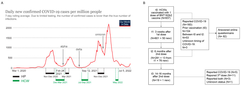
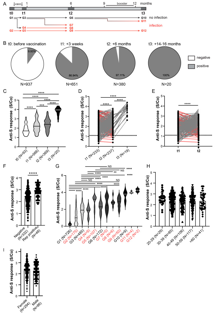
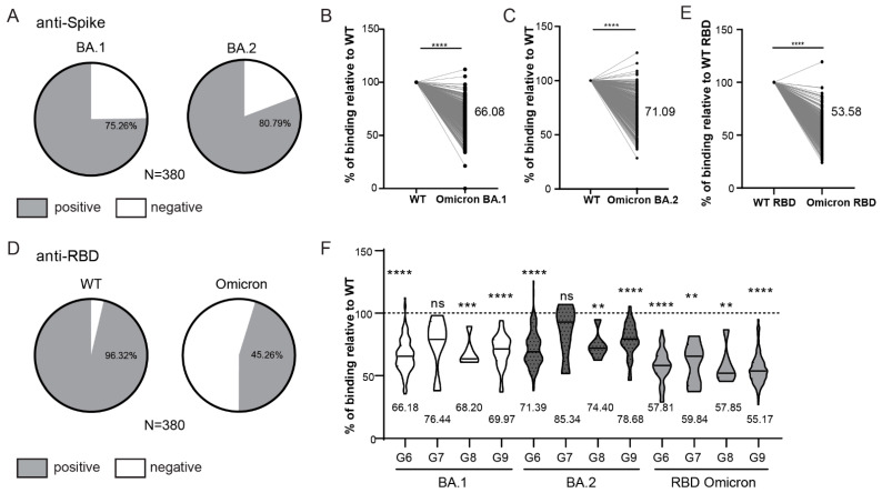
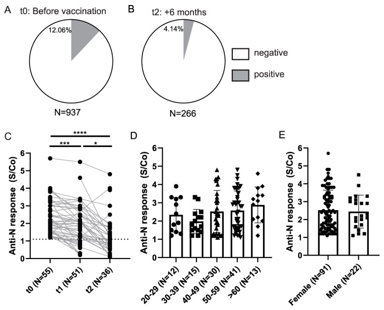
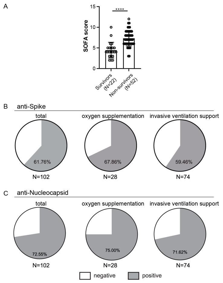
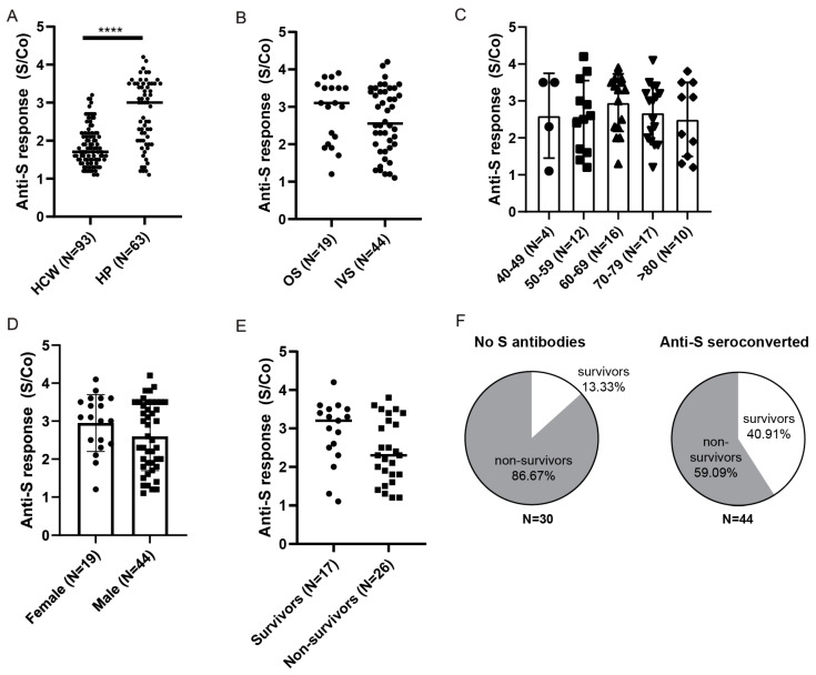
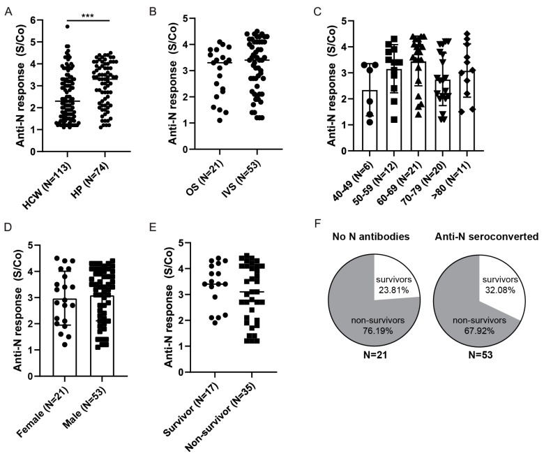
Studies assessing the dynamics and duration of antibody responses following SARS-CoV-2 infection or vaccination are an invaluable tool for vaccination schedule planning, assessment of risk groups and management of pandemics. In this study, we developed and employed ELISA assays to analyze the humoral responses to Nucleocapsid and Spike proteins in vaccinated health-care workers (HCW) and critically ill COVID-19 patients. Sera of more than 1000 HCWs and critically ill patients from the Clinical Hospital Center Rijeka were tested across a one-year period, encompassing the spread of major SARS-CoV-2 variants of concern (VOCs). We observed 97% of seroconversion in HCW cohort as well as sustained anti-Spike antibody response in vaccinees for more than 6 months. In contrast, the infection-induced anti-Nucleocapsid response was waning significantly in a six-month period. Furthermore, a substantial decrease in vaccinees' anti-Spike antibodies binding to Spike protein of Omicron VOC was also observed. Critically ill COVID-19 patients had higher levels of anti-Spike and anti-Nucleocapsid antibodies compared to HCWs. No significant differences in anti-Spike and anti-Nucleocapsid antibody levels between the critically ill COVID-19 patients that were on non-invasive oxygen supplementation and those on invasive ventilation support were observed. However, stronger anti-Spike, but not anti-Nucleocapsid, antibody response correlated with a better disease outcome in the cohort of patients on invasive ventilation support. Altogether, our results contribute to the growing pool of data on humoral responses to SARS-CoV-2 infection and vaccination.

Keywords: BNT162 vaccine; COVID-19; COVID-19 vaccine; Nucleocapsid; Omicron; SARS-CoV-2; Spike; antibody detection; healthcare workers; hospitalized patients.


Conflict of interest statement

The authors declare no conflict of interest.

Figures

Figure 1
Figure 1
Timeline of serum samples collection and flowchart of HCW cohort enrolment. (A) Intervals of sera collection for hospitalized patients (HP) and healthcare workers (HCW) in relation to COVID-19 pandemic waves in Croatia. (B) Flowchart of HCWs enrolment, follow-up and COVID-19 status. Image in (A) adjusted from Our World in Data.
Figure 2
Figure 2
Anti-Spike antibody response in HCW cohort. (A) Schematic of serum collection and groups analyzed in (G) in relation to timing of SARS-CoV-2 infection. (B) Anti-Spike (anti-S) seroconversion rates (above cut-off value) in HCWs at different sera collection time points. (C) Comparison of the anti-Spike antibody response between entire cohort of seroconverted individuals (S/Co > 1.1). (D) Comparison of the anti-Spike antibody response between all paired samples or (E) paired samples that were not Spike seroconverted prior to vaccination or Nucleocapsid protein seroconverted at any time point. Red lines samples displaying reduced anti-S response between t1 and t2 analyses. (F) Comparison of anti-Spike antibody S/Co 6-months post vaccination (t2) stratified to participants’ self-reported PCR confirmed COVID-19 infection. (G) Comparison of the anti-Spike antibody response in relation to timing of infection assessed by Nucleocapisd seroconversion and self-reported, PCR confirmed COVID-19 infection. (H) Comparison of anti-Spike antibody S/Co 6 months post vaccination (t2) stratified to participants’ age or (I) gender. S/Co, Signal to cut-off ratio; dashed line indicates cut-off value (S/Co = 1.1); red lines in D and E show samples with reduction of anti-Spike S/Co; red lines and groups in A and G indicate samples with COVID-19 infection. Statistical tests: ordinary one-way ANOVA followed by Tukey’s multiple comparison test (C,G), mixed effect analysis with Holm-Sidak’s multiple comparisons test (D), paired two-tailed t-test (E,H), p value **** p < 0.0001, p value *** and ** p < 0.05, NS-not significant.
Figure 3
Figure 3
Reactivity of vaccinated HCW’s sera to Omicron Spike variants. (A) Rates of 6-month vaccinees (t2) samples reactive to Omicron BA.1 and BA.2 Spike protein. (B) Comparison of the intensity of t2 serum binding to Omicron BA.1 and (C) BA.2 relative to binding to WT Spike protein. (D) Rates of t2 samples reactive to WT RBD and Omicron RBD protein. (E) Comparison of the intensity of t2 serum binding to Omicron RBD protein relative to binding to WT RBD protein. (F) Comparison of the relative intensity of t2 serum binding to Omicron proteins in relation to the timing of infection (see Figure 2A). S/Co, Signal to cut-off ratio. Statistical tests: paired t-test (B,C,E), p value **** p < 0.0001, p value *** and ** p < 0.05, ns-not significant.
Figure 4
Figure 4
Anti-Nucleocapsid antibody response in HCWs. (A) Anti-Nucleocapsid (anti-N) seroconversion rates in HCWs prior to vaccination and (B) 6 months after the full vaccination (t2) (S-positive individuals at t0 and N-positive at t0 and t1 were excluded). (C) Comparison of the anti-Nucleocapsid antibody S/Co between paired seroconverted samples. (D) Levels of anti-Nucleocapsid antibodies in sera of seroconverted individuals at t0 stratified on the basis of age and (E) gender. S/Co, Signal to cut-off ratio; dashed line indicates cut-off value (S/Co = 1.1). Statistical test: Mixed-effects analysis with Tukey’s multiple comparisons test (C), p value **** p < 0.0001, p value *** and * p<0.05.
Figure 5
Figure 5
Outcome in relation to SOFA scores and rates of seroconversion in hospitalized patients. (A) Comparison of the SOFA scores between survivors and non-survivors from the invasive ventilation support (IVS) cohort. (B) Presence of anti-Spike or (C) anti-Nucleocapsid antibodies above cut-off value, **** p < 0.0001.
Figure 6
Figure 6
Anti-Spike antibody response in HPs. (A) Comparison of anti-Spike antibody levels between in hospitalized patients (HP) and healthcare workers (HCW) identified prior to vaccination. (B) Hospitalized seroconverted patients were stratified based on the severity of the disease: oxygen supplementation (OS) vs. invasive ventilation support (IVS), (C) age, (D) gender, or (E) mortality outcome for non-vaccinated patients on invasive ventilation support. (F) Frequencies of survivors and non-survivors in relation to anti-Spike seroconversion in IVS cohort. S/Co, Signal to cut-off ratio. Statistical tests: unpaired t-test (A), p value **** p < 0.0001.
Figure 7
Figure 7
Anti-Nucleocapsid antibody response in HPs. (A) Comparison of anti-Nucleocapsid antibody levels between hospitalized patients (HP) and healthcare workers (HCWs) identified prior to vaccination. (B) Hospitalized patients were stratified based on the severity of the disease: oxygen supplementation (OS) vs. invasive ventilation support (IVS), (C) age, (D) gender, or (E) outcome for patients on invasive ventilation support. (F) Frequencies of survivors and non-survivors in relation to anti-Nucleocapsid seroconversion. Statistical tests: unpaired t-test (A), p value *** p < 0.0005. S/Co, Signal to cut-off ratio.

Similar articles

Cited by

References

    1. Baden L.R., El Sahly H.M., Essink B., Kotloff K., Frey S., Novak R., Diemert D., Spector S.A., Rouphael N., Creech C.B., et al. Efficacy and Safety of the mRNA-1273 SARS-CoV-2 Vaccine. N. Engl. J. Med. 2021;384:403–416. doi: 10.1056/NEJMoa2035389. - DOI - PMC - PubMed
    1. Polack F.P., Thomas S.J., Kitchin N., Absalon J., Gurtman A., Lockhart S., Perez J.L., Perez Marc G., Moreira E.D., Zerbini C., et al. Safety and Efficacy of the BNT162b2 mRNA COVID-19 Vaccine. N. Engl. J. Med. 2020;383:2603–2615. doi: 10.1056/NEJMoa2034577. - DOI - PMC - PubMed
    1. Hannah Ritchie E.M., Rodés-Guirao L., Appel C., Giattino C., Ortiz-Ospina E., Hasell J., Macdonald B., Beltekian D., Roser M. Coronavirus Pandemic (COVID-19) [(accessed on 20 June 2022)]. Available online: https://ourworldindata.org/coronavirus.
    1. Liu L., Iketani S., Guo Y., Chan J.F., Wang M., Liu L., Luo Y., Chu H., Huang Y., Nair M.S., et al. Striking antibody evasion manifested by the Omicron variant of SARS-CoV-2. Nature. 2022;602:676–681. doi: 10.1038/s41586-021-04388-0. - DOI - PubMed
    1. Odak I., Schultze-Florey C.R., Hammerschmidt S.I., Ritter C., Willenzon S., Friedrichsen M., Ravens I., Sikora R., Bayir L.M., Gutierrez Jauregui R., et al. Longitudinal Tracking of Immune Responses in COVID-19 Convalescents Reveals Absence of Neutralization Activity against Omicron and Staggered Impairment to Other SARS-CoV-2 Variants of Concern. Front. Immunol. 2022;13:863039. doi: 10.3389/fimmu.2022.863039. - DOI - PMC - PubMed

Publication types

Supplementary concepts