

A Naked-Eye Visual Reverse Transcription Loop-Mediated Isothermal Amplification with Sharp Color Changes for Potential Pen-Side Test of Foot-and-Mouth Disease Virus
- PMID: 36146788
- PMCID: PMC9504329
- DOI: 10.3390/v14091982
A Naked-Eye Visual Reverse Transcription Loop-Mediated Isothermal Amplification with Sharp Color Changes for Potential Pen-Side Test of Foot-and-Mouth Disease Virus
Abstract
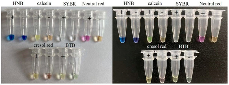
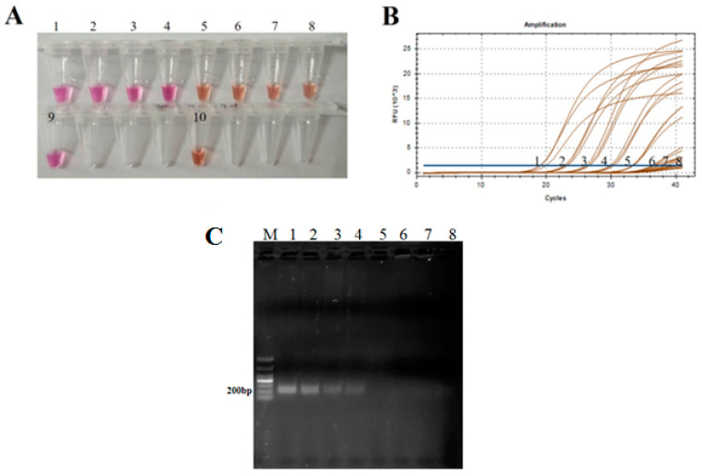
Visual loop-mediated isothermal amplification (LAMP) is qualified to be applied in the field to detect pathogens due to its simplicity, rapidity and cost saving. However, the color changes in currently reported visual reverse transcription LAMP (RT-LAMP) for foot-and-mouth disease virus (FMDV) detection are not so obvious to the naked eye, so interpretation of results is troublesome. In this study, a new naked-eye visual RT-LAMP to detect all seven distinct serotypes of FMDV was established based on the 3D genes by using pH-sensitive neutral red as the indicator, rendering a sharp contrast of color changes between the negative (light orange) and the positive (pink). Analytical sensitivity tests showed that the detection limit of the visual RT-LAMP was 104 copies/µL while those were 103 and 104 copies/µL for the RT-qPCR and conventional RT-PCR methods, respectively. Specificity tests proved that the established visual RT-LAMP assay had no cross-reactivity with other common livestock viruses. Furthermore, the analysis of 59 clinical samples showed 98.31% and 100% concordance with the RT-qPCR and the RT-PCR, respectively. The pan-serotypic FMD visual RT-LAMP assay could be suitable for a pen-side test of all seven serotypes of FMDV because the results could be easily distinguished by the naked eye without the requirement of complicated instruments and professional technicians. Hence, the novel method may have a promising prospect in field tests which exert an important role in monitoring, preventing, and controlling FMD, especially in regions with no PCR or qPCR instrument available.
Keywords: 3D gene; foot-and-mouth disease virus; loop-mediated isothermal amplification; naked-eye visualization; pen-side test.
Conflict of interest statement
The authors declare no conflict of interest.
Figures
Similar articles
-
Pan-serotype reverse transcription loop-mediated isothermal amplification (RT-LAMP) assay targeting 2B-NSP coding region for colorimetric detection of foot-and-mouth disease virus in clinical samples.Virus Genes. 2025 Aug;61(4):490-497. doi: 10.1007/s11262-025-02158-y. Epub 2025 Apr 26. Virus Genes. 2025. PMID: 40285984
-
A tailored reverse transcription loop-mediated isothermal amplification for sensitive and specific detection of serotype A foot-and-mouth disease virus circulating in pool 1 region countries.Transbound Emerg Dis. 2018 Dec;65(6):1898-1908. doi: 10.1111/tbed.12971. Epub 2018 Jul 27. Transbound Emerg Dis. 2018. PMID: 30054975
-
Defining the relative performance of isothermal assays that can be used for rapid and sensitive detection of foot-and-mouth disease virus.J Virol Methods. 2017 Nov;249:102-110. doi: 10.1016/j.jviromet.2017.08.013. Epub 2017 Aug 31. J Virol Methods. 2017. PMID: 28837842 Free PMC article.
-
Advances in the Diagnosis of Foot-and-Mouth Disease.Front Vet Sci. 2020 Aug 21;7:477. doi: 10.3389/fvets.2020.00477. eCollection 2020. Front Vet Sci. 2020. PMID: 32974392 Free PMC article. Review.
-
Visualization methods for loop mediated isothermal amplification (LAMP) assays.Analyst. 2025 Feb 10;150(4):588-599. doi: 10.1039/d4an01287a. Analyst. 2025. PMID: 39895350 Review.
Cited by
-
Designing one-step reverse transcriptase loop-mediated isothermal amplification for serotype O foot-and-mouth disease virus detection during the 2022 outbreak in East Java, Indonesia.Vet World. 2023 Sep;16(9):1889-1896. doi: 10.14202/vetworld.2023.1889-1896. Epub 2023 Sep 17. Vet World. 2023. PMID: 37859973 Free PMC article.
-
A low-cost, portable, dual-function readout device for amplification-based point-of-need diagnostics.Appl Environ Microbiol. 2023 Dec 21;89(12):e0090223. doi: 10.1128/aem.00902-23. Epub 2023 Dec 4. Appl Environ Microbiol. 2023. PMID: 38047632 Free PMC article.
-
Moving towards on-site detection of Shiga toxin-producing Escherichia coli in ready-to-eat leafy greens.Curr Res Food Sci. 2024 Mar 7;8:100716. doi: 10.1016/j.crfs.2024.100716. eCollection 2024. Curr Res Food Sci. 2024. PMID: 38511154 Free PMC article.
-
Detection of foot-and-mouth disease virus RNA using a closed loop-mediated isothermal amplification system.Front Microbiol. 2024 Jul 31;15:1429288. doi: 10.3389/fmicb.2024.1429288. eCollection 2024. Front Microbiol. 2024. PMID: 39188314 Free PMC article.
-
Pan-serotype reverse transcription loop-mediated isothermal amplification (RT-LAMP) assay targeting 2B-NSP coding region for colorimetric detection of foot-and-mouth disease virus in clinical samples.Virus Genes. 2025 Aug;61(4):490-497. doi: 10.1007/s11262-025-02158-y. Epub 2025 Apr 26. Virus Genes. 2025. PMID: 40285984
References
-
- Paton D.J., Di Nardo A., Knowles N.J., Wadsworth J., Pituco E.M., Cosivi O., Rivera A.M., Kassimi L.B., Brocchi E., de Clercq K., et al. The history of foot-and-mouth disease virus serotype C: The first known extinct serotype. Virus Evol. 2021;7:veab009. doi: 10.1093/ve/veab009. - DOI - PMC - PubMed
Publication types
MeSH terms
Substances
Supplementary concepts
LinkOut - more resources
Full Text Sources
