

SARS-CoV-2 Production, Purification Methods and UV Inactivation for Proteomics and Structural Studies
- PMID: 36146795
- PMCID: PMC9505060
- DOI: 10.3390/v14091989
SARS-CoV-2 Production, Purification Methods and UV Inactivation for Proteomics and Structural Studies
Abstract
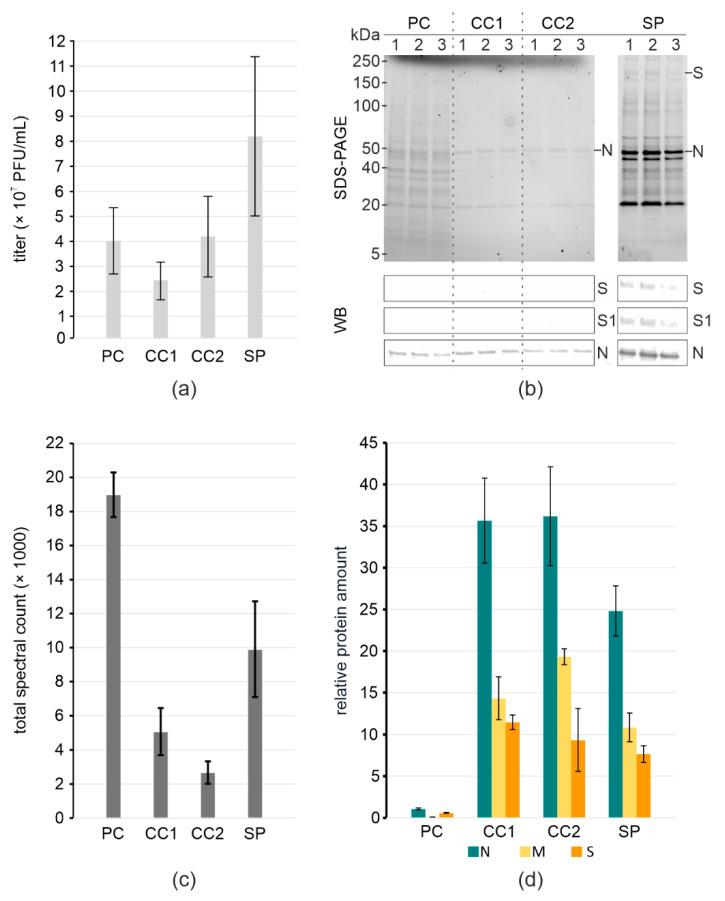
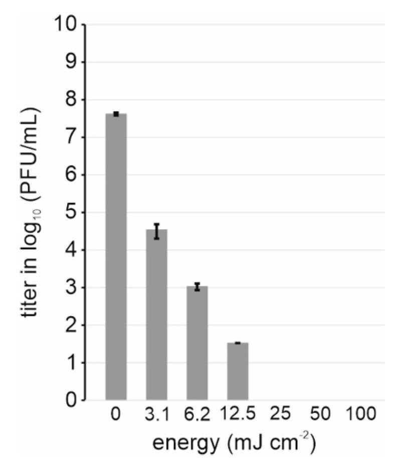
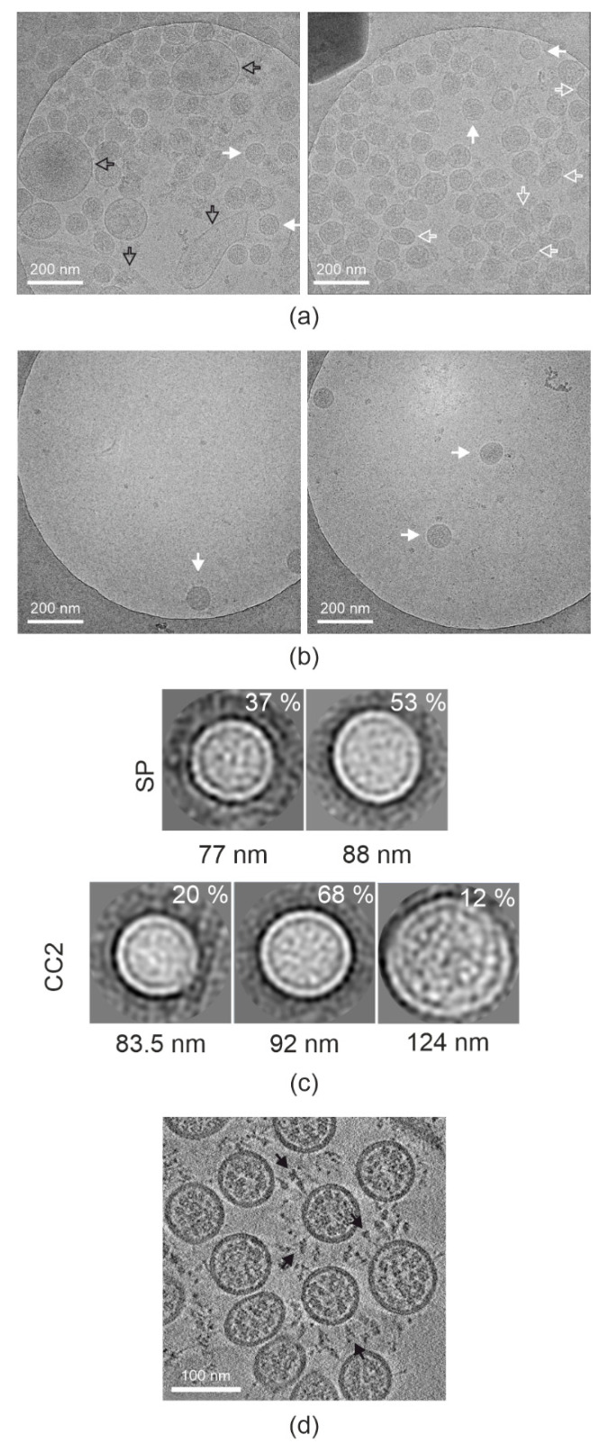
Severe acute respiratory syndrome coronavirus-2 is the causative agent of COVID-19. During the pandemic of 2019-2022, at least 500 million have been infected and over 6.3 million people have died from COVID-19. The virus is pleomorphic, and due to its pathogenicity is often handled in very restrictive biosafety containments laboratories. We developed two effective and rapid purification methods followed by UV inactivation that allow easy downstream handling of the virus. We monitored the purification through titering, sequencing, mass spectrometry and electron cryogenic microscopy. Although pelleting through a sucrose cushion, followed by gentle resuspension overnight gave the best particle recovery, infectivity decreased, and the purity was significantly worse than if using the size exclusion resin Capto Core. Capto Core can be used in batch mode, and was seven times faster than the pelleting method, obviating the need for ultracentrifugation in the containment laboratory, but resulting in a dilute virus. UV inactivation was readily optimized to allow handling of the inactivated samples under standard operating conditions. When containment laboratory space is limited, we recommend the use of Capto Core for purification and UV for inactivation as a simple, rapid workflow prior, for instance, to electron cryogenic microscopy or cell activation experiments.
Keywords: SARS-CoV-2; UV inactivation; purification; size exclusion; structural studies.
Conflict of interest statement
The authors declare no conflict of interest. The funders had no role in the design of the study; in the collection, analyses, or interpretation of data; in the writing of the manuscript, or in the decision to publish the results.
Figures
Similar articles
-
Assessment of inactivation procedures for SARS-CoV-2.J Gen Virol. 2021 Mar;102(3):001539. doi: 10.1099/jgv.0.001539. Epub 2021 Jan 8. J Gen Virol. 2021. PMID: 33416462 Free PMC article.
-
Effect of Inactivation Methods on SARS-CoV-2 Virion Protein and Structure.Viruses. 2021 Mar 26;13(4):562. doi: 10.3390/v13040562. Viruses. 2021. PMID: 33810401 Free PMC article.
-
Analysis of Inactivation of SARS-CoV-2 by Specimen Transport Media, Nucleic Acid Extraction Reagents, Detergents, and Fixatives.J Clin Microbiol. 2020 Oct 21;58(11):e01713-20. doi: 10.1128/JCM.01713-20. Print 2020 Oct 21. J Clin Microbiol. 2020. PMID: 32839250 Free PMC article.
-
Selection and application of biological safety cabinets in diagnostic and research laboratories with special emphasis on COVID-19.Rev Sci Instrum. 2021 Aug 1;92(8):081401. doi: 10.1063/5.0047716. Rev Sci Instrum. 2021. PMID: 34470433 Free PMC article. Review.
-
[Applications of separation technology in novel coronavirus research, epidemic prevention and detection].Se Pu. 2021 Jul 8;39(7):679-685. doi: 10.3724/SP.J.1123.2021.03022. Se Pu. 2021. PMID: 34227364 Free PMC article. Review. Chinese.
Cited by
-
Structural and Immunoreactivity Properties of the SARS-CoV-2 Spike Protein upon the Development of an Inactivated Vaccine.Viruses. 2023 Feb 9;15(2):480. doi: 10.3390/v15020480. Viruses. 2023. PMID: 36851694 Free PMC article.
-
Structural and Functional Characterization of Medicinal Plants as Selective Antibodies towards Therapy of COVID-19 Symptoms.Antibodies (Basel). 2024 May 7;13(2):38. doi: 10.3390/antib13020038. Antibodies (Basel). 2024. PMID: 38804306 Free PMC article.
-
Antiviral action of a functionalized plastic surface against human coronaviruses.Microbiol Spectr. 2024 Feb 6;12(2):e0300823. doi: 10.1128/spectrum.03008-23. Epub 2024 Jan 16. Microbiol Spectr. 2024. PMID: 38226803 Free PMC article.
-
β-Coronaviruses exploit ESCRT for virion assembly and egress.mBio. 2025 Jun 11;16(6):e0097925. doi: 10.1128/mbio.00979-25. Epub 2025 May 23. mBio. 2025. PMID: 40407327 Free PMC article.
-
Wavelength dependence of ultraviolet light inactivation for SARS-CoV-2 omicron variants.Sci Rep. 2023 Jun 15;13(1):9706. doi: 10.1038/s41598-023-36610-6. Sci Rep. 2023. PMID: 37322228 Free PMC article.
References
-
- Saccon E., Chen X., Mikaeloff F., Rodriguez J.E., Szekely L., Vinhas B.S., Krishnan S., Byrareddy S.N., Frisan T., Végvári Á., et al. Cell-type-resolved quantitative proteomics map of interferon response against SARS-CoV-2. iScience. 2021;24:102420. doi: 10.1016/j.isci.2021.102420. - DOI - PMC - PubMed
-
- Ogando N.S., Dalebout T.J., Zevenhoven-Dobbe J.C., Limpens R.W.A.L., van der Meer Y., Caly L., Druce J., de Vries J.J.C., Kikkert M., Bárcena M., et al. SARS-coronavirus-2 replication in Vero E6 cells: Replication kinetics, rapid adaptation and cytopathology. J. Gen. Virol. 2020;101:925–940. doi: 10.1099/jgv.0.001453. - DOI - PMC - PubMed
Publication types
MeSH terms
Substances
Grants and funding
LinkOut - more resources
Full Text Sources
Medical
Research Materials
Miscellaneous
