. 2022 Sep 9;14(9):2002.
doi: 10.3390/v14092002.

Susceptibility of Domestic Goat (Capra aegagrus hircus) to Experimental Infection with Severe Acute Respiratory Syndrome Coronavirus 2 (SARS-CoV-2) B.1.351/Beta Variant

Affiliations

Susceptibility of Domestic Goat (Capra aegagrus hircus) to Experimental Infection with Severe Acute Respiratory Syndrome Coronavirus 2 (SARS-CoV-2) B.1.351/Beta Variant

Leira Fernández-Bastit et al. Viruses. .

Abstract

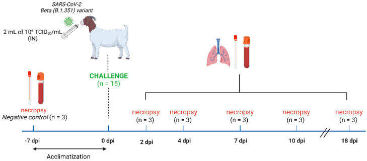
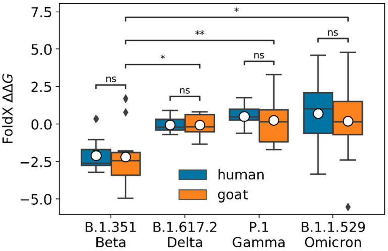
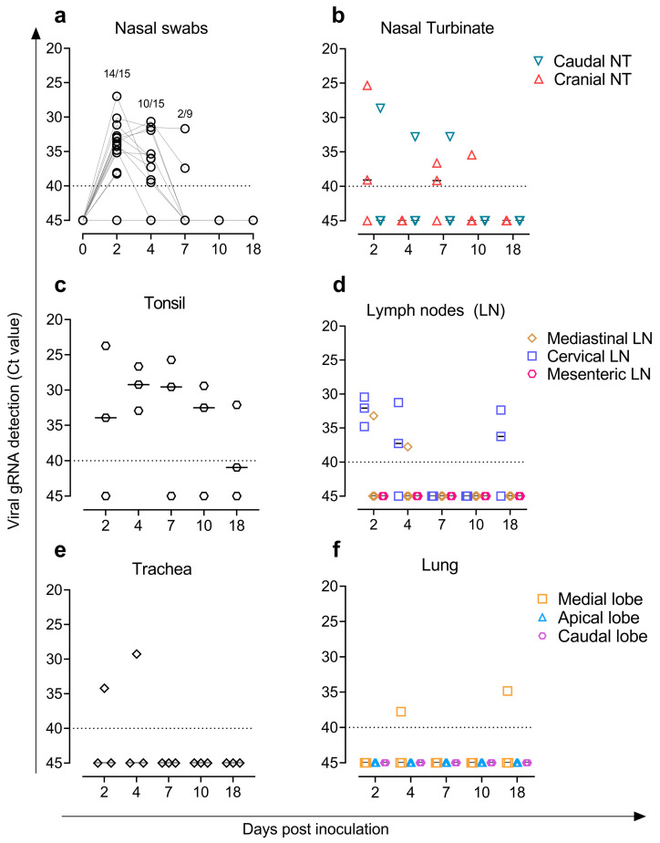
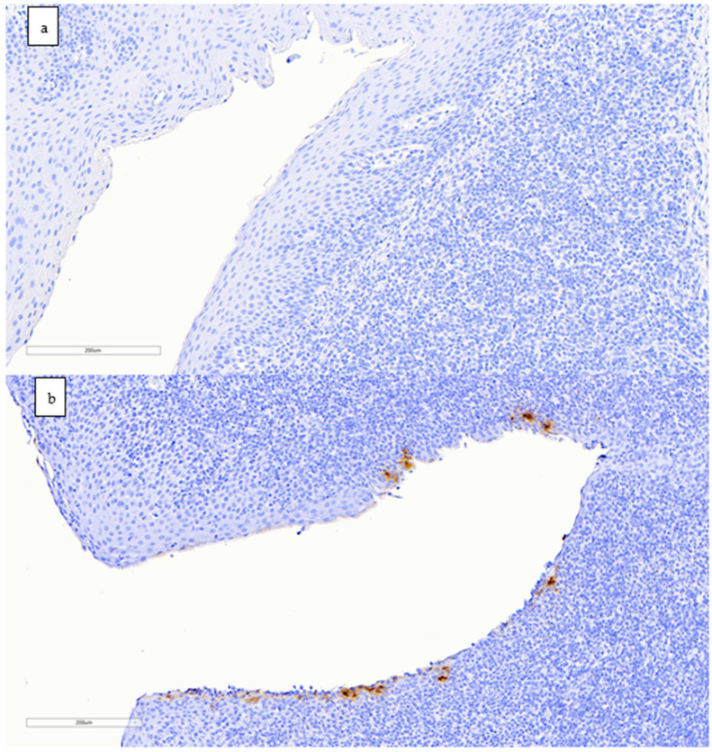
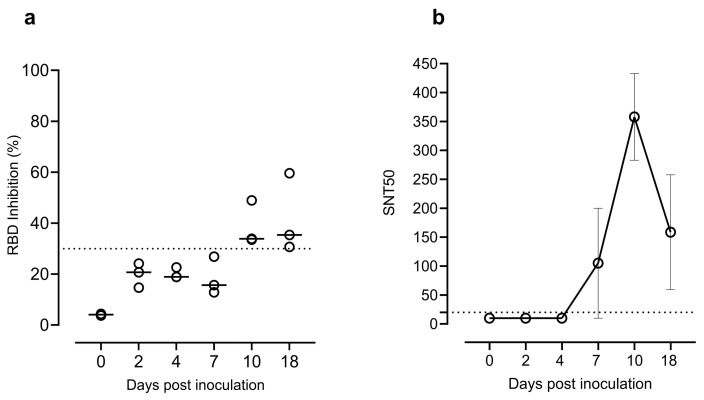
A wide range of animal species are susceptible to the severe acute respiratory syndrome coronavirus 2 (SARS-CoV-2) infection. Natural and/or experimental infections have been reported in pet, zoo, farmed and wild animals. Interestingly, some SARS-CoV-2 variants, such as B.1.1.7/Alpha, B.1.351/Beta, and B.1.1.529/Omicron, were demonstrated to infect some animal species not susceptible to classical viral variants. The present study aimed to elucidate if goats (Capra aegagrus hircus) are susceptible to the B.1.351/Beta variant. First, an in silico approach was used to predict the affinity between the receptor-binding domain of the spike protein of SARS-CoV-2 B.1.351/Beta variant and angiotensin-converting enzyme 2 from goats. Moreover, we performed an experimental inoculation with this variant in domestic goat and showed evidence of infection. SARS-CoV-2 was detected in nasal swabs and tissues by RT-qPCR and/or immunohistochemistry, and seroneutralisation was confirmed via ELISA and live virus neutralisation assays. However, the viral amount and tissue distribution suggest a low susceptibility of goats to the B.1.351/Beta variant. Therefore, although monitoring livestock is advisable, it is unlikely that goats play a role as SARS-CoV-2 reservoir species, and they are not useful surrogates to study SARS-CoV-2 infection in farmed animals.

Keywords: Beta variant; experimental infection; goat; ruminant; severe acute respiratory syndrome coronavirus 2 (SARS-CoV-2); susceptibility.


Conflict of interest statement

The authors declare no conflict of interest.

Figures

Figure 1
Figure 1
Schematic representation of the study design. Three out of eighteen goats were necropsied on the day of arrival and served as non-inoculated controls. Nasal and rectal swabs, blood, and tissue samples were collected. After acclimatisation, the remaining fifteen goats were intranasally inoculated with the SARS-CoV-2 B.1.351/Beta variant (2 mL of 1 × 10⁶ TCID50/animal). At 2, 4, 7,10, and 18 days post-infection, three goats were necropsied each day and nasal and rectal swabs, blood, and tissues were collected. Clinical signs and rectal temperature were recorded daily.
Figure 2
Figure 2
FoldX predicted ∆∆G for B.1.351/Beta, B.1.617.2/Delta, P.1/Gamma, and B.1.1.529/Omicron variants for human and goat ACE2 (blue and orange boxes, respectively). For goat, the computed ∆∆G was significantly lower for the Beta variant than for the Delta, Gamma, and Omicron variants, with Mann–Whitney–Wilcoxon test p-values of 1.06×10², 3.64×10³, and 3.20×10², respectively. The average ∆∆G predicted from the 10 Modeller models is plotted as a white dot for each variant. We found no significant differences between the predicted ∆∆G values for human and goat of the different variants (Mann–Whitney–Wilcoxon test p-values of 4.85 ×101, 5.15 × 101, 7.64 × 101, 6.61× 101 for Delta, Beta, Gamma, and Omicron, respectively). In this boxplot representation, the lower and upper ends of each box represent the first (Q1) and third (Q3) quartiles of the ∆∆G predicted values, respectively. The horizontal line, inside each box, represents the median, or second quartile (Q2), and the mean is plotted as a white dot for each variant. The box “whiskers” extend to values that are 1.5 times the size of the interquartile range (IQR = Q3 − Q1). Values that fall outside this range are displayed independently as black diamonds. Mann-Whitney-Wilcoxon test p-values are annotated according to the following criteria: ns (0.05 < p-value ≤ 1), * (0.02 < p-value ≤ 0.05), ** (0.001 < p-value ≤ 0.02).
Figure 3
Figure 3
Detection of SARS-CoV-2 genomic RNA (gRNA) by RT-qPCR. SARS-CoV-2 loads in (a) nasal swabs; (b) cranial and caudal nasal turbinate; (c) tonsil; (d) mediastinal, cervical, and mesenteric lymph nodes; (e) trachea; and (f) lung. Horizontal bars indicate median viral loads. Dotted lines reflect the limit of detection (Ct = 40).
Figure 4
Figure 4
Immunohistochemistry staining to detect the nucleocapsid protein of SARS-CoV-2 in goat tonsils (scale bar: 200 µm). (a) Negative control animal with no antigen labelling. (b) Positive result in the tonsil of a goat euthanised at 2 dpi; immunolabelling is seen as brownish staining in dendritic-like cells around a tonsillar crypt.
Figure 5
Figure 5
(a) Neutralising antibodies detected by the SARS-CoV-2 RBD Inhibition ELISA (Positive ≥ 30% RBD inhibition). (b) Neutralisation titres in sera samples from 0, 2, 4, 7, 10, and 18 dpi determined by the live virus neutralisation assay. Data reported as values of reciprocal dilution of SNT50 (mean ± SEM). The horizontal dotted lines indicate the cut-off value of the assay. Abbreviations: RBD, receptor-binding domain; SNT50, serum virus neutralisation titre (reciprocal dilution) that showed 50% protection of virus growth.

References

    1. Jayaweera M., Perera H., Gunawardana B., Manatunge J. Transmission of COVID-19 Virus by Droplets and Aerosols: A Critical Review on the Unresolved Dichotomy. Environ. Res. 2020;188:109819. doi: 10.1016/J.ENVRES.2020.109819. - DOI - PMC - PubMed
    1. Temmam S., Vongphayloth K., Salazar E.B., Munier S., Bonomi M., Regnault B., Douangboubpha B., Karami Y., Chrétien D., Sanamxay D., et al. Bat Coronaviruses Related to SARS-CoV-2 and Infectious for Human Cells. Nature. 2022;604:330–336. doi: 10.1038/s41586-022-04532-4. - DOI - PubMed
    1. Zhou P., Yang X.L., Wang X.G., Hu B., Zhang L., Zhang W., Si H.R., Zhu Y., Li B., Huang C.L., et al. A Pneumonia Outbreak Associated with a New Coronavirus of Probable Bat Origin. Nature. 2020;579:270–273. doi: 10.1038/s41586-020-2012-7. - DOI - PMC - PubMed
    1. Worobey M., Levy J.I., Serrano L.M., Crits-Christoph A., Pekar J.E., Goldstein S.A., Rasmussen A.L., Kraemer M.U.G., Newman C., Koopmans M.P.G., et al. The Huanan Seafood Wholesale Market in Wuhan Was the Early Epicenter of the COVID-19 Pandemic. Science. 2022;377:951–959. doi: 10.1126/SCIENCE.ABP8715. - DOI - PMC - PubMed
    1. Sharun K., Dhama K., Pawde A.M., Gortázar C., Tiwari R., Katterine Bonilla-Aldana D., Rodriguez-Morales A.J., De La Fuente J., Michalak I., Attia Y.A., et al. SARS-CoV-2 in Animals: Potential for Unknown Reservoir Hosts and Public Health Implications. Vet. Q. 2021;41:181–201. doi: 10.1080/01652176.2021.1921311. - DOI - PMC - PubMed

Publication types

Substances

Supplementary concepts