. 2022 Sep 10;14(9):2004.
doi: 10.3390/v14092004.

Human Cytomegalovirus Induces Vitamin-D Resistance In Vitro by Dysregulating the Transcriptional Repressor Snail

Affiliations

Human Cytomegalovirus Induces Vitamin-D Resistance In Vitro by Dysregulating the Transcriptional Repressor Snail

Carmen Stecher et al. Viruses. .

Abstract

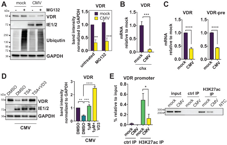
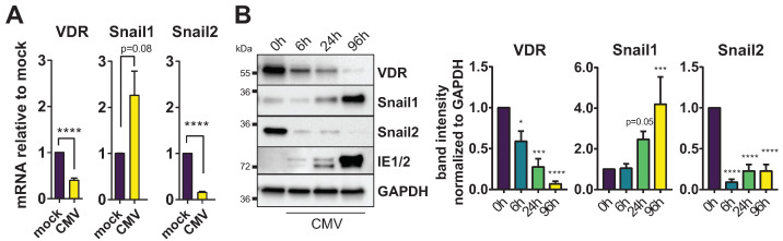
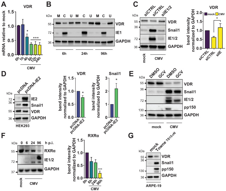
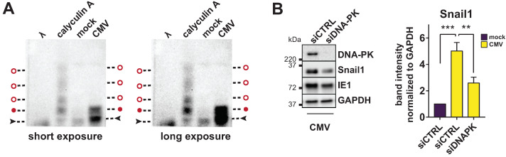
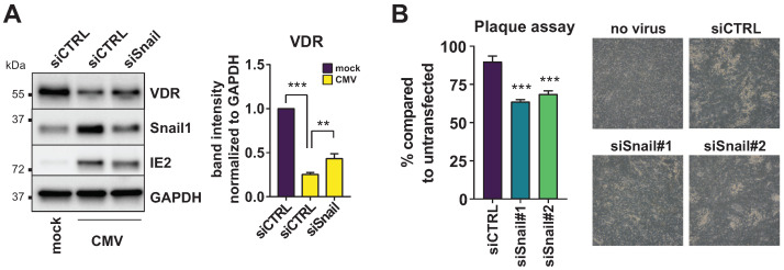
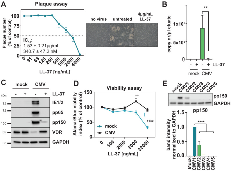
Vitamin-D supplementation is considered to play a beneficial role against multiple viruses due to its immune-regulating and direct antimicrobial effects. In contrast, the human cytomegalovirus (HCMV) has shown to be resistant to treatment with vitamin D in vitro by downregulation of the vitamin-D receptor. In this study, we aimed to elucidate the mechanism and possible biological consequences of vitamin-D resistance during HCMV infection. Mechanistically, HCMV induced vitamin-D resistance by downregulating the vitamin-D receptor (VDR) within hours of lytic infection. We found that the VDR was inhibited at the promoter level, and treatment with histone deacetylase inhibitors could restore VDR expression. VDR downregulation highly correlated with the upregulation of the transcriptional repressor Snail1, a mechanism likely contributing to the epigenetic inactivation of the VDR promoter, since siRNA-mediated knockdown of Snail partly restored levels of VDR expression. Finally, we found that direct addition of the vitamin-D-inducible antimicrobial peptide LL-37 strongly and significantly reduced viral titers in infected fibroblasts, highlighting VDR biological relevance and the potential of vitamin-D-inducible peptides for the antiviral treatment of vitamin-D deficient patients.

Keywords: HCMV; LL-37; Snail; VDR; calcitriol; cathelicidin; cytomegalovirus; vitamin D.


Conflict of interest statement

The authors declare no conflict of interest.

Figures

Figure 1
Figure 1
The VDR is epigenetically repressed during HCMV infection. (A) Immunoblot showing VDR and GAPDH expression in HFF treated with DMSO (−) or 1 μM MG132 (+) proteasome inhibitor in mock- or CMV-infected cells. Ubiquitinated proteins are shown as a control for proteasome inhibitor effectiveness. Representative blot (left) and bar graph from multiple experiments (right panel). (B) RT-qPCR analysis of VDR mRNA expression in mock- or CMV-infected HFF treated with 50 μg/mL cycloheximide (chx, n = 2) starting at 6 h p.i. and harvested 24 h p.i. Asterisks indicate significant differences to the mock control sample (unpaired Student’s t test). (C) RT-qPCR analysis of VDR and intron-containing unspliced pre-mRNA (VDR-pre) expression in mock- and HCMV-infected HFF harvested 24 h p.i. Asterisks indicate significant differences to the mock control sample using unpaired Student’s t tests. (D) Immunoblots showing VDR expression compared to GAPDH loading control in HFF infected with CMV and treated with DMSO (vehicle), 1 μM TSA, or a combination of TSA and 10 nM calcitriol (VD3), harvested 24 h p.i. A representative blot is shown on the left, the right panel shows VDR quantification from multiple experiments with significant differences to the CMV DMSO control sample as determined by ANOVA Dunnett’s multiple comparison post-hoc test. (E) Purified chromatin from mock- and CMV-infected HFF was immunoprecipitated with an anti-H3K27ac polyclonal antibody or normal goat serum (ctrl IP). The left panel shows qPCR results from three independent experiments. On the right, VDR promoter amplicons from a Taq-PCR were separated on an agarose gel. NTC = no template control.
Figure 2
Figure 2
HCMV regulates the VDR repressors Snail1 and Snail2. (A) RT-qPCR analysis of VDR, Snail1 and Snail2 mRNA expression in mock- and CMV-infected HFF is shown. Asterisks indicate significant differences to the mock control sample (unpaired Student’s t test). (B) Immunoblots showing VDR, Snail1, Snail2 and IE1/2 expression compared to GAPDH loading control in HFF infected with CMV and harvested at the indicated time point p.i. Representative blot and quantification from multiple experiments with significant differences to the mock (0 h) control sample as determined by ANOVA Dunnett’s multiple comparison post-hoc tests.
Figure 3
Figure 3
VDR holoenzyme downregulation is dependent on immediate early gene transcription. (A) Vitamin-D-receptor expression was measured in mock- or HCMV-infected HFF at the indicated time points, showing fold change (2ΔΔCt) compared to mock-infected cells (mean ± SEM). Asterisks indicate significant differences to the mock control sample as determined by ANOVA Dunnett’s multiple comparison post-hoc test. (B) VDR, IE1 (pp72) and GAPDH protein expression were determined by Western blot in HFF that were infected with mock (M), CMV (C) and UV-inactivated CMV (U) inocula and harvested at the indicated times post infection. (C) VDR and HCMV immediate early protein expression was determined in lysates of HFF transfected with a non-targeting control siRNA or IE1/2 specific siRNA harvested at 24 h p.i. Representative blot (left) and quantification of bands from multiple experiments relative to the GAPDH loading control (right). Asterisks indicate significant differences to the CMV siCTRL sample as determined by ANOVA Dunnett’s multiple comparison post-hoc test. (D) HEK293 cells were transfected with pcDNA empty vector control or pcDNA containing the IE2 open reading frame. Lysates were analyzed by Western blot at 48 h post transfection. Asterisks indicate significant differences to empty vector control (unpaired t-test). (E) VDR and Snail1 expression was determined at 96 h p.i. in lysates of HFF treated with vehicle (DMSO) or 10 μg/mL Ganciclovir (GCV). HCMV immediate early and pp150 late antigens were stained as a control. (F) RXRα protein expression in HFF was determined by specific immunoblots at the indicated time points p.i. Representative blot (left) and quantification of bands from multiple experiments relative to the GAPDH loading control (right, one-way ANOVA and Dunnett’s post-hoc test). (G) ARPE-19 cells were infected with mock supernatant or the pentamer-positive (TS15-rN) variant of the Towne strain and harvested 14 d p.i. VDR, Snail1, pp150 late antigen and a GAPDH loading control detection by Western blot is shown.
Figure 4
Figure 4
Snail phosphorylation and regulation by DNA-Protein kinase. (A) Lysates from mock- and HCMV-infected cells were harvested at 24 h p.i. and subjected to Phos-Tag SDS-PAGE. As additional controls, CMV-infected HFF were treated with a phosphatase inhibitor for 2 h before lysis (calyculin A), or with lambda phosphatase for 30 min λ). A representative Snail1-specific immunoblot shows the ratio between unphosphorylated (black arrows) and phosphorylated Snail1 (red circles). The same blot is shown with short (left) and long exposure (right). Height of phosphorylated bands also present in the CMV condition is marked with full red circles, open red circles indicate further (weak) phosphorylation bands in the calyculin A treatment control. (B) Representative Western blot showing DNA-PK and Snail protein expression determined in lysates of HCMV-infected HFF transfected with a non-targeting control siRNA or DNA-PK specific siRNAs and harvested at 24 h p.i. (representative Western blot on the left and quantification relative Snail1 band intensity of multiple independent experiments on the right; asterisks show significant differences to the CMV siCTRL sample as determined by ANOVA Dunnett’s multiple comparison post-hoc test).
Figure 5
Figure 5
Snail ablation restores VDR expression. (A) VDR and Snail protein expression was determined in lysates of HFF transfected with a non-targeting control siRNA or Snail1 specific siRNA and harvested at 24 h p.i. Representative blot (left) and quantification of bands from multiple experiments relative to the GAPDH loading control (right). Asterisks show significant differences to the CMV siCTRL control sample as determined by ANOVA Dunnett’s multiple comparison post-hoc test. (B) Plaque assay showing the percentage of plaques in siRNA-transfected cells compared to an untransfected control. Asterisks show significant differences in two different Snail-targeting siRNAs to the CMV siCTRL control sample as determined by ANOVA Dunnett’s multiple comparison post-hoc test. Light microscope pictures of representative areas are shown on the right.
Figure 6
Figure 6
Cathelicidin inhibits HCMV replication. (A) Plaque reduction assay of HFF infected with HCMV AD169 pre-treated with increasing amounts of LL-37; percentage compared to DMSO control is shown (n = 2). Light microscope pictures of representative areas for the non-infected control, virus control and maximum LL-37 concentration are shown on the right. (B) Intracellular CMV DNA was detected in lysates from infected HFF at 72 h p.i. and 4 μg/mL LL-37 (where indicated) using primers specific for the UL83 (pp65) gene. Quantification of copy numbers per measured sample were calculated by interpolation of values against a 5-point standard curve of pcDNA-pp65 plasmid dilutions. (C) Western blot showing abundance of the indicated proteins in whole cell lysate (WCL) from mock-infected or HCMV-infected HFF at 96 h p.i with or without treatment with 4 μg/mL LL-37 peptide. Representative of three independent experiments. (D) AlamarBlue viability assay showing reduction in the viability index in mock- and CMV-infected cells treated with up to 32 μg/mL LL-37 compared to the respective untreated control. (E) Western blot (left) and quantification (right) of pp150 relative protein abundance in cell lysates harvested at 96 h p.i. at MOI 0.05. HFF cells were either left untreated (mock, CMV1) or LL-37-treatment conditions (4 μg/mL, CMV2 to CMV5). LL-37 was either added to the medium 2 h after inoculation (CMV2), or incubated for 1h with the cells prior to infection and then removed before adding the inoculum (CMV3). CMV4: LL-37 was incubated for 1h with the viral supernatant before inoculating the cells, or LL-37 was added to both the cells and viral supernatant (CMV5).

Similar articles

Cited by

References

    1. Bhalla A.K., Amento E.P., Serog B., Glimcher L.H. 1,25-Dihydroxyvitamin D3 inhibits antigen-induced T cell activation. J. Immunol. 1984;133:1748–1754. - PubMed
    1. Campbell G.R., Spector S.A. Hormonally active vitamin D3 (1α,25-dihydroxycholecalciferol) triggers autophagy in human macrophages that inhibits HIV-1 infection. J. Biol. Chem. 2011;286:18890–18902. doi: 10.1074/jbc.M110.206110. - DOI - PMC - PubMed
    1. Wang T.-T., Nestel F.P., Bourdeau V., Nagai Y., Wang Q., Liao J., Tavera-Mendoza L., Lin R., Hanrahan J.W., Mader S., et al. Cutting Edge: 1,25-Dihydroxyvitamin D 3 Is a Direct Inducer of Antimicrobial Peptide Gene Expression. J. Immunol. 2004;173:2909–2912. doi: 10.4049/jimmunol.173.5.2909. - DOI - PubMed
    1. Ryan J.W., Anderson P.H., Morris H.A. Pleiotropic activities of vitamin D receptors - adequate activation for multiple health outcomes. Clin. Biochem. Rev. 2015;36:53–61. - PMC - PubMed
    1. Gordon Y.J., Huang L.C., Romanowski E.G., Yates K.A., Proske R.J., McDermott A.M. Human cathelicidin (LL-37), a multifunctional peptide, is expressed by ocular surface epithelia and has potent antibacterial and antiviral activity. Curr. Eye Res. 2005;30:385–394. doi: 10.1080/02713680590934111. - DOI - PMC - PubMed

Publication types