. 2022 Sep 11;14(9):2013.
doi: 10.3390/v14092013.

Epidemiological and Genomic Characterisation of Middelburg and Sindbis Alphaviruses Identified in Horses with Febrile and Neurological Infections, South Africa (2014-2018)

Affiliations

Epidemiological and Genomic Characterisation of Middelburg and Sindbis Alphaviruses Identified in Horses with Febrile and Neurological Infections, South Africa (2014-2018)

Isabel Fourie et al. Viruses. .

Abstract

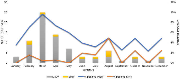
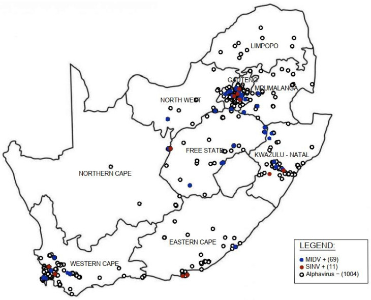
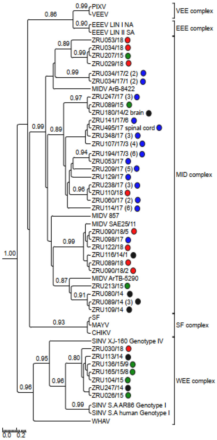
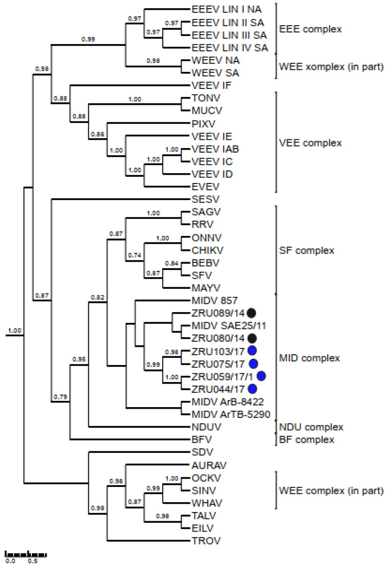
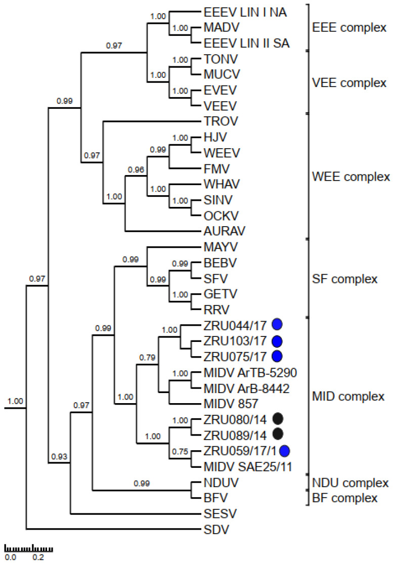
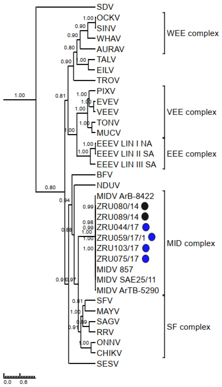
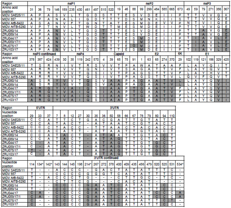
Although Old World alphaviruses, Middelburg- (MIDV) and Sindbis virus (SINV), have previously been detected in horses and wildlife with neurologic disease in South Africa, the pathogenesis and clinical presentation of MIDV and SINV infections in animals are not well documented. Clinical samples from horses across South Africa with acute or fatal neurologic and febrile infections submitted between 2014-2018 were investigated. In total, 69/1084 (6.36%) and 11/1084 (1.01%) horses tested positive for MIDV and SINV, respectively, by real-time reverse transcription (RT) PCR. Main signs/outcomes for MIDV (n = 69): 73.91% neurological, 75.36% fever, 28.99% icterus and anorexia, respectively, 8.70% fatalities; SINV (n = 11): 54.54% neurological, 72.73% fever, 36.36% anorexia and 18.18% fatalities. MIDV cases peaked in the late summer/autumn across most South African provinces while SINV cases did not show a clear seasonality and were detected in fewer South African provinces. MIDV could still be detected in blood samples via RT-PCR for up to 71,417 and 21 days after onset of signs in 4 horses respectively, suggesting prolonged replication relative to SINV which could only be detected in the initial sample. Phylogenetic analyses based on partial sequences of the nsP4 (MIDV n = 59 and SINV n = 7) and E1 (MIDV n = 45) genes, as well as full genome sequences (MIDV n = 6), clustered the MIDV and SINV strains from the present study with previously detected strains. MIDV infection appears to be more prevalent in horses than SINV infection based on RT-PCR results, however, prevalence estimates might be different when also considering serological surveillance data.

Keywords: Alphavirus; Middelburg virus; Sindbis virus; arbovirus; genome; horse; zoonotic.


Conflict of interest statement

The authors declare no conflict of interest.

Figures

Figure 1
Figure 1
Seasonal detection of Middelburg virus (MIDV) (n = 69) and Sindbis virus (SINV) (n = 11) positive infections in horses from South Africa between January 2014–December 2018. Number of MIDV positives indicated by grey bars and percent positivity indicated by blue line. Number of SINV positives indicated by yellow bars and percent positivity indicated by orange line.
Figure 2
Figure 2
Map of Middelburg (MIDV)- and Sindbis virus (SINV) PCR positive infections reported in horses tested in South Africa from January 2014–December 2018. +: positive; −: negative.
Figure 3
Figure 3
Rooted phylogenetic tree produced from Bayesian analysis using BEAST based on a 347-base pair nsP4 fragment (83 taxa, model GTR+G+I) of alphaviruses is shown. Posterior probabilities >0.7 are shown on major branches. Middelburg- and Sindbis virus positives described in current study indicated with ZRU numbers and circle colours correspond to year as follows: 2014 black, 2015 green, 2017 blue and 2018 red. All sequences obtained from whole blood or serum unless otherwise indicated. Number of sequences within the same clade for samples from the same year indicated in brackets. Alphavirus complexes indicated by brackets; tree is drawn to scale representing the number of nucleotide substitutions per site. CHIKV: Chikungunya virus; EEEV: Eastern equine encephalitis virus; MAYV: Mayaro virus; MIDV, Middelburg virus; PIXV: Pixuna virus; SFV, Semliki Forest virus; SINV: Sindbis virus; VEEV: Venezuelan equine encephalitis virus; WHAV: Whataroa virus; WEE: Western equine encephalitis.
Figure 4
Figure 4
Rooted phylogenetic tree produced from Bayesian analysis using BEAST based on a 550-base pair E1 fragment (74 taxa, model GTR+G+I) of alphaviruses is shown. Posterior probabilities >0.7 are shown on major branches. Middelburg virus positives described in the current study are indicated with ZRU numbers and circle colours correspond to year as follows: 2014 black, 2015 green, 2017 blue and 2018 red. All sequences obtained from whole blood or serum unless otherwise indicated. Number of sequences within the same clade for samples from the same year indicated in brackets. Alphavirus complexes are indicated by brackets. The tree is drawn to scale representing the number of nucleotide substitutions per site. AURAV: Aura virus; BF: Barmah Forest virus; CHIKV: Chikungunya virus; EILV: Eilat virus; EEEV: Eastern equine encephalitis virus; EVEV: Everglades virus; MAYV: Mayaro virus; MIDV, Middelburg virus; MUCV: Mucambo virus; NDUV: Ndumu virus; OCKV: Ockelbo virus; PIXV: Pixuna virus; RRV: Ross river virus; SAGV: Sagiyama virus; SDV: Sleeping disease virus; SEV: Southern elephant seal virus; SFV, Semliki Forest virus; SINV: Sindbis virus; TALV: Taï Forest alphavirus; TONV: Tonate virus; TROV: Trocara virus; VEEV: Venezuelan equine encephalitis virus; WHAV: Whataroa virus; WEE: Western equine encephalitis.
Figure 5
Figure 5
Rooted phylogenetic tree produced from Bayesian analysis using BEAST based on non-structural proteins (7236 base pairs) (33 taxa, model GTR+G+I) of alphaviruses is shown. Posterior probabilities >0.7 are shown on major branches. Sequences of horse Middelburg virus positives described in the current study are indicated with ZRU numbers and circle colours correspond to year sample was screened as follows: 2014 indicated in black (sequences obtained from whole blood samples) and 2017 (sequences obtained from culture isolates) indicated in blue. Alphavirus complexes are indicated by brackets. The tree is drawn to scale representing the number of nucleotide substitutions per site. AURAV: Aura virus; BF: Barmah Forest virus; CHIKV: Chikungunya virus; EILV: Eilat virus; EEEV: Eastern equine encephalitis virus; EVEV: Everglades virus; GETV: Getah virus; MAYV: Mayaro virus; MIDV, Middelburg virus; MUCV: Mucambo virus; NDUV: Ndumu virus; OCKV: Ockelbo virus; PIXV: Pixuna virus; RRV: Ross river virus; SDV: Sleeping disease virus; SEV: Southern elephant seal virus; SFV, Semliki Forest virus; SINV: Sindbis virus; TALV: Taï Forest alphavirus; TONV: Tonate virus; TROV: Trocara virus; VEEV: Venezuelan equine encephalitis virus; WHAV: Whataroa virus; WEE: Western equine encephalitis.
Figure 6
Figure 6
Rooted phylogenetic tree produced from Bayesian analysis using BEAST based on structural proteins (3777 base pairs) (35 taxa, model GTR+G+I) of alphaviruses is shown Posterior probabilities >0.7 are shown on major branches. Sequences of horse Middelburg virus positives described in the current study are indicated with ZRU numbers and circle colours correspond to year sample was screened as follows: 2014 indicated in black (sequences obtained from whole blood samples) and 2017 indicated in blue (sequences obtained from culture isolates). AURAV: Aura virus; BF: Barmah Forest virus; CHIKV: Chikungunya virus; EILV: Eilat virus; EEEV: Eastern equine encephalitis virus; EVEV: Everglades virus; GETV: Getah virus; MAYV: Mayaro virus; MIDV, Middelburg virus; MUCV: Mucambo virus; NDUV: Ndumu virus; OCKV: Ockelbo virus; PIXV: Pixuna virus; RRV: Ross river virus; SDV: Sleeping disease virus; SEV: Southern elephant seal virus; SFV, Semliki Forest virus; SINV: Sindbis virus; TALV: Taï Forest alphavirus; TONV: Tonate virus; TROV: Trocara virus; VEEV: Venezuelan equine encephalitis virus; WHAV: Whataroa virus; WEE: Western equine encephalitis.
Figure 7
Figure 7
Rooted phylogenetic tree produced from Bayesian analysis using BEAST based on concatenated nucleotide sequences of non-structural (7236 base pairs) and structural protein (3777 base pairs) open reading frames (35 taxa, model GTR+G+I) of alphaviruses is shown. Probabilities >0.7 are shown on major branches. Sequences of horse Middelburg virus positives described in the current study are indicated with ZRU numbers and circle colours correspond to year sample was screened as follows: 2014 indicated in black (sequences obtained from whole blood samples) and 2017 indicated in blue (sequences obtained from culture isolates). Alphavirus complexes are indicated by brackets. The tree is drawn to scale representing the number of nucleotide substitutions per site. AURAV: Aura virus; BF: Barmah Forest virus; CHIKV: Chikungunya virus; EILV: Eilat virus; EEEV: Eastern equine encephalitis virus; EVEV: Everglades virus; GETV: Getah virus; MAYV: Mayaro virus; MIDV, Middelburg virus; MUCV: Mucambo virus; NDUV: Ndumu virus; OCKV: Ockelbo virus; PIXV: Pixuna virus; RRV: Ross river virus; SDV: Sleeping disease virus; SEV: Southern elephant seal virus; SFV, Semliki Forest virus; SINV: Sindbis virus; TALV: Taï Forest alphavirus; TONV: Tonate virus; TROV: Trocara virus; VEEV: Venezuelan equine encephalitis virus; WHAV: Whataroa virus; WEE: Western equine encephalitis.
Figure 8
Figure 8
Amino acid and nucleotide sequence comparisons of the structural and non-structural proteins and UTR’s, respectively, between different Middelburg virus (MIDV) strains. Numbering refers to sequence positions of isolate SAE25/11. Changes in amino acid of ZRU080/14, ZRU089/14, ZRU044/17, ZRU059/17/1, ZRU075/17, and ZRU103/17 as compared to MIDV SAE25/11 are highlighted in grey. Deletions in MIDV SA25/11 compared to other sequences are indicated by “^”.

Similar articles

Cited by

References

    1. Carrera J.-P., Forrester N., Wang E., Vittor A.Y., Haddow A.D., López-Vergès S., Abadía I., Castaño E., Sosa N., Báez C., et al. Eastern equine encephalitis in Latin America. N. Engl. J. Med. 2013;369:732–744. doi: 10.1056/NEJMoa1212628. - DOI - PMC - PubMed
    1. Acosta-Ampudia Y., Monsalve D.M., Rodríguez Y., Pacheco Y., Anaya J.-M., Ramírez-Santana C. Mayaro: An emerging viral threat? Emerg. Microbes. Infect. 2018;7:163. doi: 10.1038/s41426-018-0163-5. - DOI - PMC - PubMed
    1. Lundstrom J.O., Turell M.J., Niklasson B. Viremia in three orders of brids (Anseriforms, Galliformes and Passeriformes) inoculated with Ockelbo virus. J. Wildl. Dis. 1993;29:189–195. doi: 10.7589/0090-3558-29.2.189. - DOI - PubMed
    1. Adouchief S., Smura T., Sane J., Vapalahti O., Kurkela S. Sindbis virus as a human pathogen-epidemiology, clinical picture and pathogenesis. Rev. Med. Virol. 2016;26:221–241. doi: 10.1002/rmv.1876. - DOI - PubMed
    1. Taylor R.M., Hurlbut H.S., Work T.H., Kingston J.R., Frothingham T.E. Sindbis Virus: A Newly Recognized Arthropod-Transmitted Virus1. Am. J. Trop. Med. Hyg. 1955;4:844–862. doi: 10.4269/ajtmh.1955.4.844. - DOI - PubMed

Publication types

LinkOut - more resources