. 2022 Sep 13;14(9):2030.
doi: 10.3390/v14092030.

Human Cytomegalovirus Modifies Placental Small Extracellular Vesicle Composition to Enhance Infection of Fetal Neural Cells In Vitro

Affiliations

Human Cytomegalovirus Modifies Placental Small Extracellular Vesicle Composition to Enhance Infection of Fetal Neural Cells In Vitro

Mathilde Bergamelli et al. Viruses. .

Abstract

Although placental small extracellular vesicles (sEVs) are extensively studied in the context of pregnancy, little is known about their role during viral congenital infection, especially at the beginning of pregnancy. In this study, we examined the consequences of human cytomegalovirus (hCMV) infection on sEVs production, composition, and function using an immortalized human cytotrophoblast cell line derived from first trimester placenta. By combining complementary approaches of biochemistry, electron microscopy, and quantitative proteomic analysis, we showed that hCMV infection increases the yield of sEVs produced by cytotrophoblasts and modifies their protein content towards a potential proviral phenotype. We further demonstrate that sEVs secreted by hCMV-infected cytotrophoblasts potentiate infection in naive recipient cells of fetal origin, including human neural stem cells. Importantly, these functional consequences are also observed with sEVs prepared from an ex vivo model of infected histocultures from early placenta. Based on these findings, we propose that placental sEVs could be important actors favoring viral dissemination to the fetal brain during hCMV congenital infection.

Keywords: congenital infection; cytotrophoblast; extracellular vesicles; hCMV; placenta.


Conflict of interest statement

The authors declare no conflict of interest. The funders had no role in the design of the study; in the collection, analyses, or interpretation of data; in the writing of the manuscript, or in the decision to publish the results.

Figures

Figure 1
Figure 1
Characterization of sEVs from NI and hCMV infected HIPECs. (a) Yield of sEV recovered from HIPECs either non-infected (NI) or infected (hCMV) at a multiplicity of infection of 10, between 48 and 72 h post-infection. *, p = 0.0464 by paired t-test for seven independent experiments. (b) Comparison of mean size (left histogram) and mode size (right histogram) between sEVs from non-infected (NI) or infected (hCMV) HIPECs. Histograms show the mean ± SEM of three independent experiments. ns: non-significant by Mann–Whitney test. (c) Electron microscopy images of sEVs (indicated by an arrow) prepared from non-infected (NI) or infected (hCMV) HIPECs. Magnification = 26,000×. Scale bar = 100 nm. Images are representative of at least three independent experiments. (d) TEM observation of sEV—isolated from non-infected (NI) or infected (hCMV) HIPECs—which were immunogold-labelled for CD9, CD81, or CD63, and revealed with Protein A-gold particle of 10 nm diameter. Scale bar = 100 nm. Magnification = 26,000×. In CD63 IEM, only one example of positive vesicle, representing around 1–5% of sEVs isolated upon infection, is shown, the other being negative (see Supplementary Figure S3 for wide field image). (e) Western blot realized on either whole cell lysates (left wells) or purified sEVs (right wells), from non-infected (NI) or infected (hCMV) HIPECs. Proteins of interest and their corresponding molecular weight are indicated on the right of the Figure, with a smear for CD63 due to the non-reducing conditions of the western blot, which preserve its rich glycosylated pattern.
Figure 2
Figure 2
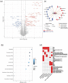
Proteomic analysis of sEV composition upon infection of HIPECs by hCMV. (a) Volcano-plot representing differences in normalized mean protein abundance in sEVs hCMV versus sEVs NI. Human and viral proteins exhibiting significant differences between the two conditions are represented by circles and triangles, respectively (Student T-test p-value ≤ 0.05 and log2 ratio ≥ 1 or ≤ −1). Red: over-represented proteins; Blue: under-represented proteins. (b) Dot plot representation of the top diseases and biological functions associated with human proteins exhibiting an absolute normalized mean abundance log2 ratio greater than 1 or lower than −1, in sEVs hCMV versus sEVs NI. Top diseases and biological functions associated with changes in the protein content of sEVs upon hCMV-infection were identified using QIAGEN Ingenuity Pathway Analysis (IPA). Size of the circles depends on the number of the proteins identified in the corresponding pathway; level of blue intensity depends on the p-value. (c) Human proteins associated with the predicted increased activation state of autophagy pathway as determined by IPA. (d) Heatmap representation of the biological functions associated with hCMV viral proteins expressed in sEVs hCMV. In red are indicated the functions attributed to the viral proteins (See Supplementary Table S2 for bibliography references).
Figure 3
Figure 3
Internalization of sEVs isolated from non-infected HIPECs in fetal MRC5 cells. (a) Confocal images of fluorescence microscopy carried out on MRC5 cells after 16 h incubation with PKH67-labelled sEVs. Blue: DAPI; Red: Phalloidin; Green: PKH67. Scale bar: 100 μm. Magnification = 63×. The right image corresponds to the orthogonal projection of the cell z-axis. (b) Histogram representing the percentage of PKH67 positive cells along time, upon incubation of MRC5 cells with sEVs. Bars represent the mean ± SEM of three independent experiments. (c) Monitoring of PKH67-labeled sEVs internalization by MRC5 cells by flow cytometry. Dot plots represent MRC5 cell fluorescence upon incubation with PKH67-stained sEVs (200 sEVs/cell) for cells that have not been incubated with sEVs (NT, non-treated), or upon 2 h or 16 h of incubation. X-axis: PKH-67 fluorescence intensity; Y-axis: FSC. Gate indicates cells positive for PKH67.
Figure 4
Figure 4
Effect of HIPEC sEVs on MRC5 cell permissiveness for hCMV. (a) Experimental procedure (NT = non-treated). (b) Immunofluorescence performed on MRC5 cells against IE viral antigen (blue: DAPI; red: IE1/2). Panel a- Non-infected control MRC5 cells upon 24 h incubation with sEVs isolated from hCMV-infected HIPECs. Panels b,c,d- MRC5 cells were either non-treated (Panel b- NT) or incubated during 2 h with sEVs prepared from non-infected (Panel c- sEV NI) or infected HIPECs (Panel d- sEV hCMV) with 50 sEV per cell, then infected during 24 h with hCMV at a MOI of 0.5 before proceeding to immunofluorescence. Scale bar: 200 μm. (c,d) MRC5 cells were incubated with sEVs prepared from non-infected (sEV NI) or hCMV-infected (sEV hCMV) HIPECs and infected by hCMV at a MOI of 0.5, concomitantly (c) or 2 h after sEV incubation (d). Three increasing doses of sEVs were used in these experiments (20, 50, or 200 sEV per MRC5 cell, from left to right). Then, 24 h later, expression of IE antigen was assessed by immunofluorescence. Quantification of the percentage of IE positive cells was carried out and normalized by the percentage of cells infected by hCMV without any sEV (NT). Each dot is an independent experiment and corresponds to the mean of the counting of 10 fields, with around 70 cells counted, i.e., around 700 cells per dot. n = 4 to 10 independent experiments. Since sEVs used in infection assays were prepared each time in parallel between non-infected and hCMV-infected HIPECs from a given batch, statistical analysis was done by pairing the results between sEV NI and sEV hCMV for each independent experiment. ns, non-significant; *, p < 0.05; ***, p < 0.001 by paired t-test. (e) MRC5 cells were incubated with sEVs prepared from non-infected (sEV NI) or hCMV-infected (sEV hCMV) HIPECs and infected by hCMV at a MOI of 0.5, 2 h after sEV incubation, with 50 sEVs per cells. At 72 h post-infection, virus titration was performed by qPCR from cell culture supernatants. n = 5 independent experiments. *, p < 0.05 by ratio paired t-test.
Figure 5
Figure 5
Effect of sEVs from different sources on neural stem cells permissiveness for hCMV. NSCs were incubated with sEVs (200 sEVs per cell) prepared from HIPECs (a) or ex vivo first trimester placental histoculture (b), then infected by hCMV at a MOI of 3. Then, 24 h upon infection, expression of IE antigen was assessed by immunofluorescence. Each dot is an independent experiment and corresponds to the mean of the counting of 10 fields, with around 70 cells counted, i.e., around 700 cells per dot. n = 3 to 7 independent experiments. HIPECs or placental explants were either non-infected (sEV NI) or hCMV-infected (sEV hCMV). Quantification of the percentage of IE positive cells was carried out and normalized by the percentage of infection of cells infected by hCMV without any sEV (NT). Since sEVs used in functional assays were prepared each time in parallel between non-infected or hCMV-infected cytotrophoblasts and placental explants from a given batch, statistical analysis was done by pairing the results between sEV NI and sEV hCMV for each independent experiment. ns, non-significant; *, p < 0.05 by paired t-test.

References

    1. Cannon M.J., Schmid D.S., Hyde T.B. Review of cytomegalovirus seroprevalence and demographic characteristics associated with infection. Rev. Med. Virol. 2010;20:202–213. doi: 10.1002/rmv.655. - DOI - PubMed
    1. Leruez-Ville M., Foulon I., Pass R., Ville Y. Cytomegalovirus infection during pregnancy: State of the science. Am. J. Obstet. Gynecol. 2020;223:330–349. doi: 10.1016/j.ajog.2020.02.018. - DOI - PubMed
    1. Pereira L., Maidji E., Fisher S.J., McDonagh S., Tabata T. HCMV persistence in the population: Potential transplacental transmission. In: Arvin A., Campadelli-Fiume G., Mocarski E., Moore P.S., Roizman B., Whitley R., Yamanishi K., editors. Human Herpesviruses: Biology, Therapy, and Immunoprophylaxis. Cambridge University Press; Cambridge, UK: 2007. - PubMed
    1. Pereira L., Tabata T., Petitt M., Fang-Hoover J. Congenital cytomegalovirus infection undermines early development and functions of the human placenta. Placenta. 2017;59:S8–S16. doi: 10.1016/j.placenta.2017.04.020. - DOI - PubMed
    1. Tabata T., Petitt M., Fang-Hoover J., Rivera J., Nozawa N., Shiboski S., Inoue N., Pereira L. Cytomegalovirus Impairs Cytotrophoblast-Induced Lymphangiogenesis and Vascular Remodeling in an In Vivo Human Placentation Model. Am. J. Pathol. 2012;181:1540–1559. doi: 10.1016/j.ajpath.2012.08.003. - DOI - PMC - PubMed

Publication types