. 2022 Sep 17;14(9):2070.
doi: 10.3390/v14092070.

COS-7 and SVGp12 Cellular Models to Study JCPyV Replication and MicroRNA Expression after Infection with Archetypal and Rearranged-NCCR Viral Strains

Affiliations

COS-7 and SVGp12 Cellular Models to Study JCPyV Replication and MicroRNA Expression after Infection with Archetypal and Rearranged-NCCR Viral Strains

Carla Prezioso et al. Viruses. .

Abstract

Since the non-coding control region (NCCR) and microRNA (miRNA) could represent two different and independent modalities of regulating JC polyomavirus (JCPyV) replication at the transcriptional and post-transcriptional levels, the interplay between JC viral load based on NCCR architecture and miRNA levels, following JCPyV infection with archetypal and rearranged (rr)-NCCR JCPyV variants, was explored in COS-7 and SVGp12 cells infected by different JCPyV strains. Specifically, the involvement of JCPyV miRNA in regulating viral replication was investigated for the archetypal CY strain-which is the transmissible form-and for the rearranged MAD-1 strain, which is the first isolated variant from patients with progressive multifocal leukoencephalopathy. The JCPyV DNA viral load was low in cells infected with CY compared with that in MAD-1-infected cells. Productive viral replication was observed in both cell lines. The expression of JCPyV miRNAs was observed from 3 days after viral infection in both cell types, and miR-J1-5p expression was inversely correlated with the JCPyV replication trend. The JCPyV miRNAs in the exosomes present in the supernatants produced by the infected cells could be carried into uninfected cells. Additional investigations of the expression of JCPyV miRNAs and their presence in exosomes are necessary to shed light on their regulatory role during viral reactivation.

Keywords: COS-7; JCPyV replication; NCCR viral strains; SVGp12; cellular models; exosomes; miRNA expression.


Conflict of interest statement

The authors declare no conflict of interest.

Figures

Figure 1
Figure 1
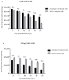
JCPyV replication in the CY- and MAD-1-infected COS-7 and SVGp12 cell lines: (a) Archetypal CY and MAD-1 strains compared to address the role of the NCCR in modulating viral replication into the COS-7 cell line at selected sampling times after infection, from 3 d.p.i until 35 d.p.i.; JCPyV DNA was quantified by qPCR. Data are expressed as the mean of three independent experiments, and error bars indicate standard deviations; ns: not significant; ** p < 0.01; *** p < 0.001. JCPyV-loads rapidly increased in MAD-1-infected cells, but only slowly in archetypal-CY-infected cultures. (b) Archetypal CY and MAD-1 strains compared to address the role of the NCCR in modulating viral replication into the SVGp12 cell line at selected sampling times after infection, from 3 d.p.i until 35 d.p.i.; JCPyV DNA was quantified by qPCR. Data are expressed as the mean of three independent experiments, and error bars indicate standard deviations. ns: not significant; *** p < 0.001. JCPyV-loads rapidly increased in MAD-1-infected SVGp12 cells, but only slowly in archetypal-CY-infected cultures.
Figure 2
Figure 2
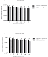
miR-J1-5p expression in the CY- and MAD-1-infected COS-7 and SVGp12 cell lines: (a) Archetypal CY and MAD-1 strains compared to address the role of the NCCR in modulating miR-J1-5p expression in the COS-7 cell line at selected sampling times after infection, from 3 d.p.i until 35 d.p.i.; JCPyV miR-J1-5p expression was quantified by quantitative TaqMan real-time PCR. Data are expressed as the mean of three independent experiments, and error bars indicate standard deviations; ns: not significant; * p < 0.05; ** p < 0.01; *** p < 0.001. The miRNA levels observed in the archetypal-CY-infected cells were lower than those observed in the MAD-1-infected cultures. (b) Archetypal CY and MAD-1 strains compared to address the role of the NCCR in modulating miR-J1-5p expression in the SVGp12 cell line at selected sampling times after infection, from 3 d.p.i until 35 d.p.i.; JCPyV miR-J1-5p expression was quantified by quantitative TaqMan real-time PCR. Data are expressed as the mean of three independent experiments, and error bars indicate standard deviations; ns: not significant; ** p < 0.01; *** p < 0.001. The miRNA levels observed in the archetypal-CY-infected cells were lower than those observed in the MAD-1-infected cultures.
Figure 3
Figure 3
Expression of miRNA levels observed in exosomes generated during archetypal and MAD-1 JCPyV infection of COS-7 and SVGp12 cell lines: (a) JCPyV miR-J1-5p expression was quantified by quantitative TaqMan real-time PCR at selected sampling times after infection, from 3 d.p.i until 35 d.p.i.; data are expressed as the mean of three independent experiments, and error bars indicate standard deviations; ns: not significant; * p < 0.05; ** p < 0.01. The results showed that JCPyV miRNA was present in the exosomes starting from 3 d.p.i. during archetypal and MAD-1 JCPyV infection of the COS-7 cell line. A higher expression of miRNA levels was observed in exosomes generated during archetypal JCPyV infection than was observed for the rr-NCCR. (b) JCPyV miR-J1-5p expression was quantified by quantitative TaqMan real-time PCR at selected sampling times after infection, from 3 d.p.i until 35 d.p.i;. data are expressed as the mean of three independent experiments, and error bars indicate standard deviations; ns: not significant; ** p < 0.01; *** p < 0.001. The results showed that JCPyV miRNA was present in the exosomes starting from 3 d.p.i. during archetypal and MAD-1 JCPyV infection of the SVGp12 cell line. A higher expression of miRNA levels was observed in exosomes generated during infection with the CY strain than was observed for the MAD-1 strain.
Figure 4
Figure 4
JCPyV replication in the CY- and MAD-1-infected COS-7 and SVGp12 cell lines after the addition of JCPyV-miRNA-5p exosome preparations to uninfected cells: (a) Viral replication into the COS-7 cell line at selected sampling times after infection, from 3 d.p.i until 35 d.p.i., was quantified by qPCR. Data are expressed as the mean of three independent experiments, and error bars indicate standard deviations; ns: not significant. (b) Viral replication into the SVGp12 cell line at selected sampling times after infection, from 3 d.p.i until 35 d.p.i., was quantified by qPCR. Data are expressed as the mean of three independent experiments, and error bars indicate standard deviations; ns: not significant; ** p < 0.01; *** p < 0.001.
Figure 5
Figure 5
WB analysis of the VP1 protein expressed in CY- and MAD-1-infected cells after infection. Cell protein extracts from uninfected (U) and infected COS-7 and SVGp12 cells were harvested at 7 d.p.i., 14 d.p.i., 21 d.p.i., 28 d.p.i., and 35 d.p.i., and analyzed by WB to evaluate the expression of the VP1 protein. Equal amounts of protein from whole-cell protein extracts were separated by SDS–PAGE, transferred to PVDF, and probed using anti-VP1 antibody. Normalization to the host cell number (VP1/GAPDH) was performed. (a) Results showing that the VP1 protein in the CY-infected COS-7 cells was expressed 7 d.p.i., and its level increased during the infection experiments, reaching the maximum expression at 21 d.p.i., although remaining detectable for up to 35 d.p.i. (b) Results showing that the VP1 protein in the MAD-1-infected SVGp12 cells was expressed 7 d.p.i., and its level increased during the infection experiments, reaching the maximum expression at 35 d.p.i.

Similar articles

Cited by

References

    1. Hirsch H.H., Kardas P., Kranz D., Leboeuf C. The Human JC Polyomavirus (JCPyV): Virological Background and Clinical Implications. APMIS. 2013;121:685–727. doi: 10.1111/apm.12128. - DOI - PubMed
    1. Atkinson A.L., Atwood W.J. Fifty Years of JC Polyomavirus: A Brief Overview and Remaining Questions. Viruses. 2020;12:969. doi: 10.3390/v12090969. - DOI - PMC - PubMed
    1. Ranganathan P.N., Khalili K. The Transcriptional Enhancer Element, Kappa B, Regulates Promoter Activity of the Human Neurotropic Virus, JCV, in Cells Derived from the CNS. Nucleic Acids Res. 1993;21:1959–1964. doi: 10.1093/nar/21.8.1959. - DOI - PMC - PubMed
    1. Romagnoli L., Wollebo H.S., Deshmane S.L., Mukerjee R., Del Valle L., Safak M., Khalili K., White M.K. Modulation of JC Virus Transcription by C/EBPbeta. Virus Res. 2009;146:97–106. doi: 10.1016/j.virusres.2009.09.005. - DOI - PMC - PubMed
    1. Wollebo H.S., Melis S., Khalili K., Safak M., White M.K. Cooperative Roles of NF-κB and NFAT4 in Polyomavirus JC Regulation at the KB Control Element. Virology. 2012;432:146–154. doi: 10.1016/j.virol.2012.06.010. - DOI - PMC - PubMed

Publication types

LinkOut - more resources