

Obstruction of ventriculoperitoneal shunt by air bubble: A case report
- PMID: 36147100
- PMCID: PMC9486626
- DOI: 10.1016/j.amsu.2022.104351
Obstruction of ventriculoperitoneal shunt by air bubble: A case report
Abstract
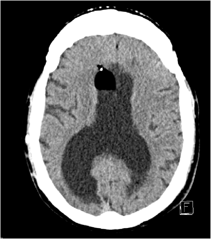
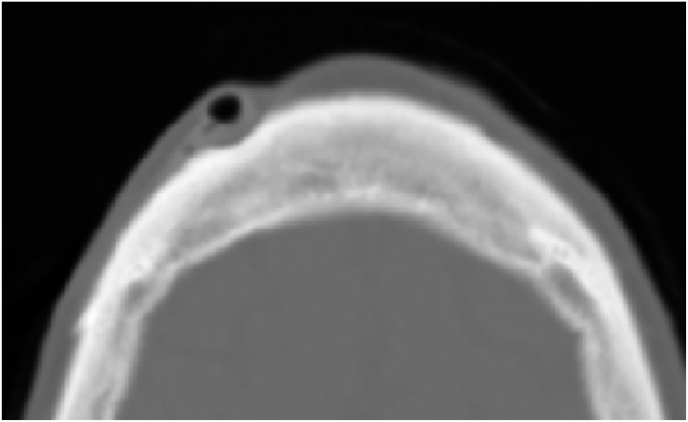
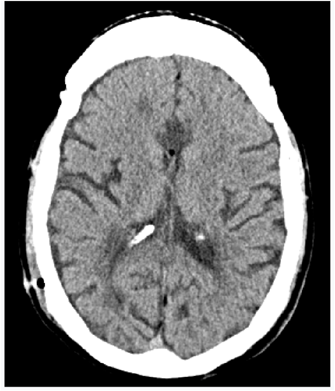
Complications related to Ventriculoperitoneal shunt placement are common, and multiple. Among them blockage and infection. We report a case of 44 years old man admitted to our hospital after an obstruction of his ventriculo-peritoneal shunt by an air bubble that caused behavioral problems and confusion. The patient was operated twice, the last time the puncture point had to be changed. The follow up was marked by a clear clinical improvement. Shunt malfunction continues to be a common neurosurgical problem in patients with shunted hydrocephalus, often leading to frequent and sometimes lengthy hospital stays. This case illustrates the management of this rare situation causing air bubble shunt obstruction.
Keywords: CT SCAN; Case report; Hydrocephalus; Pneumocephalus; Shunt failure.
© 2022 Published by Elsevier Ltd on behalf of IJS Publishing Group Ltd.
Conflict of interest statement
The authors declare having no conflicts of interest for this article.
Figures


References
-
- Paff Michelle, Alexandru-Abrams Daniela, Muhonen Michael, Loudon William. Ventriculoperitoneal shunt complications: a review. Interdisciplinary Neurosurg. September 2018;13:66–70.
-
- Agha R.A., Franchi T., Sohrabi C., Mathew G., Kerwan A., SCARE Group The SCARE 2020 guideline: updating consensus surgical CAse REport (SCARE) guidelines. Int. J. Surg. 2020 S1743-9191(20)30771-30778. - PubMed
-
- Kourosh Tavanaiepour and George J. Kaptain. Non-surgical Approach to Ventriculo-Peritoneal Shunt Failure and Pneumoventricle. JSM Neurosurgery and Spine.
-
- Perrin Richard G., Bernstein Mark. Tension pneumoventricle after placement of a ventriculoperitoneal shunt: a novel treatment strategy. J. Neurosurg. 2005;102:386–388. - PubMed
Publication types
LinkOut - more resources
Full Text Sources
