

Bilateral Vision Loss in an Adult Patient with Woakes' Syndrome: An Unprecedented Case
- PMID: 36147257
- PMCID: PMC9487011
- DOI: 10.4103/joco.joco_302_21
Bilateral Vision Loss in an Adult Patient with Woakes' Syndrome: An Unprecedented Case
Abstract
Purpose: To report a rare case of Woakes' syndrome presented with bilateral vision loss.
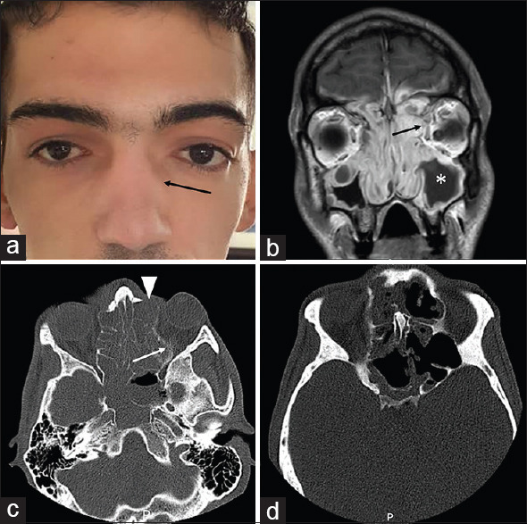
Methods: A 28-year-old male with a 1-year history of vision loss in the left eye was referred to the neuro-ophthalmology clinic after sudden vision loss in his right eye. A detailed review of clinical findings and the presumed pathophysiological basis of vision loss was performed.
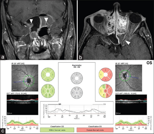
Results: Neuroimaging revealed bilateral massive nasal polyps, sphenoid sinus mucocele formation, and optic nerve dehiscence inside the sphenoid sinus. The vision in the right eye was restored after pulse corticosteroid therapy; however, the left eye remained severely visually compromised even after nasal polypectomy and mucocele drainage.
Conclusion: Sinonasal disorders should be sought for patients with unexplained vision loss, as prompt intervention could be vision-saving in these patients.
Keywords: Sphenoid sinus mucocele; Vision loss; Woakes’ syndrome.
Copyright: © 2022 Journal of Current Ophthalmology.
Conflict of interest statement
There are no conflicts of interest.
Figures
Similar articles
-
A Case of Woakes' Syndrome With A Bilateral, Large Nasal Polyp.Ear Nose Throat J. 2023 Jul 11:1455613231186473. doi: 10.1177/01455613231186473. Online ahead of print. Ear Nose Throat J. 2023. PMID: 37431670
-
Adult-onset woakes' syndrome: Report of two cases.Ann Med Surg (Lond). 2021 Aug 9;69:102695. doi: 10.1016/j.amsu.2021.102695. eCollection 2021 Sep. Ann Med Surg (Lond). 2021. PMID: 34457252 Free PMC article.
-
Woakes' syndrome.BMJ Case Rep. 2019 Mar 31;12(3):e229021. doi: 10.1136/bcr-2018-229021. BMJ Case Rep. 2019. PMID: 30936362 Free PMC article.
-
[Clinical analysis of patients with sphenoid sinus mucocele and literature review].Lin Chuang Er Bi Yan Hou Tou Jing Wai Ke Za Zhi. 2015 Nov;29(21):1850-2. Lin Chuang Er Bi Yan Hou Tou Jing Wai Ke Za Zhi. 2015. PMID: 26930903 Review. Chinese.
-
Large sphenoid mucocele presenting with cranial neuropathies in a 10-year-old boy: case report and literature review.Childs Nerv Syst. 2022 May;38(5):1035-1039. doi: 10.1007/s00381-021-05314-5. Epub 2021 Aug 4. Childs Nerv Syst. 2022. PMID: 34347143 Review.
References
Publication types
LinkOut - more resources
Full Text Sources