

Mother's physical activity during pregnancy and newborn's brain cortical development
- PMID: 36147297
- PMCID: PMC9486075
- DOI: 10.3389/fnhum.2022.943341
Mother's physical activity during pregnancy and newborn's brain cortical development
Abstract
Background: Physical activity is known to improve mental health, and is regarded as safe and desirable for uncomplicated pregnancy. In this novel study, we aim to evaluate whether there are associations between maternal physical activity during pregnancy and neonatal brain cortical development.
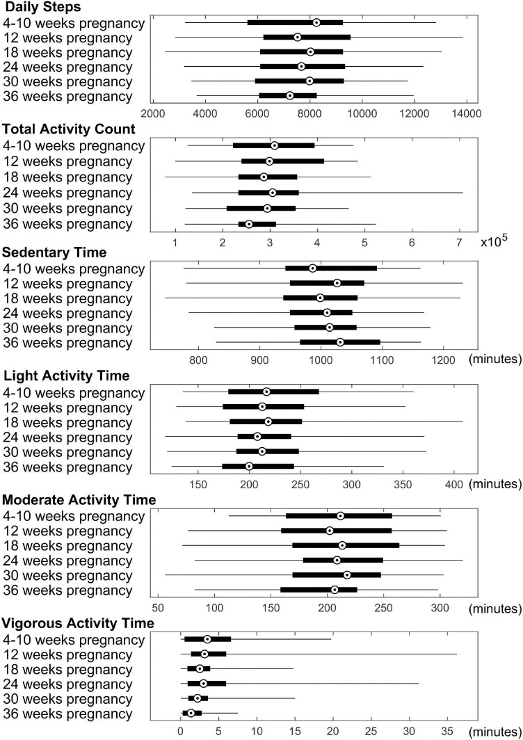
Methods: Forty-four mother/newborn dyads were included in this longitudinal study. Healthy pregnant women were recruited and their physical activity throughout pregnancy were documented using accelerometers worn for 3-7 days for each of the 6 time points at 4-10, ∼12, ∼18, ∼24, ∼30, and ∼36 weeks of pregnancy. Average daily total steps and daily total activity count as well as daily minutes spent in sedentary/light/moderate/vigorous activity modes were extracted from the accelerometers for each time point. At ∼2 weeks of postnatal age, their newborns underwent an MRI examination of the brain without sedation, and 3D T1-weighted brain structural images were post-processed by the iBEAT2.0 software utilizing advanced deep learning approaches. Cortical surface maps were reconstructed from the segmented brain images and parcellated to 34 regions in each brain hemisphere, and mean cortical thickness for each region was computed for partial correlation analyses with physical activity measures, with appropriate multiple comparison corrections and potential confounders controlled.
Results: At 4-10 weeks of pregnancy, mother's daily total activity count positively correlated (FDR corrected P ≤ 0.05) with newborn's cortical thickness in the left caudal middle frontal gyrus (rho = 0.48, P = 0.04), right medial orbital frontal gyrus (rho = 0.48, P = 0.04), and right transverse temporal gyrus (rho = 0.48, P = 0.04); mother's daily time in moderate activity mode positively correlated with newborn's cortical thickness in the right transverse temporal gyrus (rho = 0.53, P = 0.03). At ∼24 weeks of pregnancy, mother's daily total activity count positively correlated (FDR corrected P ≤ 0.05) with newborn's cortical thickness in the left (rho = 0.56, P = 0.02) and right isthmus cingulate gyrus (rho = 0.50, P = 0.05).
Conclusion: We identified significant relationships between physical activity in healthy pregnant women during the 1st and 2nd trimester and brain cortical development in newborns. Higher maternal physical activity level is associated with greater neonatal brain cortical thickness, presumably indicating better cortical development.
Keywords: cortical thickness; exercise in pregnant women; neonatal brain development; physical activity during pregnancy; structural MRI.
Copyright © 2022 Na, Raja, Phelan, Tadros, Moore, Wu, Wang, Li, Glasier, Ramakrishnaiah, Andres and Ou.
Conflict of interest statement
The authors declare that the research was conducted in the absence of any commercial or financial relationships that could be construed as a potential conflict of interest.
Figures


References
-
- ACOG (2020). Physical Activity and Exercise During Pregnancy and the Postpartum Period. Washington, DC: ACOG.
-
- Akhavan M., Miladi-Gorji H., Emami-Abarghoie M., Safari M., Sadighi-Moghaddam B., Vafaei A., et al. (2013). Maternal voluntary exercise during pregnancy enhances the spatial learning acquisition but not the retention of memory in rat pups via a trkb-mediated mechanism: the role of hippocampal BDNF expression. Iran J. Basic Med. Sci. 16 955–961. - PMC - PubMed
Grants and funding
LinkOut - more resources
Full Text Sources