. 2022 Sep 6:13:939780.
doi: 10.3389/fphar.2022.939780. eCollection 2022.

Muscarinic receptor M3 activation promotes fibrocytes contraction

Affiliations

Muscarinic receptor M3 activation promotes fibrocytes contraction

Pauline Henrot et al. Front Pharmacol. .

Abstract

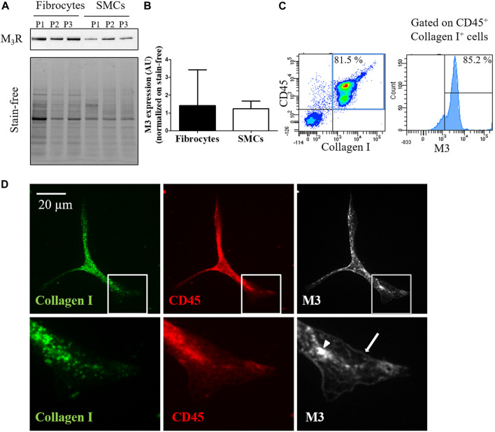
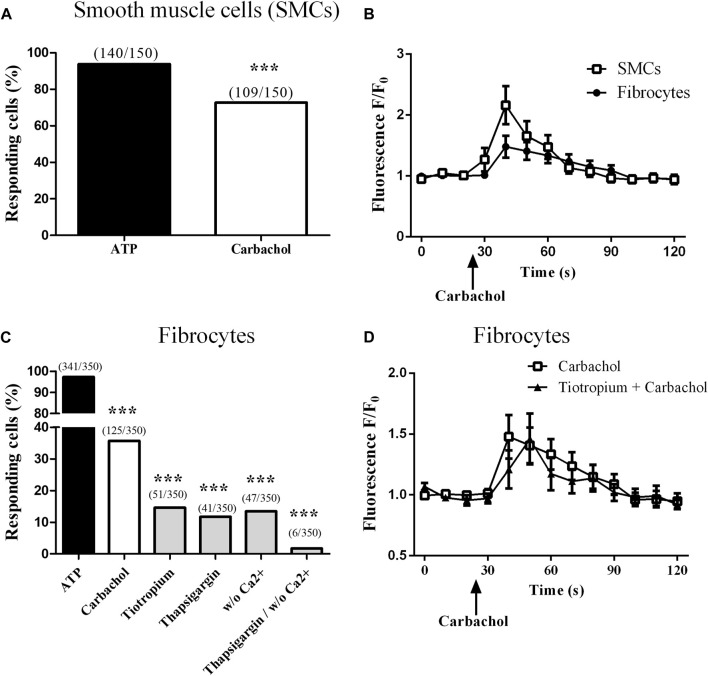
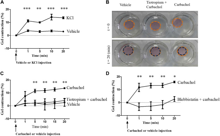
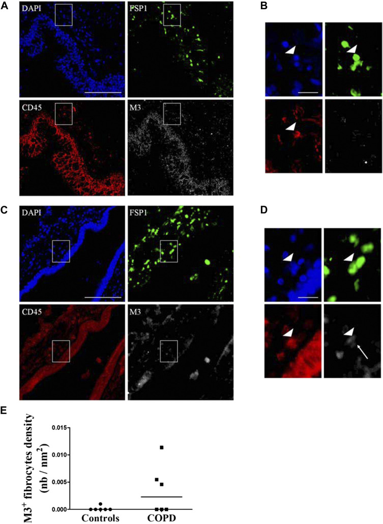
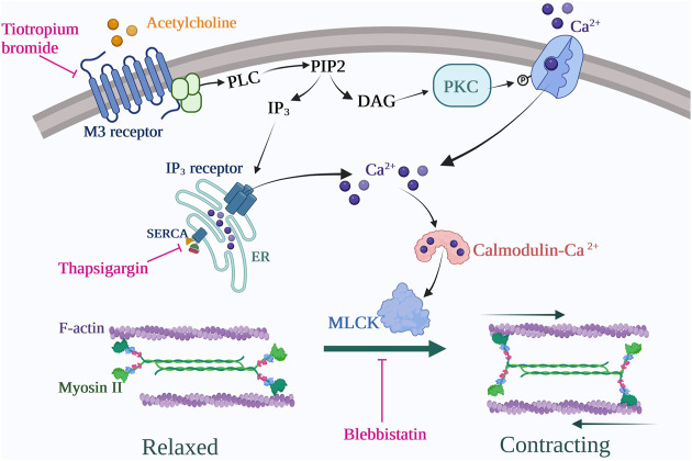
Fibrocytes are monocyte-derived cells able to differentiate into myofibroblasts-like cells. We have previously shown that they are increased in the bronchi of Chronic Obstructive Pulmonary Disease (COPD) patients and associated to worse lung function. COPD is characterized by irreversible airflow obstruction, partly due to an increased cholinergic environment. Our goal was to investigate muscarinic signalling in COPD fibrocytes. Fibrocytes were isolated from 16 patients with COPD's blood and presence of muscarinic M3 receptor was assessed at the transcriptional and protein levels. Calcium signalling and collagen gels contraction experiments were performed in presence of carbachol (cholinergic agonist) ± tiotropium bromide (antimuscarinic). Expression of M3 receptor was confirmed by Western blot and flow cytometry in differentiated fibrocytes. Immunocytochemistry showed the presence of cytoplasmic and membrane-associated pools of M3. Stimulation with carbachol elicited an intracellular calcium response in 35.7% of fibrocytes. This response was significantly blunted by the presence of tiotropium bromide: 14.6% of responding cells (p < 0.0001). Carbachol induced a significant contraction of fibrocytes embedded in collagen gels (13.6 ± 0.3% versus 2.5 ± 4.1%; p < 0.0001), which was prevented by prior tiotropium bromide addition (4.1 ± 2.7% of gel contraction; p < 0.0001). Finally, M3-expressing fibrocytes were also identified in situ in the peri-bronchial area of COPD patients' lungs, and there was a tendency to an increased density compared to healthy patient's lungs. In conclusion, around 1/3 of COPD patients' fibrocytes express a functional muscarinic M3 receptor. Cholinergic-induced fibrocyte contraction might participate in airway diameter reduction and subsequent increase of airflow resistance in patients with COPD. The inhibition of these processes could participate to the beneficial effects of muscarinic antagonists for COPD treatment.

Keywords: COPD; M3; cholinergic; contraction; fibrocyte.


Conflict of interest statement

PH, EE, EM, SP, GC, JFQ, PE, TT and RM report no relevant conflict of interest; ID, P-OG and PB report a patent (EP N3050574: Use of plerixafor for treating and/or preventing acute exacerbations of chronic obstructive pulmonary disease (Berger et al., 2019)) (granted); POG reports grants, personal fees and non-financial support from AstraZeneca, personal fees and non-financial support from Chiesi, personal fees and non-financial support from GlaxoSmithKline, personal fees and non-financial support from Novartis, personal fees and non-financial support from Sanofi, outside the submitted work; MZ reports grants from AstraZeneca; grants “Fondation Bordeaux Université,” with funding from “Assistance Ventilatoire à Domicile” (AVAD) and “Fédération Girondine de Lutte contre les Maladies Respiratoires” (FGLMR) and personal fees from AstraZeneca, Boehringer Ingelheim, Novartis, Chiesi, GlaxoSmithKline and non-financial support Lilly outside the submitted work; PB reports grants from AstraZeneca, Glaxo-Smith-Kline, Novartis, Chiesi, which support COBRA during the conduct of the study; grants and personal fees from AstraZeneca, BoehringerIngelheim, Novartis, personal fees and non-financial support from Chiesi, Sanofi, Menarini, outside the submitted work; ID reports grant from the “Fondation Bordeaux Université,” with funding from “Assistance Ventilatoire à Domicile” (AVAD) and “Fédération Girondine de Lutte contre les Maladies Respiratoires” (FGLMR) during the conduct of the study. The remaining authors declare that the research was conducted in the absence of any commercial or financial relationships that could be construed as a potential conflict of interest.

Figures

FIGURE 1
FIGURE 1
Fibrocytes from patients with COPD express the muscarinic M3 receptor. (A). Presence of M3 receptor (M3R) is assessed in cultured fibrocytes from patients with COPD or in bronchial smooth muscle cells (SMCs) by Western blot. Total protein load is shown with the stain-free image. (B). quantification of M3R expression normalized on stain-free. No significant difference is found between fibrocytes and SMCs (p = 0.40). (C). M3 receptor is identified by flow cytometry in fibrocytes isolated from the blood of COPD patients. In the represented experiment, 85.2% of fibrocytes (double-positive CD45-Collagen I cells, blue square) express M3. (D). Representative confocal photographs show the expression of M3 receptor (white) evidenced in the cytoplasm (arrow head) and at the surface (arrow) of a fibrocyte (CD45+ Collagen I+ fusiform cell).
FIGURE 2
FIGURE 2
Muscarinic M3 receptor expressed on cultured fibrocytes from patients with COPD is functional. Variations of relative cytosolic Ca2+ concentration were monitored by fluorescence video microscopy in fluo4-loaded fibrocytes and smooth muscle cells (SMCs). (A). Proportion of responding cells to 10−4 M carbachol for control SMCs. (B). Variations of relative cytosolic Ca2+ concentration against time (mean ± SD) in responding cells, for SMCs (squares) and fibrocytes (circles). (C). Proportion of responding cells for COPD fibrocytes stimulated with 10−4 M ATP and with 10−4 M carbachol, in the absence or the presence of tiotropium bromide 10−6 M, thapsigargin 10 µM and with or without extracellular Ca2+. (D). Variations of relative cytosolic Ca2+ concentration against time (mean ± SD) in responding cells, for fibrocytes stimulated with carbachol with (triangles) or without (squares) prior addition of tiotropium bromide 10−6 M. Significant difference ***p < 0.0001, otherwise no significant difference.
FIGURE 3
FIGURE 3
Muscarinic M3 receptor activation promotes COPD fibrocytes contraction. (A). Significant contraction of fibrocytes-embedded collagen gels stimulated with KCl 80 mM (squares) compared to control vehicle (circles). Percentage of gel contraction is presented against time. (B). Aspect of fibrocytes-embedded collagen gels stimulated with control vehicle or with carbachol 10−4 M, with or without prior tiotropium bromide 10−6 M addition, before and 20 min after the beginning of the experiment. (C). Significant contraction of fibrocytes-embedded collagen gels stimulated with carbachol 10−4 M (squares) compared to control vehicle (circles) or tiotropium bromide 10−6 M + carbachol 10−4 M (triangles). (D). Significant inhibition of carbachol-induced gel contraction (squares) when incubating collagen gels with blebbistatin 10 µM prior to carbachol stimulation (triangles). Significant difference *p < 0.05, **p < 0.01, ***p < 0.001, otherwise no significant difference.
FIGURE 4
FIGURE 4
Identification of M3-expressing fibrocytes in the lungs of COPD patients and healthy subjects. Peri-bronchial fibrocytes are identified as double-positive FSP1+ (green) CD45+ (red) cells. M3 staining is represented in white and nuclei are stained with DAPI (blue). (A) representative triple immunostaining of a healthy subject’s lung tissue. Scale bar, 100 µm. (B). Close-up view of the sub-epithelial region indicated in A (white square). Arrowhead indicate a fibrocyte, identified as a double positive FSP1-CD45 cell, without M3 expression. Scale bar, 20 µm. (C) representative triple immunostaining of a COPD patient’s lung tissue. Scale bar, 100 µm. (D). Close-up view of the sub-epithelial region indicated in C (white square). Arrowhead indicate a fibrocyte, identified as a double positive FSP1-CD45 cell, expressing M3 (white arrow). Scale bar, 20 µm. (E). Quantification of M3+ fibrocytes densities as represented by the number of peri-bronchial triple-positive cells divided by the area of lamina propria analyzed.
FIGURE 5
FIGURE 5
Proposed signalling pathway for COPD fibrocytes stimulated with cholinergic/muscarinic agonists. Muscarinic M3 receptor activation leads to intracellular Ca2+ increase via emptying of endoplasmic reticulum (ER) stores as well as secondary extracellular Ca2+ uptake (store-operator calcium entry). Cytosolic Ca2+ rise leads to acto-myosin dimerization via myosin light chain kinase (MLCK) activation, thus leading to cell contraction. These processes are inhibited at the receptor level by tiotropium bromide (M2/M3 antagonist) use, at the endoplasmic reticulum level by thapsigargin use and at the contraction level by blebbistatin use. Created with BioRender.com.

Similar articles

Cited by

References

    1. Afroj T., Mitsuhashi A., Ogino H., Saijo A., Otsuka K., Yoneda H., et al. (2021). Blockade of PD-1/PD-L1 pathway enhances the antigen-presenting capacity of fibrocytes. J. Immunol. 206, 1204–1214. 10.4049/jimmunol.2000909 - DOI - PMC - PubMed
    1. Agustí A., Hogg J. C. (2019). Update on the pathogenesis of chronic obstructive pulmonary disease. N. Engl. J. Med. 381, 1248–1256. 10.1056/NEJMra1900475 - DOI - PubMed
    1. Arai N., Kondo M., Izumo T., Tamaoki J., Nagai A. (2010). Inhibition of neutrophil elastase-induced goblet cell metaplasia by tiotropium in mice. Eur. Respir. J. 35, 1164–1171. 10.1183/09031936.00040709 - DOI - PubMed
    1. Barnes P. J., Belvisi M. G., Mak J. C., Haddad E. B., O’Connor B. (1995). Tiotropium bromide (Ba 679 BR), a novel long-acting muscarinic antagonist for the treatment of obstructive airways disease. Life Sci. 56, 853–859. 10.1016/0024-3205(95)00020-7 - DOI - PubMed
    1. Barnes P. J. (1993). Muscarinic receptor subtypes in airways. Eur. Respir. J. 6, 328–331. - PubMed

LinkOut - more resources