. 2022 Sep 6:13:956886.
doi: 10.3389/fphar.2022.956886. eCollection 2022.

Functional characterization of cannabidiol effect on the serotonergic neurons of the dorsal raphe nucleus in rat brain slices

Affiliations

Functional characterization of cannabidiol effect on the serotonergic neurons of the dorsal raphe nucleus in rat brain slices

Aitziber Mendiguren et al. Front Pharmacol. .

Abstract

Cannabidiol (CBD), the main non-psychoactive cannabinoid found in the cannabis plant, elicits several pharmacological effects via the 5-HT1A receptor. The dorsal raphe nucleus (DRN) is the main serotonergic cluster in the brain that expresses the 5-HT1A receptor. To date, the effect of CBD on the neuronal activity of DRN 5-HT cells and its interaction with somatodendritic 5-HT1A autoreceptors have not been characterized. Our aim was to study the effect of CBD on the firing activity of DRN 5-HT cells and the 5-HT1A autoreceptor activation by electrophysiological and calcium imaging techniques in male Sprague-Dawley rat brain slices. Perfusion with CBD (30 μM, 10 min) did not significantly change the firing rate of DRN 5-HT cells or the inhibitory effect of 5-HT (50-100 μM, 1 min). However, in the presence of CBD (30 μM, 10 min), the inhibitory effects of 8-OH-DPAT (10 nM) and ipsapirone (100 nM) were reduced by 66% and 53%, respectively. CBD failed to reverse ipsapirone-induced inhibition, whereas perfusion with the 5-HT1A receptor antagonist WAY100635 (30 nM) completely restored by 97.05 ± 14.63% the firing activity of 5-HT cells. Administration of AM251 (1 µM), MDL100907 (30 nM), or picrotoxin (20 μM) did not change the blockade produced by CBD (30 μM) on ipsapirone-induced inhibition. Our study also shows that CBD failed to modify the KCl (15 mM, 4 min)-evoked increase in [Ca2+]i or the inhibitory effect of ipsapirone (1 μM, 4 min) on KCl-evoked [Ca2+]i. In conclusion, CBD does not activate 5-HT1A autoreceptors, but it hindered the inhibitory effect produced by selective 5-HT1A receptor agonists on the firing activity of DRN 5-HT cells through a mechanism that does not involve CB1, 5-HT2A, or GABAA receptors. Our data support a negative allosteric modulation of DRN somatodendritic 5-HT1A receptor by CBD.

Keywords: 5-HT1A receptor; calcium; cannabidiol; dorsal raphe nucleus; firing; rat; slice.


Conflict of interest statement

The authors declare that the research was conducted in the absence of any commercial or financial relationships that could be construed as a potential conflict of interest.

Figures

FIGURE 1
FIGURE 1
Effect of CBD on the firing rate of DRN 5-HT cells and on 5-HT-induced inhibition. (A,B) Representative examples of firing rate recordings from two DRN cells, which show the effect of CBD (30 μM) on the firing activity of 5-HT neurons (A) and the effect of CBD (30 μM) on 5-HT (100 μM)-induced inhibition (B). Vertical lines refer to the integrated firing rate values (spikes per 10 s) and the horizontal lines represent the time scale. Drugs were perfused at the concentration and for the time indicated by the horizontal bars. ACSF was applied in the continuous presence of the vehicle (DMSO 0.08%). (C) Bar histograms showing the firing rate of 5-HT cells (mean ± SEM) before and after perfusion with the vehicle (DMSO 0.08%, n = 5), 5-HT (100 μM) (n = 5), CBD (30 μM) (n = 5), and CBD (30 μM) + 5-HT (100 μM) (n = 5). (D) Bar histograms showing the percentage of 5-HT (100 µM)-induced inhibition from the basal firing rate (mean ± SEM) in the absence and presence of CBD (30 μM) (n = 5). *p < 0.005 compared with the firing rate before drug application by paired Student’s t-test. ΦP < 0.005 compared with the firing rate after administration of CBD (30 μM) in the absence of 5-HT by paired Student’s t-test.
FIGURE 2
FIGURE 2
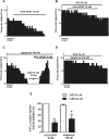
Effect of CBD on the selective 5-HT1A receptor agonist-induced inhibition of the firing activity of DRN 5-HT cells. (A–D) Representative examples of firing rate recordings from DRN cells, which show the inhibition of the firing activity of 5-HT neurons by 8-OH-DPAT (10 nM) (A), the blockade of 8-OH-DPAT (10 nM)-induced inhibition by CBD (B), the inhibition of the firing rate of DRN 5-HT cells by ipsapirone (100 nM) and the effect of CBD (30 μM) or the 5-HT1A receptor antagonist WAY100635 (30 nM) on that inhibition (C), and the blockade of the effect of CBD on the inhibition of the firing activity of DRN 5-HT neurons induced by ipsapirone (100 nM) (D). Vertical lines refer to the integrated firing rate values (spikes per 10 s), and the horizontal lines represent the time scale. Drugs were perfused at the concentration and for the time indicated by the horizontal bars. (E) Bar histograms showing the percentage of inhibition from the basal firing rate (mean ± SEM) induced by 8-OH-DPAT (10 nM) and ipsapirone (100 nM) in the absence (n = 5 and n = 13, respectively) and presence of CBD (30 μM, n = 6 and n = 10, respectively). *p < 0.05 compared with the inhibition induced by 8-OH-DPAT (10 nM) or ipsapirone (100 nM) in the absence of CBD (30 μM) by unpaired Student’s t-test.
FIGURE 3
FIGURE 3
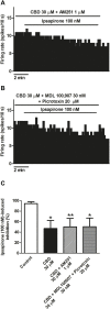
Mechanisms involved in the blockade by CBD of the ipsapirone-induced inhibitory effect on DRN 5-HT cells. (A,B) Representative examples of firing rate recordings from two DRN 5-HT neurons, which represent the effect of CBD on the ipsapirone (100 nM)-induced inhibition in the presence of the CB1 receptor antagonist AM251 (1 μM) (A) or in the presence of the GABAA receptor antagonist picrotoxin (20 μM) and the 5-HT2A receptor antagonist MDL100907 (30 nM) (B). (C) Bar histograms showing the percentage of inhibition from the basal firing rate (mean ± SEM) induced by ipsapirone (100 nM) in the absence (n = 13) and presence of CBD (30 μM, n = 10), CBD (30 μM) + AM251 (1 µM) (n = 9) and CBD (30 μM) + MDL100907 (30 nM) + picrotoxin (20 μM) (n = 9). *p < 0.05, **p < 0.01 compared with ipsapirone (100 nM) group (control) by unpaired Student’s t-test.
FIGURE 4
FIGURE 4
Effect of ipsapirone and CBD on KCl-evoked calcium influx in DRN. (A,B) Representative recordings of KCl-evoked (15 mM, 4 min) [Ca2+]i in DRN 5-HT cells in the absence (A) or presence of ipsapirone (1 µM) (B). Vertical lines refer to the change in KCl-evoked 340/380 ratio, and the horizontal lines represent the time scale. (C) Bar histograms showing mean ± SEM of the AUC for KCl (15 mM)-evoked [Ca2+]i in DRN 5-HT cells in the absence (n = 29 cells, three slices from three rats) and presence of ipsapirone (1 µM) (n = 26 cells, three slices from three rats), CBD (30 μM, n = 21 cells, three slices from three rats), and CBD (30 µM) + ipsapirone (1 µM) (n = 17 cells, three slices from two rats). Each histogram represents the mean ± SEM of the percentage from the first AUC for KCl-evoked [Ca2+]i. *p < 0.001 compared with the corresponding groups without ipsapirone (1 µM) by two-way ANOVA (with CBD and ipsapirone as the main factors) followed by Bonferroni´s post-hoc test.

References

    1. Abigail M., Xiaohua Li. (2010). 5-HT1A receptor-regulated signal transduction pathways in brain. Cell. Signal. 22 (10), 1406–1412. 10.1016/j.cellsig.2010.03.019 - DOI - PMC - PubMed
    1. Adell A., Celada P., Artigas F. (2001). The role of 5-HT1B receptors in the regulation of serotonin cell firing and release in the rat brain. J. Neurochem. 79, 172–182. 10.1046/j.1471-4159.2001.00550.x - DOI - PubMed
    1. Aghajanian G. K., Lakoski J. M. (1984). Hyperpolarization of serotonergic neurons by serotonin and LSD: Studies in brain slices showing increased K+ conductance. Brain Res. 305, 181–185. 10.1016/0006-8993(84)91137-5 - DOI - PubMed
    1. Albert P. R., Vahid-Ansari F. (2019). The 5-HT1A receptor: Signaling to behavior. Biochimie 161, 34–45. 10.1016/j.biochi.2018.10.015 - DOI - PubMed
    1. Andrade R., Huereca D., Lyons J. G., Andrad e. E. M., McGregor K. M. (2015). 5-HT1A receptor-mediated Autoinhibition and the control of serotonergic cell firing. ACS Chem. Neurosci. 6 (7), 1110–1115. 10.1021/acschemneuro.5b00034 - DOI - PMC - PubMed