. 2022 Aug 16;18(14):5291-5308.
doi: 10.7150/ijbs.73438. eCollection 2022.

SNHG16 upregulation-induced positive feedback loop with YAP1/TEAD1 complex in Colorectal Cancer cell lines facilitates liver metastasis of colorectal cancer by modulating CTCs epithelial-mesenchymal transition

Affiliations

SNHG16 upregulation-induced positive feedback loop with YAP1/TEAD1 complex in Colorectal Cancer cell lines facilitates liver metastasis of colorectal cancer by modulating CTCs epithelial-mesenchymal transition

Zhenxian Xiang et al. Int J Biol Sci. .

Abstract

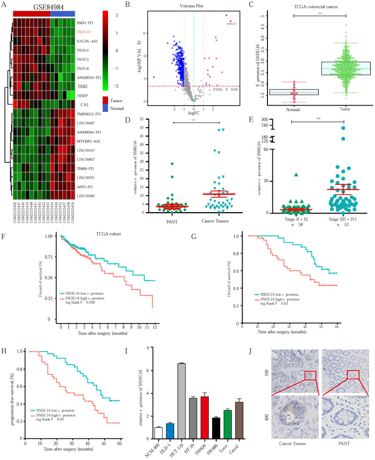
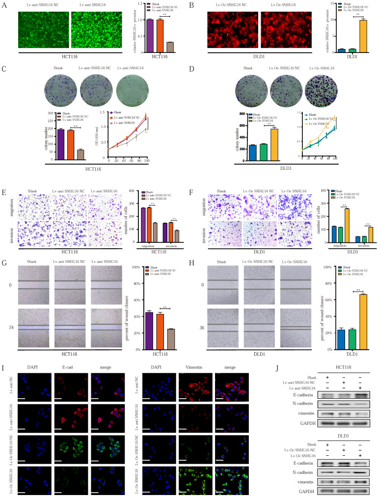
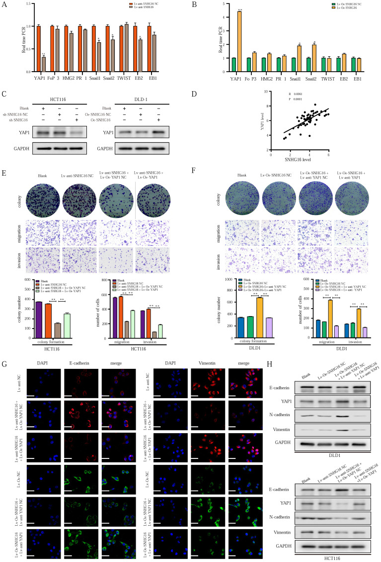
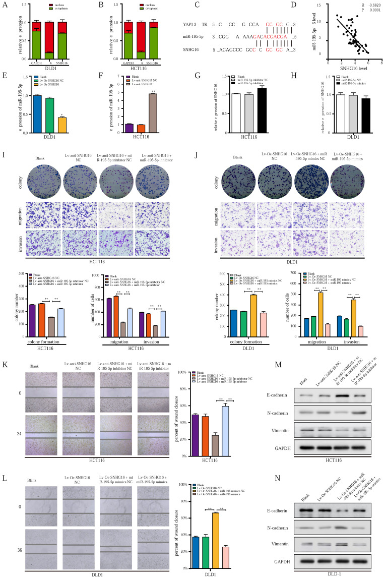
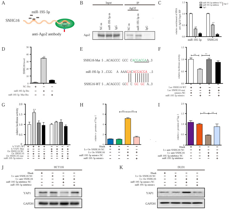
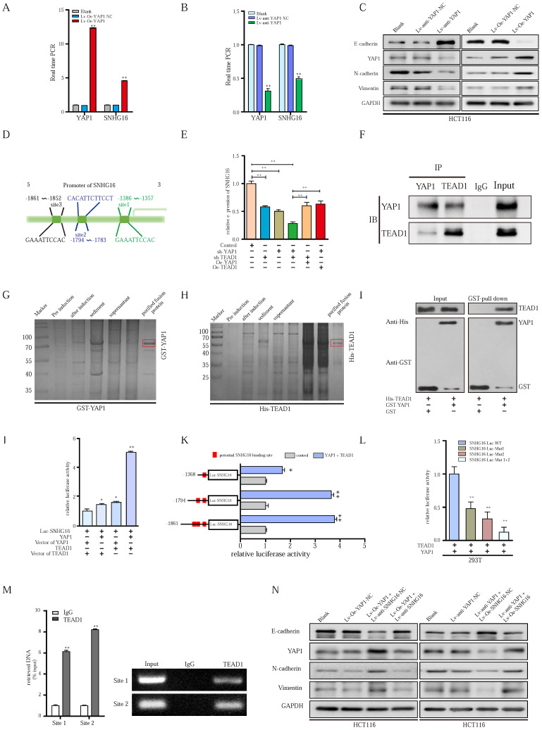
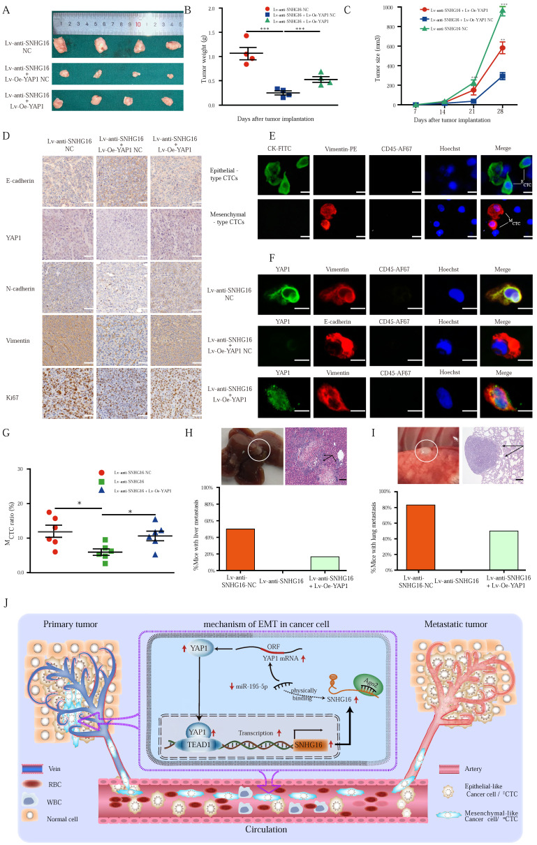
Circulating tumor cells (CTCs) are important precursors of colorectal cancer (CRC) metastasis. The epithelial-mesenchymal transition (EMT) process facilitates CTC invasion by allowing these cells to evade antimetastatic checkpoints to mediate distant metastasis. However, the specific molecular mechanism of tumor EMT remains largely unknown. Based on our previous research on the YAP1 pathway, we further studied the upstream molecule small nucleolar RNA host gene 16 (SNHG16), whose expression was correlated with advanced TNM stage, distant metastasis, and poor prognosis in CRC patients. Furthermore, loss- and gain-of-function assays revealed that SNHG16 promoted CRC colony formation, proliferation, migration, invasion, EMT, mesenchymal-like CTC generation, and liver metastasis through YAP1. Mechanistically, SNHG16 acted as a miRNA sponge to sequester miR-195-5p on Ago2, thereby protecting YAP1 from repression. Moreover, YAP1 bound TEA domain transcription factor 1 (TEAD1) to form a YAP1/TEAD1 complex, which in turn bound two sites in the promoter of SNHG16 and regulate SNHG16 transcription. Finally, in vivo experiments showed that the inhibition of SNHG16 suppressed tumor progression, and that YAP1 rescued the effect of SNHG16 on tumor progression. Herein, we have clarified a hitherto unexplored SNHG16-YAP1/TEAD1 positive feedback loop, that may be a candidate target for CRC treatment.

Keywords: CTCs; EMT; SNHG16; TEAD1; YAP1.


Conflict of interest statement

Competing Interests: We declare that we do not have any commercial or associative interest that represents a conflict of interest in connection with this work.

Figures

Figure 1
Figure 1
SNHG16 is upregulated in colorectal cancer tissues and indicates poor prognosis of colorectal cancer patients. A: Heatmap of most differentially expressed lncRNAs in GSE84984. B: Differential expressions of lncRNAs were shown in volcano plots. C: SNHG16 expression was validated by qRT-PCR in TCGA datasets, mean ± SD is shown. D: qRT-PCR analyses of SNHG16 expression in CRC tumor tissue (n = 45) and paired non-tumor tissue (n = 45), mean ± SEM is shown. E: qRT-PCR analyses of SNHG16 expression in different clinical stages, mean ± SEM is shown. F: Kaplan-Meier analyses of the correlation between SNHG16 expression level and OS in TCGA cohort. G-H: Kaplan-Meier analyses of the correlation between SNHG16 expression and OS or PFS of CRC patients. I: Relative expression of SNHG16 in CRC cell lines and colon epithelial cell line. J: ISH analyses of SNHG16 expression in cancer tissues and adjacent normal tissues. All representative data are from three independent experiments. Statistical analysis was conducted using Student's t-test. Error bars, SEM. *p < 0.05, **p < 0.01.
Figure 2
Figure 2
The ectopic expression of lncRNA-SNHG16 affects proliferation, migration, invasion, and EMT of CRC cells. A: Micrographs of lentivirus-mediated SNHG16 knockdown in HCT116 and the knockdown effect were validated. B: Micrographs of lentivirus-mediated SNHG16 overexpression in DLD1 and the overexpression effect were validated. C-D: Colony formation and proliferation assays were performed after SNHG16 knockdown (C) or overexpression (D) in HCT116 or DLD1, respectively. E-F: Migration and invasion assays of CRC cell line after SNHG16 knockdown (E) or overexpression (F), respectively. G-H: Wound healing assay was performed in CRC cell lines following SNHG16 knockdown or overexpression. I-J: EMT-associated markers were detected by immunofluorescence (I) and WB (J) in CRC cell lines with SNHG16 knockdown or overexpression. All representative data are from three independent experiments. Statistical analysis was conducted using one-way ANOVA. Error bars, SEM. *p < 0.05, **p < 0.01.
Figure 3
Figure 3
LncRNA-SNHG16 facilitates CRC cellular proliferation, migration, invasion, and EMT in a YAP1-dependent manner. A-B: Relative expression levels of representative EMT-related transcription factors were detected following SNHG16 knockdown (A) or overexpression (B), respectively. C: YAP1 protein levels were assessed after SNHG16 knockdown or overexpression in CRC cells, respectively. D: The correlations between the expression SNHG16 and YAP1 in CRC tissues were analyzed by Pearson's correlation. E: Colony formation and Transwell assay were performed to determine the colony formation, migration, and invasion ability of HCT116 co-transfected with Lv-anti-SNHG16 and Lv-Oe-YAP1. F: Colony formation and Transwell assay were performed to determine the colony formation, migration, and invasion ability of DLD1 co-transfected with Lv-Oe-SNHG16 and Lv-anti-YAP1. G: The expression level of EMT markers in HCT116 (HCT116 Blank, HCT116 Lv-anti-SNHG16-NC, HCT116 Lv-anti-SNHG16 + Lv-Oe-YAP1-NC, HCT116 Lv-anti-SNHG16 + Lv-Oe-YAP1) and DLD1 (DLD1 Blank, DLD1 Lv-Oe-SNHG16-NC, DLD1 Lv-Oe-SNHG16 + Lv-anti-YAP1 NC, DLD1 Lv-Oe-SNHG16 + Lv-anti-YAP1) was detected by immunofluorescence. H: The expression level of EMT markers and YAP1 in HCT116 (HCT116 Blank, HCT116 Lv-anti-SNHG16-NC, HCT116 Lv-anti-SNHG16 + Lv-Oe-YAP1-NC, HCT116 Lv-anti-SNHG16 + Lv-Oe-YAP1) and DLD1 (DLD1 Blank, DLD1 Lv-Oe-SNHG16-NC, DLD1 Lv-Oe-SNHG16 + Lv-anti-YAP1 NC, DLD1 Lv-Oe-SNHG16 + Lv-anti-YAP1) was detected by WB. All representative data are from three independent experiments. Statistical analysis was conducted using Student's t-test of one-way ANOVA. Error bars, SEM. *p < 0.05, **p < 0.01.
Figure 4
Figure 4
miR-195-5p could potently abrogate the effect of SNHG16/YAP1 axis on tumor progression. A-B: The cellular localization of SNHG16 in HCT116 (A) and DLD1 (B) was identified by subcellular fractionation assay. C: The putative miR-195-5p binding sites with SNHG16 and YAP1 3′-UTR were shown. D: The correlations between SNHG16 and YAP1 expression in CRC tissues were analyzed by Pearson's correlation. E: MiR-195-5p expression levels in DLD1 after SNHG16 overexpression. F: MiR-195-5p expression levels in HCT116 after SNHG16 overexpression. G: SNHG16 expression levels in HCT116 after the miR-195-5p knockdown. H: SNHG16 expression levels in DLD1 after miR-195-5p overexpression. I: The colony formation, migration, and invasion of HCT116 Blank, HCT116 Lv-anti-SNHG16-NC, HCT116 Lv-anti-SNHG16 + miR-195-5p inhibitor NC, and HCT116 Lv-anti-SNHG16 + miR-195-5p inhibitor was detected by colony formation, and Transwell assay. J: The colony formation, migration and invasion of DLD1 (DLD1 Blank, DLD1 Lv-Oe-SNHG16-NC, DLD1 Lv-Oe-SNHG16 + miR-195-5p mimics NC, DLD1 Lv-Oe-SNHG16 + miR-195-5p mimics) was detected by colony formation and transwell assay. K: Wound healing assays were performed to determine the migration ability of HCT116 co-transfected with Lv-anti-SNHG16 and miR-195-5p inhibitor. L: Wound healing assays were performed to determine the migration ability of HCT116 co-transfected with Lv-Oe-SNHG16 and miR-195-5p mimics. M: The protein expression level of EMT markers in SNHG16 knockdown-HCT116 cells with or without miR-195-5p inhibitor. N: The protein expression level of EMT markers in SNHG16 overexpression-DLD1 cells with or without miR-195-5p mimics. All representative data are from three independent experiments. Statistical analysis was conducted using one-way ANOVA. Error bars, SEM. *p < 0.05, **p < 0.01.
Figure 5
Figure 5
lncRNA-SNHG16 functions as a ceRNA and sponges miR-195-5p, further regulating YAP1 expression and facilitating tumor progression. A: Schematic illustration of anti-Ago2 RIP strategy. B: Ago2 protein was immunoprecipitated and purified from cell extracts and was further detected by WB. C: In the presence of miR-195-5p inhibitor or negative control, the relative expression of SNHG16 and miR-195-5p bound to Ago2 or IgG was measured by RT-qPCR. D: Interaction between miR-195-5p and SNHG16 was confirmed by RNA pulldown assay. E: The position of the miR-195-5p binding site in SNHG16 is shown. Mutation (underlined) was introduced into SNHG16 to disrupt base-pairing with miR-195-5p seed sequence. F: A dual-luciferase reporter assay was performed in 293T to reveal the binding of miR-195-5p to SNHG16. G: Luciferase assay among SNHG16 (SNHG16-WT and SNHG16-mut), miR-195-5p, and dual-luciferase vector YAP1 (WT and mut) were performed in 293T to confirm the interplay of these three factors. H: The relative expression of YAP1 mRNA in SNHG16-overexpressed-DLD1 cells with or without miR-195-5p mimics. I: The relative expression of YAP1 mRNA in SNHG16 knockdown-HCT116 cells with or without miR-195-5p inhibitor. J: The YAP1 protein expression in SNHG16 knockdown-HCT116 cells with or without miR-195-5p inhibitor. K: The YAP1 protein expression in SNHG16 overexpressed-DLD1 cells with or without miR-195-5p mimics. All representative data are from three independent experiments. Statistical analysis was conducted using one-way ANOVA. Error bars, SEM. *p < 0.05, **p < 0.01.
Figure 6
Figure 6
YAP1 could combine with TEAD1, forming a complex that binds to the promoter region of SNHG16 and activates its transcription. A-B: Relative expression of SNHG16 was detected following YAP1 overexpression (A) or knockdown (B). C: The protein expression levels of EMT markers following YAP1 knockdown or overexpression. D: Schematic diagram showing the human SNHG16 upstream promoter region (top), including the predicted TEAD1-binding regions. E: Expression levels of SNHG16 were detected when we changed the level of either YAP1 or TEAD1. F: Co-IP experiments were used to detect the interplay between YAP1 and TEAD1. G: The GST-tagged YAP1 was successfully induced and purified from Escherichia coli. H: The His-tagged TEAD1 was successfully induced and purified from Escherichia coli. I: GST pull down assay between YAP1 and TEAD1. J: A dual-luciferase reporter assay driven by the SNHG16 promoter was co-transfected in the presence or absence of YAP1 or TEAD1. K: Selective mutation analyses to detect YAP1/TEAD1-complex-responsive-regions in the SNHG16 promoter in 293T. L: Dual-luciferase reporter assay was performed to detect YAP1/TEAD1-complex-responsive-regions in the SNHG16 promoter. M: ChIP assay was performed to detect the binding site of TEAD1 to the SNHG16 promoter, including CHIP1 and CHIP2 in HCT116 cells. Input, 10% of total lysate. N: The protein expression of E-cadherin, N-cadherin, vimentin, and YAP1 in eight different groups were detected by WB. All representative data are from three independent experiments. Statistical analysis was conducted using one-way ANOVA or Student's t-test. Error bars, SEM. *p < 0.05, **p < 0.01.
Figure 7
Figure 7
The alteration of SNHG16 expression influenced the CRC tumorigenesis and CTC generation in vivo. A-C: The morphological characteristics (A), weight (B), and size (C) of tumor xenograft in different group (HCT116 Lv-anti-SNHG16-NC, HCT116 Lv-anti-SNHG16 + Lv-Oe-YAP1-NC, HCT116 Lv-anti-SNHG16 + Lv-Oe-YAP1) injected nude mice. Error bars, SEM. D: IHC analyzed the expression of E-cadherin, Vimentin, N-cadherin, Ki67, and YAP1 in different group (HCT116 Lv-anti-SNHG16-NC, HCT116 Lv-anti-SNHG16 + Lv-Oe-YAP1-NC, HCT116 Lv-anti-SNHG16 + Lv-Oe-YAP1). Scale bar, 50 µm. E: Representative images of CTC isolated from two mice, respectively. Epithelial-like CTC (ECTC) means CTC is accompanied by overexpression of CK. Mesenchymal-like CTC (MCTC) means CTC is accompanied by overexpression of vimentin. Scale bar, 10 µm. F: The expression of YAP1 and EMT marker on CTCs that isolated from different group (HCT116 Lv-anti-SNHG16-NC, HCT116 Lv-anti-SNHG16 + Lv-Oe-YAP1-NC, HCT116 Lv-anti-SNHG16 + Lv-Oe-YAP1). Scale bar, 10 µm. G: The MCTC ratio of mouse blood collected from different group (HCT116 Lv-anti-SNHG16-NC, HCT116 Lv-anti-SNHG16 + Lv-Oe-YAP1-NC, HCT116 Lv-anti-SNHG16 + Lv-Oe-YAP1) Error bars, SEM. H-I: Representative images of metastatic lesions in the liver of mice in the HCT116/Lv-anti-SNHG16 NC group, and representative images of metastatic lesions in the lung of mice in the HCT116 Lv-anti-SNHG16 + Lv-Oe-YAP1 group. Representative hematoxylin and eosin-stained sections of metastatic nodules in the liver and lung are shown. Scale bar, 100 µm. J: A schematic diagram illustrated the mechanism by which LncRNA SNHG16 promoted CRC progression and liver metastasis. All representative data are from three independent experiments. Statistical analysis was conducted using one-way ANOVA. Error bars, SEM. *p < 0.05, **p < 0.01.

Similar articles

Cited by

References

    1. Siegel RL, Miller KD, Fuchs HE, Jemal A. Cancer statistics, 2022. CA: a cancer journal for clinicians. 2022;72:7–33. - PubMed
    1. Xu L, Li X, Cai M, Chen J, Li X, Wu WK. et al. Increased expression of Solute carrier family 12 member 5 via gene amplification contributes to tumour progression and metastasis and associates with poor survival in colorectal cancer. Gut. 2016;65:635–46. - PMC - PubMed
    1. Miller KD, Nogueira L, Mariotto AB, Rowland JH, Yabroff KR, Alfano CM. et al. Cancer treatment and survivorship statistics, 2019. CA: a cancer journal for clinicians. 2019;69:363–85. - PubMed
    1. Torre LA, Bray F, Siegel RL, Ferlay J, Lortet-Tieulent J, Jemal A. Global cancer statistics, 2012. CA: a cancer journal for clinicians. 2015;65:87–108. - PubMed
    1. van de Velde CJ, Boelens PG, Borras JM, Coebergh JW, Cervantes A, Blomqvist L. et al. EURECCA colorectal: multidisciplinary management: European consensus conference colon & rectum. Eur J Cancer. 2014;50:1. e- e34. - PubMed

Publication types

MeSH terms