. 2022 Sep 16:17:4321-4337.
doi: 10.2147/IJN.S364862. eCollection 2022.

Glucose-Functionalized Silver Nanoparticles as a Potential New Therapy Agent Targeting Hormone-Resistant Prostate Cancer cells

Affiliations

Glucose-Functionalized Silver Nanoparticles as a Potential New Therapy Agent Targeting Hormone-Resistant Prostate Cancer cells

Mariana Morais et al. Int J Nanomedicine. .

Abstract

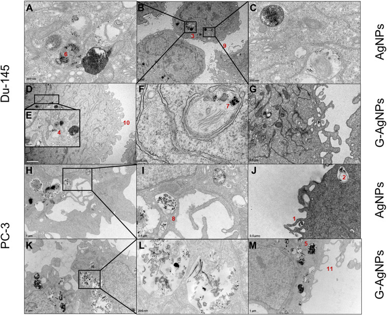
Purpose: Silver nanoparticles (AgNPs) have shown great potential as anticancer agents, namely in therapies' resistant forms of cancer. The progression of prostate cancer (PCa) to resistant forms of the disease (castration-resistant PCa, CRPC) is associated with poor prognosis and life quality, with current limited therapeutic options. CRPC is characterized by a high glucose consumption, which poses as an opportunity to direct AgNPs to these cancer cells. Thus, this study explores the effect of glucose functionalization of AgNPs in PCa and CRPC cell lines (LNCaP, Du-145 and PC-3).

Methods: AgNPs were synthesized, further functionalized, and their physical and chemical composition was characterized both in water and in culture medium, through UV-visible spectrum, dynamic light scattering (DLS), transmission electron microscopy (TEM) and Fourier-transform infrared spectroscopy (FTIR). Their effect was assessed in the cell lines regarding AgNPs' entering pathway, cellular proliferation capacity, ROS production, mitochondrial membrane depolarization, cell cycle analysis and apoptosis evaluation.

Results: AgNPs displayed an average size of 61nm and moderate monodispersity with a slight increase after functionalization, and a round shape. These characteristics remained stable when redispersed in culture medium. Both AgNPs and G-AgNPs were cytotoxic only to CRPC cells and not to hormone-sensitive ones and their effect was higher after functionalization showing the potential of glucose to favor AgNPs' uptake by cancer cells. Entering through endocytosis and being encapsulated in lysosomes, the NPs increased the ROS, inducing mitochondrial damage, and arresting cell cycle in S Phase, therefore blocking proliferation, and inducing apoptosis.

Conclusion: The nanoparticles synthesized in the present study revealed good characteristics and stability for administration to cancer cells. Their uptake through endocytosis leads to promising cytotoxic effects towards CRPC cells, revealing the potential of G-AgNPs as a future therapeutic approach to improve the management of patients with PCa resistant to hormone therapy or metastatic disease.

Keywords: Warburg Effect; castration-resistant prostate cancer; hormonal therapy; therapy resistance.


Conflict of interest statement

The authors report no conflicts of interest in this work.

Figures

Figure 1
Figure 1
Characterization of AgNPs and G-AgNPs by means of (A) Size and PDI; (B) ζ-potential, and (C) UV-Vis spectra. (D) TEM images of AgNPs and G-AgNPs (Scale bars = 100 nm). (E) ATR-FTIR spectra of AgNPs and G-AgNPs. Error bars represent mean ± SD (n ≥ 3).
Figure 2
Figure 2
Stability of AgNPs in RPMI supplemented with 10% of FBS after 2 h incubation, at 37 °C: effect on the size (A), PDI (B) and ζ-potential (C) of AgNPs. Errors bars represent mean ± SD (n = 3).
Figure 3
Figure 3
Evaluation of cell viability, by Resazurin Assay, upon treatment with AgNPs and G-AgNPs for 24 hours in LNCaP cell line (A) Du-145 cell line (B) and PC-3 cell line (C). Results are expressed as percentage of control (untreated cells), as mean ± SEM.
Figure 4
Figure 4
TEM images of Du-145 cells treated with IC25 of AgNPs (AC) or G-AgNPs (DG) and of PC-3 cells treated with IC25 of AgNPs (HJ) and G-AgNPs (KM). From each sample, three pictures were taken with different ampliations. Thus, scale bars in B and D are 2μm, in H, K and M are 1μm, in G, I and J are 0.5 μm, in A, C, E and L are 200 nm and in F is 100 nm. 1 – Nanoparticles entering the cells through endocitosis; 2 – Nanoparticles trapped in lysosomes; 3 – Nanoparticles dispersed throughout the cytoplasm; 4 – Nanoparticles localized in the nucleus; 5 – Nanoparticles localized in the mitochondria; 6–8 – Nanoparticles maintain the monodispersed nature inside the cell; 9–11 – Membrane blebbing of the cells.
Figure 5
Figure 5
Evaluation of cell proliferation, by BrDu Assay, upon treatment with AgNPs and G-AgNPs in the Du-145 cell line using the determined IC25 (A) and the determined IC50 (B); and in the PC-3 cell line using the determined IC25 (C) and the determined IC50 (D). Results are expressed as percentage of control (untreated cells), as mean ± SEM.
Figure 6
Figure 6
(A) Representative DNA histograms of cell cycle analysis assessed using a PI stain and flow cytometry of Du-145 cells treated with AgNPs and G-AgNPs in the determined IC25 and IC50; (B) quantitative analysis of cell cycle disruptions of Du-145 cells treated with AgNPs and G-AgNPs in the determined IC25 and IC50; (C) Representative DNA histograms of cell cycle analysis assessed using a PI stain and flow cytometry of PC-3 cells treated with AgNPs and G-AgNPs in the determined IC25 and IC50; (D) Quantitative analysis of cell cycle disruptions of PC-3 cells treated with AgNPs and G-AgNPs in the determined IC25 and IC50; (E) Representative flow cytometry images of apoptosis evaluation in Du-145 cells treated with AgNPs and G-AgNPs in the determined IC25 and IC50; (F) Quantitative analysis of live, early apoptotic and dead cells of Du-145 cells treated with AgNPs and G-AgNPs in the determined IC25 and IC50; (G) Representative flow cytometry images of apoptosis evaluation in PC-3 cells treated with AgNPs and G-AgNPs in the determined IC25 and IC50; (H) Quantitative analysis of live, early apoptotic and dead cells of PC-3 cells treated with AgNPs and G-AgNPs in the determined IC25 and IC50. Data are expressed as mean ± SEM.
Figure 7
Figure 7
Evaluation of ROS production, by DCFH2-DA Assay, in the Du-145 cell line upon treatment with the determined IC25 and IC50 AgNPs (A and B) and upon treatment with the determined IC25 and IC50 G-AgNPs (C and D); and in the PC-3 cell line upon treatment with the determined IC25 and IC50 AgNPs (E and F) and upon treatment with the determined IC25 and IC50 G-AgNPs (G and H). (A, C, E and G) Representative images (10×) of DCFH2-DA labeled mitochondria (Photographs taken using an Olympus IX51 microscope). (B, D, F and H) Graphical representation of the DCFH2-DA fluorescence signal intensity in the different tested conditions. T-bhp is used as positive control. Results are expressed as percentage of control (untreated cells), as mean ± SEM.
Figure 8
Figure 8
Evaluation of mitochondria membrane depolarization, by TMRE Assay, upon treatment with AgNPs and G-AgNPs in the Du-145 cell line (C) using the determined IC25 (A) and the determined IC50 (B); and in the PC-3 cell line (F) using the determined IC25 (D) and the determined IC50 (E). Representative images (10×) of TMRE labeled mitochondria (Photographs taken using an Olympus IX51 microscope). Results are expressed as percentage of control (untreated cells), as mean ± SEM.

Similar articles

Cited by

References

    1. Rawla P. Epidemiology of prostate cancer. World J Oncol. 2019;10(2):63–89. doi:10.14740/wjon1191 - DOI - PMC - PubMed
    1. Sung H, Ferlay J, Siegel RL, et al. Global cancer statistics 2020: GLOBOCAN estimates of incidence and mortality worldwide for 36 cancers in 185 countries. CA Cancer J Clin. 2021;71:209–249. doi:10.3322/caac.21660 - DOI - PubMed
    1. Lim MCJ, Baird A-M, Greene J, et al. hsa_circ_0001275 is one of a number of circRNAs dysregulated in enzalutamide resistant prostate cancer and confers enzalutamide resistance in vitro. Cancers. 2021;13(24):6383. doi:10.3390/cancers13246383 - DOI - PMC - PubMed
    1. Mottet N, Bellmunt J, Bolla M, et al. EAU-ESTRO-SIOG guidelines on prostate cancer. part 1: screening, diagnosis, and local treatment with curative intent. Eur Urol. 2017;71(4):618–629. doi:10.1016/j.eururo.2016.08.003 - DOI - PubMed
    1. Gandhi J, Afridi A, Vatsia S, et al. The molecular biology of prostate cancer: current understanding and clinical implications. Prostate Cancer Prostatic Dis. 2018;21(1):22–36. doi:10.1038/s41391-017-0023-8 - DOI - PubMed