Case Reports
. 2022 Aug 15;12(4):156-162.
eCollection 2022.

Rheumatological picture of a patient having multifocal osteonecrosis associated with sickle cell anemia: a case study

Affiliations
Case Reports

Rheumatological picture of a patient having multifocal osteonecrosis associated with sickle cell anemia: a case study

Albader Hamza Hussein et al. Am J Blood Res. .

Abstract

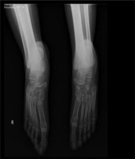
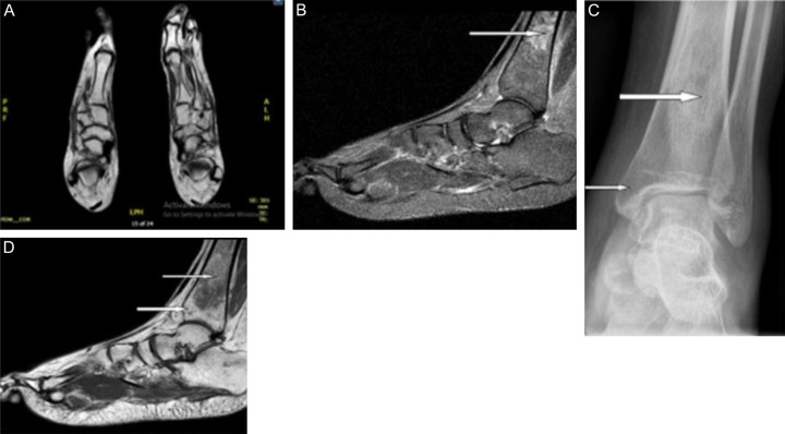
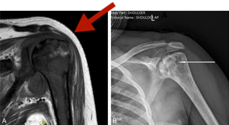
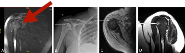
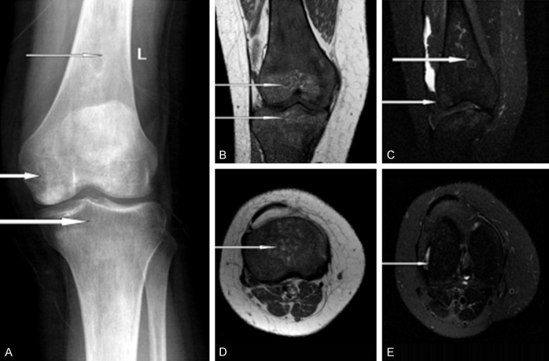
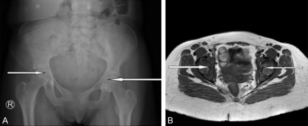
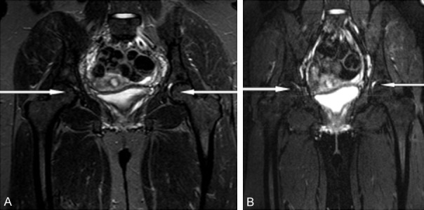
Avascular necrosis (AVN) is a critical health condition associated with local death of the bone tissue resulting in multifocal osteonecrosis (MFON). After a prior patient's consent, we present a case of sickle cell anemia associated with severe MFON that affected both long bones and short bones. She had a positive history of DVT. Initially, she presented with generalized severe bone pain with fever for seven days that got worse on the day of admission, a picture suggestive of sickle cell anemia-induced vaso-occlusive crisis. She was treated with adequate hydration, morphine, enoxaparin (a low molecular weight heparin), paracetamol and ceftriaxone. She got improved on treatment. On 5th day after admission, she developed sudden severe local tenderness at the distal tibia above the medial malleoli in both legs and she was unable to put a weight on her feet and could not stand up or walk. Plain X-ray films were not diagnostic. Complete liver function tests and kidney function tests were normal. The patient had leukocytosis, high serum urate and high serum LDH (may reflect cellular damage in bone cells). MRI scans revealed an evidence of bilateral multiple avascular necrosis in both femoral heads, left shoulder, left knee, and pelvic bones were evident. The patient's condition was evaluated and the diagnosis of MFON associated with sickle cell crisis was established. This patient responded well to same treatments and her condition got improved. In conclusion, MFON should be considered after vaso-occlusive crisis of sickle cell anemia. Plain X-ray is non-conclusive in diagnosing bony lesions induced by AVN while MRI is diagnostic.

Keywords: LDH; MFON; enoxaparin; feet AVN; leukocytosis; sickle cell anemia; uric acid.


Conflict of interest statement

None.

Figures

Figure 1
Figure 1
Chest X-ray of the patient showing normal findings.
Figure 2
Figure 2
X-ray of both feet and legs of the patient showing normal findings. X-ray was not conclusive during the severe pain attacks of vaso-occlusive crisis of sickle cell anemia.
Figure 3
Figure 3
Bilateral distal tibial avascular necrosis. X-ray of the ankle region revealing sclerotic intramedullary density. MRI axial and sagittal planes revealed abnormal area of bone marrow signal intensity low in T1WI and high in T2WI fat saturation due to the intramedullary bone infarction.
Figure 4
Figure 4
Manifestations of AVN of the humeral head. Left shoulder X-ray reveled heterogeneous sclerosis of the left humeral head due to avascular necrosis. Left shoulder coronal MRI revealing evidence of bone marrow edema and “double line sign” denoting avascular necrosis of the head of the humerus (denoting recent infarction new crisis on top of the old ones). (So, recent crisis of the bone infarction and avascular necrosis are diagnosed by MRI while plain X ray can detect the sclerosis related to the sequelae of the old ones).
Figure 5
Figure 5
Right humeral medullary infarction and avascular necrosis. A, C and D. Coronal, axial and sagittal MRI views of the shoulder revealing “Double line sign” in the humerus head with a rim of edema denoting a recent attack of avascular necrosis and intramedullary areas of high T2WI area of bone marrow edema (denoting recent bone infarction). B. X ray shoulder revealed humerus having heterogeneous areas of sclerosis denoting old avascular necrosis.
Figure 6
Figure 6
A. Plain X-ray showing left upper tibial intramedullary, distal femoral intramedullary and medial condylar areas of heterogeneous sclerosis due to the old bone infarction. B-E. Axial and coronal MRI sequences revealing areas of bone marrow edema at the distal femur intramedullary region and the medial condyle related to the new crisis of bone infarction.
Figure 7
Figure 7
Bilateral pelvic bone avascular necrosis. X-ray of both hip regions reveled linear sclerosis of avascular necrosis affecting both femoral heads. MRI of both femur heads showed linear high signal intensity of edema due to avascular necrosis.
Figure 8
Figure 8
Bilateral T2WI sequences with and without fat saturation. Coronal view showed femoral head linear high signal intensity denoting stage II avascular necrosis.

References

    1. Herman K, Pękala P, Szwedowski D, Grabowski R, Cholewiński J. Joint Function Preservation. Springer; 2022. Avascular Necrosis; pp. 161–171.
    1. Goodman SB, Maruyama M. Inflammation, bone healing and osteonecrosis: from bedside to bench. J Inflamm Res. 2020;13:913–923. - PMC - PubMed
    1. Adesina O, Brunson A, Keegan THM, Wun T. Osteonecrosis of the femoral head in sickle cell disease: prevalence, comorbidities, and surgical outcomes in California. Blood Adv. 2017;1:1287–1295. - PMC - PubMed
    1. Sun W, Shi ZC, Gao FQ, Wang BL, Li ZR. The pathogenesis of multifocal osteonecrosis. Sci Rep. 2016;6:29576. - PMC - PubMed
    1. Torgashin AN, Rodionova SS. Avascular necrosis in patients after COVID-19: development and treatment (literature review) Traumatology and Orthopedics of Russia. 2022

Publication types

LinkOut - more resources