. 2022 Aug 19:20:4717-4732.
doi: 10.1016/j.csbj.2022.08.021. eCollection 2022.

Integrating knowledge of protein sequence with protein function for the prediction and validation of new MALT1 substrates

Affiliations

Integrating knowledge of protein sequence with protein function for the prediction and validation of new MALT1 substrates

Peter A Bell et al. Comput Struct Biotechnol J. .

Abstract

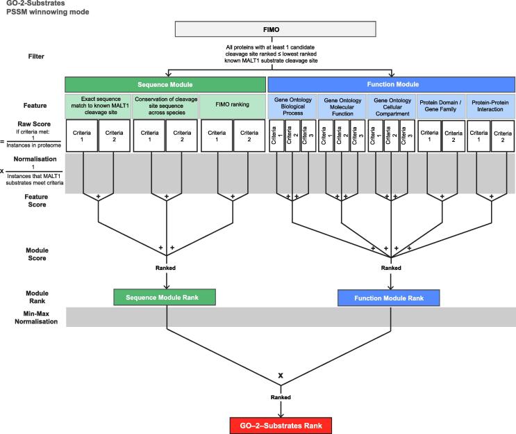
We developed a bioinformatics-led substrate discovery workflow to expand the known substrate repertoire of MALT1. Our approach, termed GO-2-Substrates, integrates protein function information, including GO terms from known substrates, with protein sequences to rank substrate candidates by similarity. We applied GO-2-Substrates to MALT1, a paracaspase and master regulator of NF-κB signalling in adaptive immune responses. With only 12 known substrates, the evolutionarily conserved paracaspase functions and phenotypes of Malt1 -/- mice strongly implicate the existence of undiscovered substrates. We tested the ranked predictions from GO-2-Substrates of new MALT1 human substrates by co-expression of candidates transfected with the oncogenic constitutively active cIAP2-MALT1 fusion protein or CARD11/BCL10/MALT1 active signalosome. We identified seven new MALT1 substrates by the co-transfection screen: TANK, TAB3, CASP10, ZC3H12D, ZC3H12B, CILK1 and ILDR2. Using catalytically inactive cIAP2-MALT1 (Cys464Ala), a MALT1 inhibitor, MLT-748, and noncleavable P1-Arg to Ala mutant versions of each substrate in dual transfections, we validated the seven new substrates in vitro. We confirmed the cleavage of endogenous TANK and the RNase ZC3H12D in B cells by Western blotting and mining TAILS N-terminomics datasets, where we also uncovered evidence for these and 12 other candidate substrates by endogenous MALT1. Thus, protein function information improves substrate predictions. The new substrates and other high-ranked MALT1 candidate substrates should open new biological frontiers for further validation and exploration of the function of MALT1 within and beyond NF-κB regulation.

Keywords: CASP10; CBM; CILK1; GO-2-Substrates; ILDR2; MALT1; Mucosa-associated lymphoid tissue lymphoma translocation protein 1; NF-kB; Prediction; Protease; Proteolysis; Proteolytic processing; Signalling; TAB3; TANK; ZC3H12B; ZC3H12D.


Conflict of interest statement

The authors declare that they have no known competing financial interests or personal relationships that could have appeared to influence the work reported in this paper.

Figures

None
Graphical abstract
Fig. 1
Fig. 1
MALT1 cleavage site analysis and FIMO proteome analyses. a Alignment of human and mouse P5 – P5′ cleavage site sequences of MALT1 substrates reported in the literature. Downward arrow indicates the site of the scissile bond. The relevant PubMed Identification (PMID) reference for each experimentally determined cleavage site is shown. Red shading indicates species-specific homology in the human and mouse cleavage site sequences. b, c Sequence logo representation of MALT1 cleavage sites in human (b) and mouse (c) substrates, generated using the ggseqlogo package in R. d Schematic of the workflow we developed to generate the Position Specific Scoring Matrix (PSSM) from the published MALT1 substrate cleavage sites. The PSSM was inputted into FIMO (Find Individual Motif Occurrences) to identify sequences in all human proteins that most closely matched the PSSM. e PSSM derived from human and mouse MALT1 substrate cleavage sites. Light to dark red colour range represents the increasing relative frequency of each amino acid from P4 – P1′. f Scatter plot showing the ranking of FIMO scores of known MALT1 P4 – P1′ cleavage site sequences found in the human protein substrates versus sensitivity. Locally weighted scatterplot smoothing was used to generate the line of best fit. Red: proteins reported to be cleaved by the CARD–BCL10–MALT1 (CBM) complex; green: proteins reported to be cleaved by cIAP2-MALT1 without evidence of CBM cleavage yet published. (For interpretation of the references to colour in this figure legend, the reader is referred to the web version of this article.)
Fig. 2
Fig. 2
GO-2-Substrates integrates sequence, GO and protein features to rank candidate substrates. Schematic of the GO-2-Substrates bioinformatic workflow we developed to predict substrate cleavage sites. Features that we considered valuable in predicting cleavage were assigned to two modules, either the Sequence Module or the Function Module. Feature-specific criteria were used to derive raw scores for each protein that were normalized to the proportion of published MALT1 substrates meeting each criterion. Criteria scores were summed to yield Feature Scores, which in turn were summed to yield Module scores, which were then ranked and min–max normalized. Finally, the ranked product of normalized Sequence and Function Module ranks was derived and designated as the GO-2-Substrates rank.
Fig. 3
Fig. 3
A cell-based screen for validation of GO-2-Substrates predictions. a The top 30 GO-2-Substrates ranked proteins, their corresponding FIMO, Function and Sequence Module ranks and best ranked candidate cleavage site. Known substrates are coloured. Proteins of boxed gene names were tested for MALT1 cleavage in co-transfection assays. b, c Scatter plots visualizing the distribution of proteins selected (red) for co-transfection screen in terms of their Sequence and Function rank (b), or GO-2-Substrates rank (c). d Schematic of cDNA expression constructs used in the co-transfection screen encoding: constitutively active cIAP2-MALT1; inactive cIAP2-MALT1 (C464A), and e CARD11 (L251P), BCL10 and MALT1 that assemble active CBM. Myc, FLAG and 6 × His C-terminal tags are as indicated. f Schematic of HOIL1 (RBCK1) positive control cDNA expression construct used for co-transfection with the previously reported cut site and molecular weights of MALT1 cleavage products shown. g Western blot analysis of lysates from HEK293 cells co-transfected with RBCK1. Full-length proteins are indicated with a black arrow; red arrow indicates C-terminal cleavage product of RBCK1 (C-RBCK1). β-actin, loading control was detected by rabbit β-actin antibody. Positions of electrophoretic mobility of molecular weight markers are as shown. (For interpretation of the references to colour in this figure legend, the reader is referred to the web version of this article.)
Fig. 4
Fig. 4
RNases ZC3H12D and ZC3H12B are novel MALT1 substrates. a, d Schematic of ZC3H12B and ZC3H12D cDNA expression constructs used for co-transfection with the predicted cut sites and molecular weights of MALT1 cleavage products shown. Myc and FLAG C-terminal tags are as indicated. c-f, Western blot analysis of lysates from HEK293 cells co-transfected with either ZC3H12D (b,c) or ZC3H12B (e, f), together with active or inactive forms of MALT1, or active MALT1 in the presence of a specific MALT1 inhibitor (MLT-748). Full-length proteins are indicated with a black arrow; red arrows indicate C-terminal cleavage products of ZC3H12D and ZC3H12B. c, f Cleavage of ZC3H12D and ZC3H12B are virtually eliminated where the MALT1 cleavage site is mutated to (c) ZC3H12D (R64A) and (f) ZC3H12B (R165A). * Denotes minor cleavage products of ZC3H12B generated at a different site to MALT1 by unknown protease. Mouse α-FLAG antibody was used to detect the proteins as indicated. β-actin, loading control was detected by rabbit β-actin antibody. Positions of electrophoretic mobility of molecular weight markers are as shown. (For interpretation of the references to colour in this figure legend, the reader is referred to the web version of this article.)
Fig. 5
Fig. 5
Immune signalling proteins TAB3 and CASP10 are cleaved by MALT1. a, e Schematic of TAB3 (a) and CASP10 (e) cDNA expression constructs used for co-transfection showing the predicted cut sites and molecular weights of MALT1 cleavage products. Myc and FLAG C-terminal tags are as indicated. b-d, f-h Western blot analysis HEK293 cells lysates co-transfected with either TAB3 (b) or CASP10 (f); together with active or inactive forms of MALT1, or with active MALT1 and the allosteric MALT1 inhibitor (MLT-748). N = 3 independent biological experiments are shown; g, h cleavage by MALT1 in the CBM is shown in triplicate n = 3. Full-length proteins are indicated with a black arrow; C-terminal cleavage product of TAB3 (C-TAB3) is indicated by red arrow. c C-TAB3 was only detected upon proteasome inhibition with MG-132. d, h Cleavage of TAB3 and CASP10 are eliminated where the predicted MALT1 cleavage site was mutated to TAB3 (R605A) (d) and CASP10 (R254A) (h). Mouse α-FLAG antibody was used to detect the proteins as indicated.β-actin, loading control was detected by rabbit β-actin antibody. The positions of electrophoretic mobility of molecular weight markers are as shown. Imaged immunoblots are displayed at K = 0, except as indicated when K = 1. K value refers to the curve applied to the pixel intensity histogram in Image Studio Lite. (For interpretation of the references to colour in this figure legend, the reader is referred to the web version of this article.)
Fig. 6
Fig. 6
Validation of GO-2-Substrates prediction of CILK1 and ILDR2 as novel MALT1 substrates. Schematic of CILK1 (a) and ILDR2 (e) used for co-transfection with the predicted cut sites and molecular weights of MALT1 cleavage products shown. Myc and FLAG C-terminal tags are as indicated. b-g Western blot analysis of lysates from HEK293 cells co-transfected with either CILK1 (b) or ILDR2 (f), together with active (cIAP2-MALT1, CBM) or inactive (cIAP2-MALT1 C464A) forms of MALT1, or with active MALT1 in the presence of a specific MALT1 inhibitor (MLT-748). Full-length proteins are indicated with a black arrow; C-terminal cleavage products of CILK1 (C-CILK1) and ILDR2 (C-ILDR2) are indicated by red arrows. c Inhibition of CILK1 cleavage is greater in cells treated with the irreversible active site inhibitor of MALT1 (z-VRPR-fmk) compared with a reversible allosteric inhibitor (MLT-748). d Cleavage of CILK1 is eliminated where the MALT1 cleavage site is mutated to CILK1 (R407A). g ILDR2 is cleaved by CBM proportional to increased MALT1 cDNA transfected from 1.25 µg to 3.25 μg as indicated by a black-filled triangle. Mouse α-FLAG antibody was used to detect the proteins as indicated. β-actin, loading control was detected by rabbit β-actin antibody. Positions of electrophoretic mobility of molecular weight markers are as shown. Imaged immunoblots are displayed at K = 0, except as indicated when K = 1. K value refers to the curve applied to the pixel intensity histogram in Image Studio Lite. (For interpretation of the references to colour in this figure legend, the reader is referred to the web version of this article.)
Fig. 7
Fig. 7
GO-2-Substrates rank is predictive of cleavage by MALT1. a The equations used to generate receiver operating characteristic (ROC) curves. True positives (TP), false positives (FP), true negatives (TN) and false negatives (FN). b-e ROC curves and associated area under the curve (AUC) were calculated using precrec package in R to measure the performance of FIMO (b), the Function Module (c), the Sequence Module (d), or GO-2-Substrates (e) in the classification of outcomes from the co-transfection screen. Outcomes were defined as 'positive’ (n = 6) or ‘negative’ (n = 24) where evidence of cleavage was reproducibly detected or absent, respectively. In terms of AUC, the Function and Sequence Modules both surpassed the performance of FIMO for classification. GO-2-Substrates was the best classifier overall. f Definition of precision used to generate Precision-Recall (PR) curves. g-j PR curves and associated PR-AUC were calculated using the precrec package in R to measure the performance of FIMO (g), the Function Module (h), the Sequence Module (i), or GO-2-Substrates (j) in the classification of outcomes from the co-transfection screen. When applied alone or combined with the Substrate Module as part of GO-2-Substrates, the Function Module increased precision at most sensitivity thresholds versus FIMO or the Sequence Module, respectively. k Scatterplot showing the experimentally derived classification precision of GO-2-Substrates, at the thresholds of all proteins included in the co-transfection screen, up to GO-2-Substrates rank 196 (sensitivity = 1). l Precision at the GO-2-Substrates rank thresholds of all positive outcomes from the co-transfection screen. A precision of 0.5 was determined for classifications at the GO-2-Substrates rank threshold of 40.
Fig. 8
Fig. 8
Expansion of analysis identifies TANK as a novel MALT1 substrate. GO-2-Substrates can be analyzed in two modes, which rank a different range of proteins according to their potential for cleavage by MALT1. a 'PSSM winnowing mode' considers only proteins containing a candidate cleavage site with FIMO ranking better than the last known MALT1 substrate cleavage site, or b 'Whole proteome mode' that includes every human protein. c TANK was identified as one of the top 15 functionally ranked candidate substrates of GO-2-Substrates analyzed in Whole proteome mode. d, Schematic of TANK cDNA expression construct used in the MALT1 co-transfection screen, together with the predicted cut-site and MALT1 cleavage products if cut. e Western blot showing TANK cleavage by cIAP2-MALT1, which was not observed where TANK was co-transfected with inactive cIAP2-MALT1 (C464A) or using noncleavable MALT1 (R215K). f TANK was cleaved by co-transfected active CBM. g Apparent molecular weights of TANK cleavage products predicted from cleavage at the indicated cut-site in TANK. h TANK in vitro cleavage by MALT in 0.8 M Na-citrate, 0.1 mM EGTA, 0.05 % CHAPS, 1 mM DTT, 200 mM Tris-HCl, pH 7.4, for 2 h at 37 °C; cleavage was proportional to MALT1 concentration. i Mutagenesis of TANK (R125K) eliminated MALT1 cleavage by the CBM in the co-transfection assay. j TBK1 was not cleaved by co-transfected CBM. k Endogenous TANK cleavage in SSK41 and RAJI cells stimulated with PMA/ionomycin for the times shown and in the presence of MG-132. Positive controls, cleavage fragments (Δ) of RELB and CYLD. β-actin and β-tubulin loading controls were detected by rabbit β-actin and mouse β-tubulin antibodies. Positions of electrophoretic mobility of molecular weight markers are shown.
Fig. 9
Fig. 9
Endogenous ZC3H12D is processed by MALT1 in two independent normal human B lymphocyte cell lines. a Raw mass spectrometry data from proteomic and TMT-TAILS analysis of PMA/ionomycin-stimulated human B lymphocytes were searched against the human proteome using MSFragger. TMT-labelled neo-N-termini generated by protease activity were identified by PSMs, which were compared in sequence with the predicted neo-N-termini of GO-2-Substrates candidate substrates. Annotated fragment spectrum of a ZC3H12D PSM at the predicted MALT1 cleavage site is shown. b Schematic of human ZC3H12D, with the validated and candidate MALT1 cut sites (solid / dashed arrows, respectively) shown. The location of the TMT-labelled neoN-terminal peptide identified by TAILS N-terminomics is shown in red. The peptide spanning the MALT1 cleavage site that was used to raise the 24991–1-AP antibody to ZC3H12D is depicted by a blue line. c, d Cleavage of endogenous ZC3H12D in normal human B lymphocyte cell lines derived from two different donors after stimulation with PMA/ionomycin, 2 h, performed in triplicate (n = 3) for each donor. Cleavage of ZC3H12D was not detected by loss of intact protein when the cells were treated with the MLT-748 inhibitor. The antibody was raised to a peptide spanning one of the two cleavage sites of MALT1, and so it was not unexpected that cleavage fragments of ZC3H12D were not detected. β-tubulin loading control was detected by mouse β-tubulin antibody. Positions of electrophoretic mobility of molecular weight markers are shown. (For interpretation of the references to colour in this figure legend, the reader is referred to the web version of this article.)

References

    1. Overall C.M., Blobel C.P. In search of partners: linking extracellular proteases to substrates. Nat Rev Mol Cell Biol. 2007;8:245–257. doi: 10.1038/nrm2120. - DOI - PubMed
    1. Li F., et al. DeepCleave: a deep learning predictor for caspase and matrix metalloprotease substrates and cleavage sites. Bioinformatics. 2020;36:1057–1065. doi: 10.1093/bioinformatics/btz721. - DOI - PMC - PubMed
    1. Scott B.M., Lacasse V., Blom D.G., Tonner P.D., Blom N.S. Predicted coronavirus Nsp5 protease cleavage sites in the human proteome. BMC Genom Data. 2022;23:25. doi: 10.1186/s12863-022-01044-y. - DOI - PMC - PubMed
    1. Li F., et al. Procleave: Predicting Protease-specific Substrate Cleavage Sites by Combining Sequence and Structural Information. Genomics Proteomics Bioinformatics. 2020;18:52–64. doi: 10.1016/j.gpb.2019.08.002. - DOI - PMC - PubMed
    1. Schauperl M., et al. Characterizing Protease Specificity: How Many Substrates Do We Need? PLoS ONE. 2015;10:e0142658. - PMC - PubMed