

Biochemical characterization of Pectin Methylesterase Inhibitor 3 from Arabidopsis thaliana
- PMID: 36147700
- PMCID: PMC9486134
- DOI: 10.1016/j.tcsw.2022.100080
Biochemical characterization of Pectin Methylesterase Inhibitor 3 from Arabidopsis thaliana
Abstract
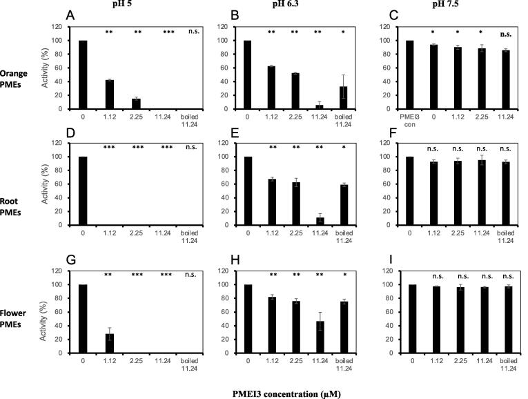
The de-methylesterification of the pectic polysaccharide homogalacturonan (HG) by pectin methylesterases (PMEs) is a critical step in the control of plant cell expansion and morphogenesis. Plants have large gene families encoding PMEs but also PME inhibitors (PMEIs) with differ in their biochemical properties. The Arabidopsis thaliana PECTIN METHYLESTERASE INHIBITOR 3 (PMEI3) gene is frequently used as a tool to manipulate pectin methylesterase activity in studies assessing its role in the control of morphogenesis. One limitation of these studies is that the exact biochemical activity of this protein has not yet been determined. In this manuscript we produced the protein in Pichia pastoris and characterized its activity in vitro. Like other PMEIs, PMEI3 inhibits PME activity at acidic pH in a variety of cell wall extracts and in purified PME preparations, but does not affect the much stronger PME activity at neutral pH. The protein is remarkable heat stable and shows higher activity against PME3 than against PME2, illustrating how different members of the large PMEI family can differ in their specificities towards PME targets. Finally, growing Arabidopsis thaliana seedlings in the presence of purified PMEI3 caused a dose-dependent inhibition of root growth associated with the overall inhibition of HG de-methylesterification of the root surface. This suggests an essential in vivo role for PME activity at acidic pH in HG de-methylesterification and growth control. These results show that purified recombinant PMEI3 is a powerful tool to study the connection between pectin de-methylesterification and cell expansion.
Keywords: Cell expansion; GalA, galacturonic acid; HG, homogalacturonan; PME inhibitor; PME, pectin methylesterase; PMEI, PME inhibitor; Pectin; Pectin methylesterase (PME); Pichia pastoris; Root.
© 2022 The Authors. Published by Elsevier B.V.
Conflict of interest statement
The authors declare that they have no known competing financial interests or personal relationships that could have appeared to influence the work reported in this paper.
Figures


References
-
- Andres-Robin A., Reymond M.C., Dupire A., Battu V., Dubrulle N., Mouille G., Lefebvre V., Pelloux J., Boudaoud A., Traas J., Scutt C.P., Monéger F. Evidence for the regulation of gynoecium morphogenesis by ETTIN via cell wall dynamics. Plant Physiol. 2018;178:1222–1232. doi: 10.1104/pp.18.00745. - DOI - PMC - PubMed
-
- Cosgrove D.J. Growth of the plant cell wall. Nat Rev Mol Cell Biol. 2005;6(11):850–861. - PubMed
-
- Downie B., Dirk L.M., Hadfield K.A., Bennett A.B., Bradford K.J. A gel diffusion assay for quantification of pectin methylesterase activity. Anal. Biochem. 1998;264:149–157. - PubMed
LinkOut - more resources
Full Text Sources
Molecular Biology Databases
Research Materials
