Review
. 2022 Sep 6:10:993257.
doi: 10.3389/fcell.2022.993257. eCollection 2022.

Regulation and coordination of the different DNA damage responses in Drosophila

Affiliations
Review

Regulation and coordination of the different DNA damage responses in Drosophila

Antonio Baonza et al. Front Cell Dev Biol. .

Abstract

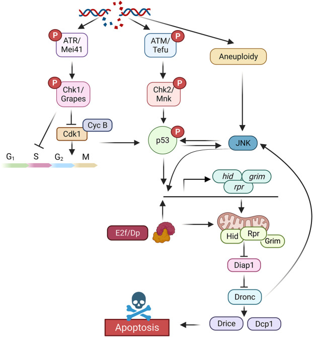
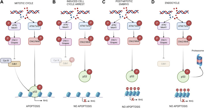
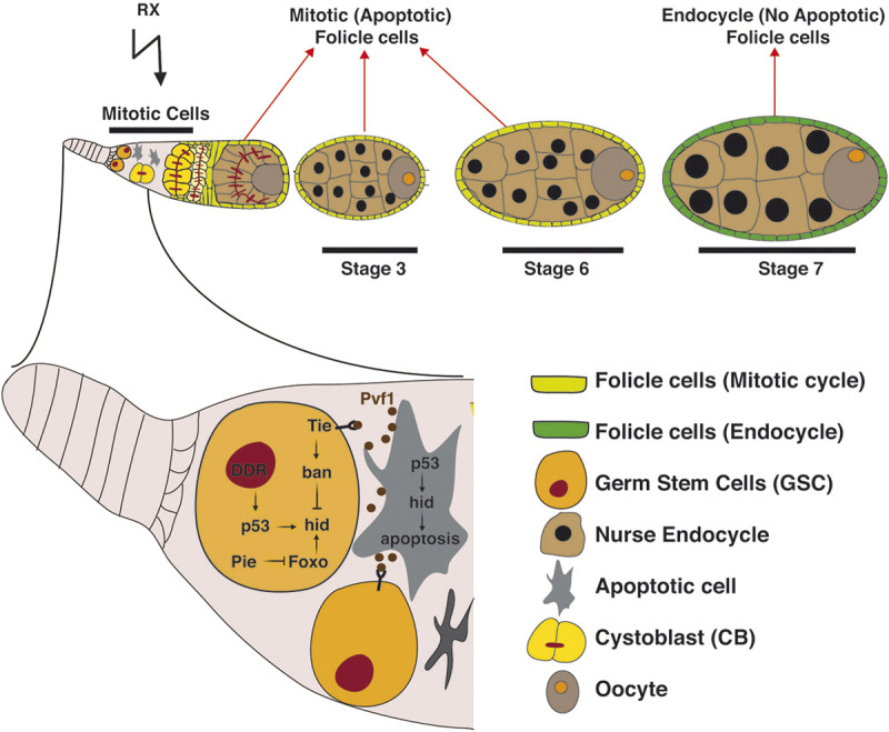
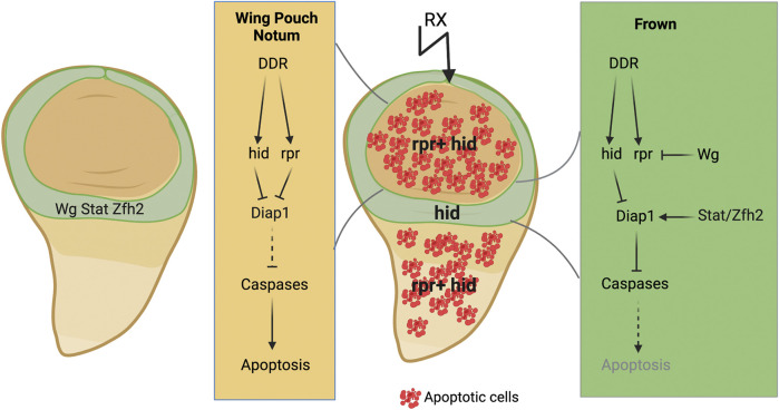
Cells have evolved mechanisms that allow them to respond to DNA damage to preserve genomic integrity and maintain tissue homeostasis. These responses include the activation of the cell cycle checkpoints and the repair mechanisms or the induction of apoptosis that eventually will eliminate damaged cells. These "life" vs. "death" decisions differ depending on the cell type, stages of development, and the proliferation status of the cell. The apoptotic response after DNA damage is of special interest as defects in its induction could contribute to tumorigenesis or the resistance of cancer cells to therapeutic agents such as radiotherapy. Multiples studies have elucidated the molecular mechanisms that mediate the activation of the DNA damage response pathway (DDR) and specifically the role of p53. However, much less is known about how the different cellular responses such as cell proliferation control and apoptosis are coordinated to maintain tissue homeostasis. Another interesting question is how the differential apoptotic response to DNA damage is regulated in distinct cell types. The use of Drosophila melanogaster as a model organism has been fundamental to understand the molecular and cellular mechanisms triggered by genotoxic stress. Here, we review the current knowledge regarding the cellular responses to ionizing radiation as the cause of DNA damage with special attention to apoptosis in Drosophila: how these responses are regulated and coordinated in different cellular contexts and in different tissues. The existence of intrinsic mechanisms that might attenuate the apoptotic pathway in response to this sort of DNA damage may well be informative for the differences in the clinical responsiveness of tumor cells after radiation therapy.

Keywords: DNA damage response; Drosophila; apoptosis; cell cycle; cellular context; ionizing radiation; p53; tissue homeostasis.


Conflict of interest statement

The authors declare that the research was conducted in the absence of any commercial or financial relationships that could be construed as a potential conflict of interest.

Figures

FIGURE 1
FIGURE 1
Simplified representation of the different components of the DNA damage response pathway that leads to cell cycle arrest and apoptotic induction in Drosophila. Created with BioRender.com.
FIGURE 2
FIGURE 2
DNA damage can lead to different outcomes depending on the cell cycle stage of the cell. (A) In cycling cells, IR activates p53 through the DDR pathway. The presence of an active CycB/Cdk1complex facilitates the binding of p53 to the proapoptotic genes and therefore the induction of apoptosis. (B) In induced cell cycle arrested cells p53 is initially activated by DDR pathway, however since these cells do not have active Cdk1, p53 fails to induce the expression of rpr and hid. (C) In embryonic post-mitotic cells, the epigenetic silencing of the IRER interferes with the ability of p53 to regulate the expression of the proapoptotic genes. (D) In endocycle cells even though the DDR pathway is active in response to IR, apoptosis is not induced. This is due to at least two mechanisms of control. On one hand, an epigenetic silencing at the regulatory region of the proapoptotic genes blocks the ability of p53 to induce their expression. Secondly, the levels of p53 in these cells are significatively lower that in mitotic tissues through p53 targeted degradation by the proteasome. Created with BioRender.com.
FIGURE 3
FIGURE 3
A schematic representation showing the germarium of the Drosophila ovary. Proposed mechanism of how female GSCs are protected against apoptosis through the collaborative contribution of multiple signalling pathways. Dying cells send Pvf1 as a survival signal that activates the Tie receptor in GSCs. Tie activation leads to ban microRNA upregulation that represses Foxo and p53 mediated hid activation in response to DNA damage. Created with BioRender.com.
FIGURE 4
FIGURE 4
A schematic diagram showing the different responses after IR in the three main parts of the wing disc. In green is indicated the frown (hinge). In this region Wg and Stat signalling are strongly activated. The function of these signalling pathways in this domain prevents the induction of rpr in response to IR, and therefore apoptosis. Created with BioRender.com.

References

    1. Adams B. R., Golding S. E., Rao R. R., Valerie K. (2010). Dynamic dependence on atr and atm for double-strand break repair in human embryonic stem cells and neural descendants. Plos One 5, E10001. 10.1371/journal.pone.0010001 - DOI - PMC - PubMed
    1. Akdemir F., Christich A., Sogame N., Chapo J., Abrams J. M. (2007). P53 directs focused genomic responses in Drosophila. Oncogene 26, 5184–5193. 10.1038/sj.onc.1210328 - DOI - PubMed
    1. Ambrus A. M., Islam A. B., Holmes K. B., Moon N. S., Lopez-Bigas N., Benevolenskaya E. V., et al. (2013). Loss of De2f compromises mitochondrial function. Dev. Cell 27, 438–451. 10.1016/j.devcel.2013.10.002 - DOI - PMC - PubMed
    1. Ayeni J. O., Varadarajan R., Mukherjee O., Stuart D. T., Sprenger F., Srayko M., et al. (2014). Dual phosphorylation of Cdk1 coordinates cell proliferation with key developmental processes in Drosophila. Genetics 196, 197–210. 10.1534/genetics.113.156281 - DOI - PMC - PubMed
    1. Ayyaz A., Jasper H. (2013). Intestinal inflammation and stem cell homeostasis in aging Drosophila melanogaster. Front. Cell. Infect. Microbiol. 3, 98. 10.3389/fcimb.2013.00098 - DOI - PMC - PubMed

LinkOut - more resources