. 2022 Aug 31;25(10):105037.
doi: 10.1016/j.isci.2022.105037. eCollection 2022 Oct 21.

Cervical transcutaneous spinal stimulation for spinal motor mapping

Affiliations

Cervical transcutaneous spinal stimulation for spinal motor mapping

Jeonghoon Oh et al. iScience. .

Abstract

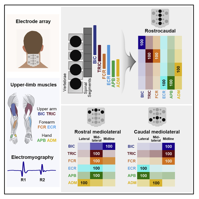
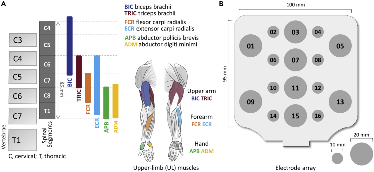
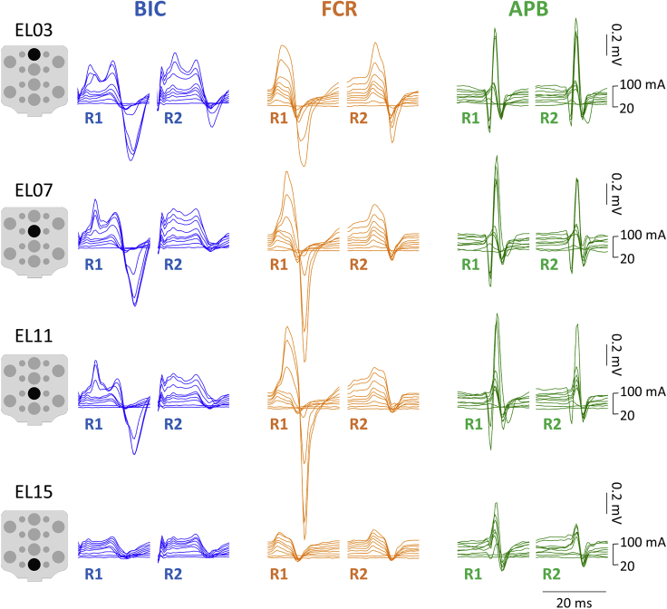
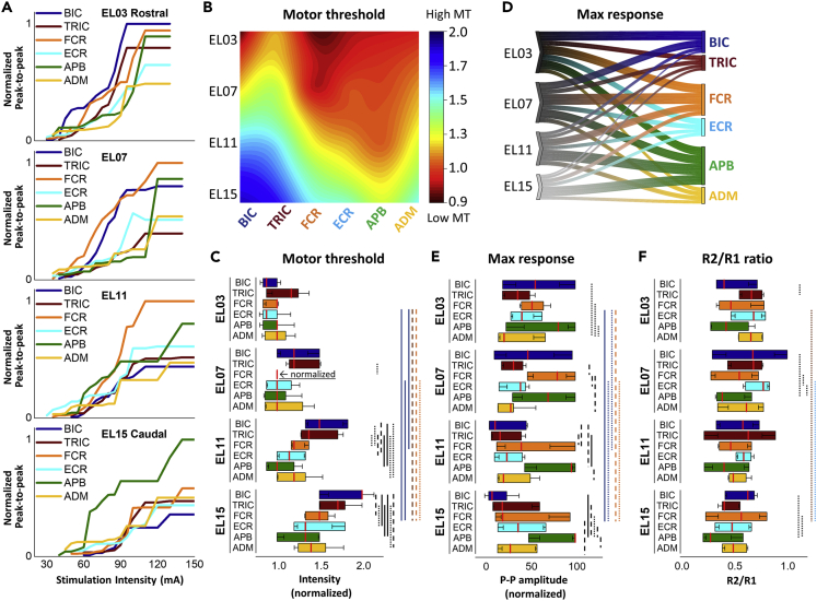
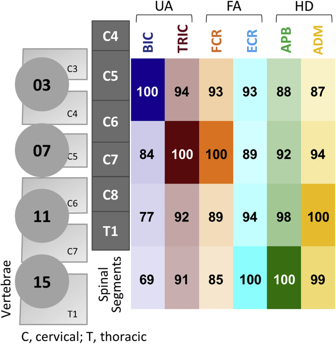
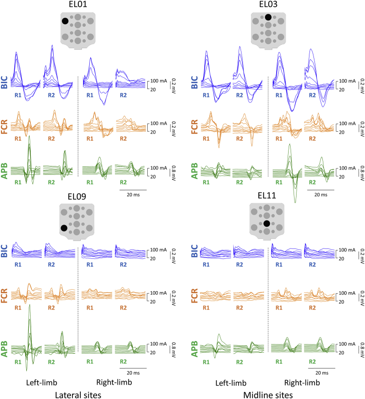
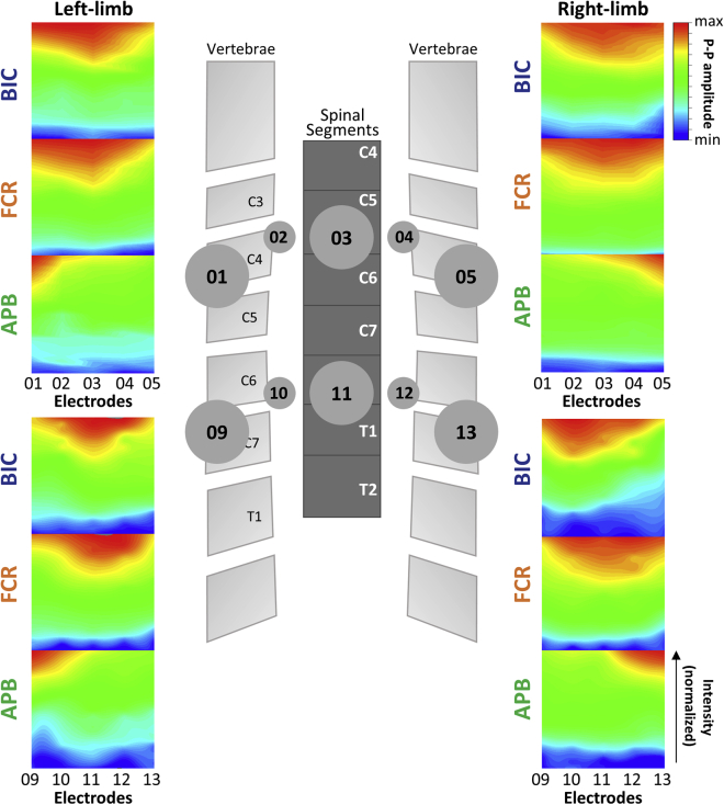
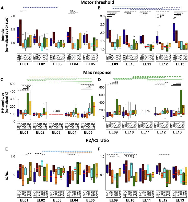
Transcutaneous spinal stimulation (TSS) is a promising approach to restore upper-limb (UL) functions after spinal cord injury (SCI) in humans. We sought to demonstrate the selectivity of recruitment of individual UL motor pools during cervical TSS using different electrode placements. We demonstrated that TSS delivered over the rostrocaudal and mediolateral axes of the cervical spine resulted in a preferential activation of proximal, distal, and ipsilateral UL muscles. This was revealed by changes in motor threshold intensity, maximum amplitude, and the amount of post-activation depression of the evoked responses. We propose that an arrangement of electrodes targeting specific UL motor pools may result in superior efficacy, restoring more diverse motor activities after neurological injuries and disorders, including severe SCI.

Keywords: Biological sciences; Clinical neuroscience; Neuroscience; Techniques in neuroscience.


Conflict of interest statement

Y.-K.L. holds shareholder interest in Aneuvo.

Figures

None
Graphical abstract
Figure 1
Figure 1
Cervical spinal map by epidurally evoked motor pools and cathode electrodes of transcutaneous electrical spinal stimulation (A) Distribution of motor pools projecting to upper-limb muscles based on the segmental charts provided by (McIntyre et al., 2002). BIC: biceps brachii, TRI: triceps brachii, FCR: flexor carpi radialis, ECR: extensor carpi radialis, APB: abductor pollicis brevis, ADM: abductor digiti minimi. (B) Electrode array with sixteen stimulation sites.
Figure 2
Figure 2
Examples of responses of the upper-limb muscles to double-pulse TSS along the rostrocaudal stimulation sites The first (R1) and second responses (R2) elicited with the stimulation placed over EL03, EL07, EL11, and EL15 are shown for BIC, FCR, and APB for a representative participant (see pooled data for the R2/R1 ratio in Figure 3F). Stimulation intensity ranged from 20 to 100 mA for each site. TSS: transcutaneous spinal stimulation, BIC: biceps brachii; FCR: flexor carpi radialis; APB: abductor pollicis brevis muscles.
Figure 3
Figure 3
Responses of upper-limb muscles during TSS delivered along the rostrocaudal axis (A) Recruitment curves of left BIC, TRIC, FCR, ECR, APB, and ADM during stimulation delivered at EL03, EL07, EL11, and EL15. Responses were normalized for each muscle using the maximum value (MaxR) across all amplitudes and sites. (B) Heatmap of motor thresholds (MT) of the UL muscles. The MT values for each muscle were normalized to the MT of FCR at EL07. (C) Averaged data of the MT intensity at rostrocaudal stimulation sites (see also Table S3). (D) Parallel coordinates plots between rostrocaudal stimulation sites to the upper limb muscles derived from maximum response. Individual muscles are grouped along the right of the plot. Thickness of connections denotes the amplitude of the maximum motor response. (E) Averaged data of the MaxR at rostrocaudal stimulation sites. (F) Averaged data of the R2/R1 ratio at rostrocaudal stimulation sites. Box range was set as percentage 25%–75%, bold vertical red lines in boxplots present the median values, and whiskers indicate the 95% confidence interval. A post-hoc Holm-Bonferroni correction was carried out for multiple comparison within figures. Significant differences are indicated with vertical lines. Dotted lines: p < 0.05; dashed lines: p < 0.01; and solid lines: p < 0.001. TSS: transcutaneous spinal stimulation, BIC: biceps brachii; TRIC: triceps brachii; FCR: flexor carpi radialis; ECR: extensor carpi radialis; APB: abductor pollicis brevis; ADM: abductor digiti minimi.
Figure 4
Figure 4
Probability of the upper-limb muscle response during stimulation along the rostrocaudal axis The values in the box are calculated based on the motor threshold (MT), maximum response (MaxR), and amount of post-activation depression (R2/R1 ratio). Probabilities of muscle activation are illustrated by the opacity of different colors aligned with the stimulation electrodes. UA: upper arm; FA: forearm; HD: hand, BIC: biceps brachii; TRIC: triceps brachii; FCR: flexor carpi radialis; ECR: extensor carpi radialis; APB: abductor pollicis brevis; ADM: abductor digiti minimi.
Figure 5
Figure 5
Examples of responses of the left and right upper-limb muscles to double-pulse TSS at lateral (EL01 and EL09) and midline stimulation sites (EL03 and EL11) The first (R1) and second responses (R2) elicited with the stimulation placed each electrode are shown for BIC, FCR, and APB for a representative participant. Stimulation intensity ranged from 20 to 100 mA for each site. TSS: transcutaneous spinal stimulation, BIC: biceps brachii; FCR: flexor carpi radialis; APB: abductor pollicis brevis muscles.
Figure 6
Figure 6
Ipsilateral upper-limb muscle activation during TSS delivered along the mediolateral axis of the cervical spinal cord Heatmap pattern of motor evoked responses with ipsilateral versus contralateral stimulation sites using normalized MaxR for left and right BIC, FCR, and APB for the rostral mediolateral (EL01, EL02, EL03, EL04, and EL05) and caudal mediolateral (EL09, EL10, EL11, EL12, and EL13) stimulation sties.
Figure 7
Figure 7
Statistical comparison between six of upper-limb muscles during TSS delivered along the mediolateral axes (A) Averaged data of the MT intensity at rostral mediolateral stimulation sites (see also Table S3). (B) Averaged data of the MT at caudal mediolateral stimulation sites (see also Table S3). (C) Averaged data of the MaxR at rostral mediolateral stimulation sites. (D) Averaged data of the MaxR at caudal mediolateral stimulation sites. (E) Averaged data of the R2/R1 ratio at rostral mediolateral stimulation sites. (F) Averaged data of the R2/R1 ratio at caudal mediolateral stimulation sites. Box range was set as percentage 25%–75%, bold horizontal red lines in boxplots present the median values, and whiskers indicate the 95% confidence interval. A post-hoc Holm-Bonferroni correction was carried out for multiple comparison within figures. Significant differences are indicated with horizontal lines. Dotted lines: p < 0.05; dashed lines: p < 0.01; and solid lines: p < 0.001. Rostral mediolateral stimulation sites include EL01, EL02, EL03, EL4, and EL05; caudal mediolateral stimulation sites include EL09, EL10, EL11, EL12, and EL13. TSS: transcutaneous spinal stimulation, BIC: biceps brachii; TRIC: triceps brachii; FCR: flexor carpi radialis; ECR: extensor carpi radialis; APB: abductor pollicis brevis; ADM: abductor digiti minimi.
Figure 8
Figure 8
Probability of the upper-limb muscle response during stimulation along the mediolateral axes (A) Probability of motor response for BIC, TRI, FCR, ECR, APB, and ADM at rostral mediolateral stimulation site. Stimulation via lateral (EL01 and EL05), mid-lateral (EL02 and EL04), and midline (EL03) portion of array was averaged. (B) Probability of motor response for BIC, TRI, FCR, ECR, APB, and ADM at each caudal mediolateral stimulation site. Stimulation via lateral (EL09 and EL13), mid-lateral (EL10 and EL12), and midline (EL13) portion of array was averaged.

Similar articles

Cited by

References

    1. Alstermark B., Isa T. Circuits for skilled reaching and grasping. Annu. Rev. Neurosci. 2012;35:559–578. - PubMed
    1. Andrews J.C., Stein R.B., Roy F.D. Post-activation depression in the human soleus muscle using peripheral nerve and transcutaneous spinal stimulation. Neurosci. Lett. 2015;589:144–149. - PubMed
    1. Angeli C.A., Boakye M., Morton R.A., Vogt J., Benton K., Chen Y., Ferreira C.K., Harkema S.J. Recovery of over-ground walking after chronic motor complete spinal cord injury. N. Engl. J. Med. 2018;379:1244–1250. - PubMed
    1. Atkinson D.A., Steele A.G., Manson G.A., Sheynin J., Oh J., Gerasimenko Y.P., Sayenko D.G. Characterization of interlimb interaction via transcutaneous spinal stimulation of cervical and lumbar spinal enlargements. J. Neurophysiol. 2022;127:1075–1085. - PMC - PubMed
    1. Barra B., Conti S., Perich M.G., Zhuang K., Schiavone G., Fallegger F., Galan K., James N.D., Barraud Q., Delacombaz M., et al. Epidural electrical stimulation of the cervical dorsal roots restores voluntary arm control in paralyzed monkeys. bioRxiv. 2021 doi: 10.1101/2020.11.13.379750. Preprint at. - DOI - PubMed

LinkOut - more resources