Review
. 2022 Sep 6:9:886549.
doi: 10.3389/fcvm.2022.886549. eCollection 2022.

SlicerHeart: An open-source computing platform for cardiac image analysis and modeling

Affiliations
Review

SlicerHeart: An open-source computing platform for cardiac image analysis and modeling

Andras Lasso et al. Front Cardiovasc Med. .

Abstract

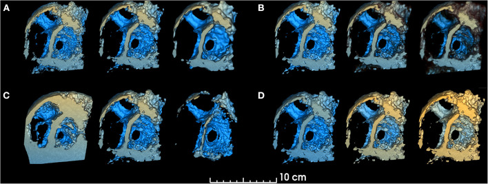
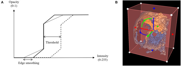
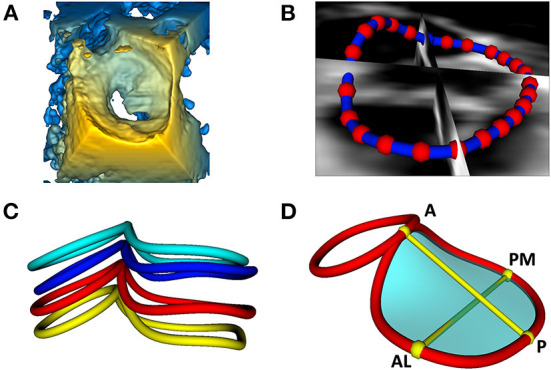
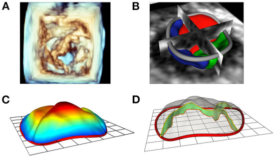
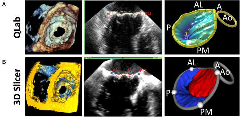
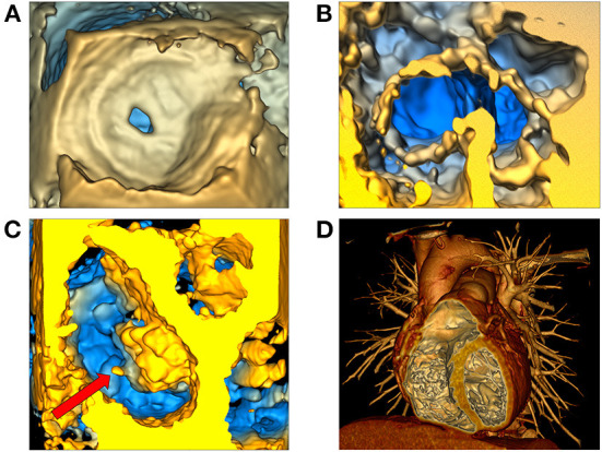
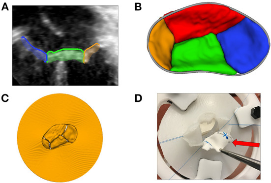
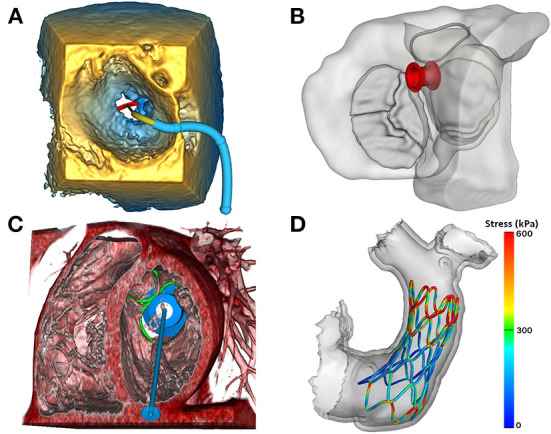
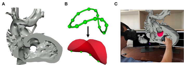
Cardiovascular disease is a significant cause of morbidity and mortality in the developed world. 3D imaging of the heart's structure is critical to the understanding and treatment of cardiovascular disease. However, open-source tools for image analysis of cardiac images, particularly 3D echocardiographic (3DE) data, are limited. We describe the rationale, development, implementation, and application of SlicerHeart, a cardiac-focused toolkit for image analysis built upon 3D Slicer, an open-source image computing platform. We designed and implemented multiple Python scripted modules within 3D Slicer to import, register, and view 3DE data, including new code to volume render and crop 3DE. In addition, we developed dedicated workflows for the modeling and quantitative analysis of multi-modality image-derived heart models, including heart valves. Finally, we created and integrated new functionality to facilitate the planning of cardiac interventions and surgery. We demonstrate application of SlicerHeart to a diverse range of cardiovascular modeling and simulation including volume rendering of 3DE images, mitral valve modeling, transcatheter device modeling, and planning of complex surgical intervention such as cardiac baffle creation. SlicerHeart is an evolving open-source image processing platform based on 3D Slicer initiated to support the investigation and treatment of congenital heart disease. The technology in SlicerHeart provides a robust foundation for 3D image-based investigation in cardiovascular medicine.

Keywords: 3D echocardiography (3DE); cardiac valves; computer modeling (simulation); image-based modeling; open-source; pediatric cardiology and surgery.


Conflict of interest statement

CP is a contracted developer employed by Pixel Medical. SP is a software architect at Isomics, Inc. Both Pixel and Isomics are companies which support open-source software. The remaining authors declare that the research was conducted in the absence of any commercial or financial relationships that could be construed as a potential conflict of interest.

Figures

Figure 1
Figure 1
Smoothing, threshold, edge smoothing and depth range parameters. changes in volume rendered model for different values of (A). Smoothing (left: 0.25, center: 1.0, right: 1.75), (B) Threshold (left: 30%, center: 50%, right: 70%), (C) Edge smoothing (left: 0%, center: 10%, right: 20%), (D) Depth range [left: (−150, +10), center: (−103, +40), right: (−103, +80)]. Unchanged parameters have remained constant at the following value: Smoothing factor = 1.00, Threshold = 50.9%, Edge smoothing = 2.0%, Depth range = [−103, 10], Depth darkening = 30.0%, Depth coloring = [−24.35, 23.55%], Brightness = 165%, Saturation = 200%.
Figure 2
Figure 2
Threshold and edge smoothing parameters for changes in opacity. (A) Opacity is shown as a function of voxel intensity, where voxels having an intensity value greater than the upper limit appear completely opaque and voxels with an intensity value inferior to the lower limit appear invisible, having a zero-opacity value (see Appendix); (B) Selection of a Region of Interest (ROI) to enable cropping of the rendered 3D echocardiographic volume.
Figure 3
Figure 3
Visualization and modeling of mitral valve annuli. (A) Volume rendering of a 3DE of a mitral valve using the echo volume render module in SlicerHeart. (B) Curve creation of annular model of a mitral valve visualized through 2D slice intersections using valve annulus analysis module in SlicerHeart. (C) Visualization of mitral and aortic annular curves in all four phases of the cardiac cycle (top to bottom: End Diastole, Mid Diastole, Mid Systole, End Systole). (D) Quantification of mitral and aortic annuli using valve quantification module in SlicerHeart. 3DE, 3D echocardiogram; A, Anterior; P, Posterior; AL, Anterolateral; PM, posterior medial.
Figure 4
Figure 4
Modeling of valve leaflets and quantification of annular and leaflet structure. (A) Volume rendering of a tricuspid valve using Phillips Qlab Software; (B) Visualization of a tricuspid valve (right atrial view) segmentation with slice intersections (blue = septal leaflet, red = anterior leaflet, green = posterior leaflet) in SlicerHeart; (C) Color intensity map depicting quantitative degree of billow in a tricuspid valve with 2D annular plane visible in SlicerHeart; (D) Visualization of leaflet coaptation regions with 2D annular plane visible in SlicerHeart.
Figure 5
Figure 5
Comparison of 3D Slicer vs. QLab Mitral Valve Quantification (MVN). (Left) volume rendering of a mitral valve viewed from the left ventricle in Qlab (A) and 3D Slicer (B); 2D Transesophageal view of the mitral valve highlighting the AL and PM points in Qlab (A) and 3D Slicer (B); 3D Model generated in Qlab (A) and 3D Slicer (B). Qlab, Philips Medical, Andover, MA.
Figure 6
Figure 6
Volume rendering of 3D echocardiographic and tomographic data. (A) Visualization of an atrial septal defect in the echo volume render module in SlicerHeart from a right atrial view; (B) Visualization of a complete common atrioventricular canal valve in diastole (ventricular view in SlicerHeart); (C) Left atrial view of an aortic valve and a mitral valve with anterior leaflet prolapse and a ruptured chord which is visible as indicated by the red arrow; (D) CT image of a normal anatomic heart cropped anteriorly to visualize the right atrium and ventricle and the left ventricle using volume rendering in 3D Slicer. CT, Computed Tomography.
Figure 7
Figure 7
3D printing and novel visualization technologies. (A) A 2D transthoracic apical view of a CAVC valve (from image left to right: right mural leaflet in blue, inferior bridging leaflet in green, and left mural leaflet in orange) segmented using the Valve Segmentation module in SlicerHeart; (B) An 3D atrial view of a segmented CAVC valve with the annular curve visible in gray; (C) A virtual model of the segmented CAVC valve with a skirt used as a template for 3D printing; (D) A surgeon practices a patch repair on a AVC valve created from a 3D echocardiogram with the dividing ventricular septal defect patch indicated by the red arrow. CAVC, complete common atrioventricular canal.
Figure 8
Figure 8
Image-based modeling of transcatheter devices. (A) Demonstration of the ValveClip delivery simulator module in SlicerHeart, to model a transcatheter edge-to-edge repair within a 3DE image of a mitral valve rendered in the echo volume rendering module; (B) Visualization of an ASD closure device in segmented myocardium and valves viewed from the atrium using the ASD/VSD Device simulator module; (C) An apical tether transcatheter mitral valve device positioned in a mitral valve in a CT volume rendering using the transcatheter atrioventricular valve simulator module in SlicerHeart; (D) Visualization of a finite element model of a self-expanding valve deployment simulation in SlicerHeart. 3DE, 3D Echocardiography; ASD, Atrial septal defect; VSD, Ventricular septal defect; CT, Computed Tomography.
Figure 9
Figure 9
Image-based planning of surgical procedures. (A) 3D virtual model of a heart with double outlet right ventricle anatomy cut to visualize the Ventricular Septal Defect that will be baffled to allow for continuous blood flow; (B) Closed baffle perimeter contour created by placing points in the 3D heart model which becomes a baffle model onto which surface points may be placed to customize the shape of the baffle; (C) Rendering demonstration visualization of the baffle model placed within the 3D heart model in SlicerHeart using the SlicerVR module.

References

    1. Mahmood F, Matyal R. A quantitative approach to the intraoperative echocardiographic assessment of the mitral valve for repair. Anesth Analg. (2015) 121:34–58. 10.1213/ANE.0000000000000726 - DOI - PubMed
    1. Sacks M, Drach A, Lee CH, Khalighi A, Rego B, Zhang W, et al. . On the simulation of mitral valve function in health, disease, and treatment. J Biomech Eng. (2019) 141:0708041–07080422. 10.1115/1.4043552 - DOI - PMC - PubMed
    1. Lee AP, Hsiung MC, Salgo IS, Fang F, Xie JM, Zhang YC, et al. . Quantitative analysis of mitral valve morphology in mitral valve prolapse with real-time 3-dimensional echocardiography: importance of annular saddle shape in the pathogenesis of mitral regurgitation. Circulation. (2013) 127:832–41. 10.1161/CIRCULATIONAHA.112.118083 - DOI - PubMed
    1. Kasel AM, Cassese S, Bleiziffer S, Amaki M, Hahn RT, Kastrati A, et al. . Standardized imaging for aortic annular sizing: implications for transcatheter valve selection. JACC Cardiovasc Imaging. (2013) 6:249–62. 10.1016/j.jcmg.2012.12.005 - DOI - PubMed
    1. Nguyen KL, Moriarty JM, Plotnik AN, Aksoy O, Yoshida T, Shemin RJ, et al. . Ferumoxytol-enhanced Mr angiography for vascular access mapping before transcatheter aortic valve replacement in patients with renal impairment: a step toward patient-specific care. Radiolog. (2018) 286:326–37. 10.1148/radiol.2017162899 - DOI - PMC - PubMed

LinkOut - more resources