

Proteostasis unbalance in prion diseases: Mechanisms of neurodegeneration and therapeutic targets
- PMID: 36148145
- PMCID: PMC9485628
- DOI: 10.3389/fnins.2022.966019
Proteostasis unbalance in prion diseases: Mechanisms of neurodegeneration and therapeutic targets
Abstract
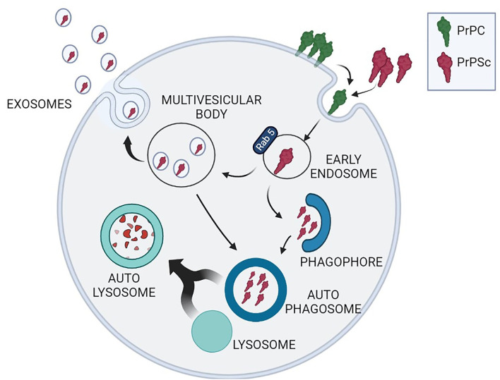
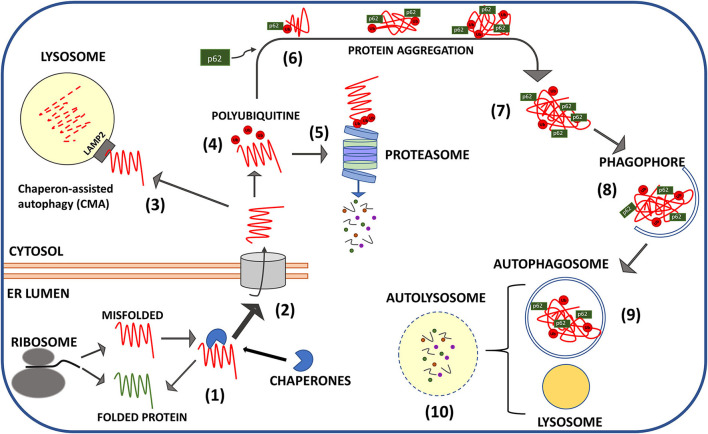
Transmissible spongiform encephalopathies (TSEs), or prion diseases, are progressive neurodegenerative disorders of the central nervous system that affect humans and animals as sporadic, inherited, and infectious forms. Similarly to Alzheimer's disease and other neurodegenerative disorders, any attempt to reduce TSEs' lethality or increase the life expectancy of affected individuals has been unsuccessful. Typically, the onset of symptoms anticipates the fatal outcome of less than 1 year, although it is believed to be the consequence of a decades-long process of neuronal death. The duration of the symptoms-free period represents by itself a major obstacle to carry out effective neuroprotective therapies. Prions, the infectious entities of TSEs, are composed of a protease-resistant protein named prion protein scrapie (PrPSc) from the prototypical TSE form that afflicts ovines. PrPSc misfolding from its physiological counterpart, cellular prion protein (PrPC), is the unifying pathogenic trait of all TSEs. PrPSc is resistant to intracellular turnover and undergoes amyloid-like fibrillation passing through the formation of soluble dimers and oligomers, which are likely the effective neurotoxic entities. The failure of PrPSc removal is a key pathogenic event that defines TSEs as proteopathies, likewise other neurodegenerative disorders, including Alzheimer's, Parkinson's, and Huntington's disease, characterized by alteration of proteostasis. Under physiological conditions, protein quality control, led by the ubiquitin-proteasome system, and macroautophagy clears cytoplasm from improperly folded, redundant, or aggregation-prone proteins. There is evidence that both of these crucial homeostatic pathways are impaired during the development of TSEs, although it is still unclear whether proteostasis alteration facilitates prion protein misfolding or, rather, PrPSc protease resistance hampers cytoplasmic protein quality control. This review is aimed to critically analyze the most recent advancements in the cause-effect correlation between PrPC misfolding and proteostasis alterations and to discuss the possibility that pharmacological restoring of ubiquitin-proteasomal competence and stimulation of autophagy could reduce the intracellular burden of PrPSc and ameliorate the severity of prion-associated neurodegeneration.
Keywords: autophagy; neurodegeneration; prion protein; proteasome; protein misfolding.
Copyright © 2022 Thellung, Corsaro, Dellacasagrande, Nizzari, Zambito and Florio.
Conflict of interest statement
The authors declare that the research was conducted in the absence of any commercial or financial relationships that could be construed as a potential conflict of interest.
Figures
Similar articles
-
In Vitro Approach To Identify Key Amino Acids in Low Susceptibility of Rabbit Prion Protein to Misfolding.J Virol. 2017 Nov 30;91(24):e01543-17. doi: 10.1128/JVI.01543-17. Print 2017 Dec 15. J Virol. 2017. PMID: 28978705 Free PMC article.
-
Pathogenic mechanisms of prion protein, amyloid-β and α-synuclein misfolding: the prion concept and neurotoxicity of protein oligomers.J Neurochem. 2016 Oct;139(2):162-180. doi: 10.1111/jnc.13772. Epub 2016 Sep 15. J Neurochem. 2016. PMID: 27529376 Review.
-
Inducing prion protein shedding as a neuroprotective and regenerative approach in pathological conditions of the brain: from theory to facts.Neural Regen Res. 2023 Sep;18(9):1869-1875. doi: 10.4103/1673-5374.366496. Neural Regen Res. 2023. PMID: 36926701 Free PMC article. Review.
-
Prion protein misfolding, strains, and neurotoxicity: an update from studies on Mammalian prions.Int J Cell Biol. 2013;2013:910314. doi: 10.1155/2013/910314. Epub 2013 Dec 24. Int J Cell Biol. 2013. PMID: 24454379 Free PMC article. Review.
-
The transmissible spongiform encephalopathies of livestock.ILAR J. 2015;56(1):7-25. doi: 10.1093/ilar/ilv008. ILAR J. 2015. PMID: 25991695 Review.
Cited by
-
Linear poly-ubiquitin remodels the proteome and influences hundreds of regulators in Drosophila.G3 (Bethesda). 2024 Nov 6;14(11):jkae209. doi: 10.1093/g3journal/jkae209. G3 (Bethesda). 2024. PMID: 39325835 Free PMC article.
-
Molecular signatures in prion disease: altered death receptor pathways in a mouse model.J Transl Med. 2024 May 27;22(1):503. doi: 10.1186/s12967-024-05121-x. J Transl Med. 2024. PMID: 38802941 Free PMC article.
-
Drosophila melanogaster as a model to study autophagy in neurodegenerative diseases induced by proteinopathies.Front Neurosci. 2023 May 18;17:1082047. doi: 10.3389/fnins.2023.1082047. eCollection 2023. Front Neurosci. 2023. PMID: 37274187 Free PMC article. Review.
-
Amyloids and brain cancer: molecular linkages and crossovers.Biosci Rep. 2023 Oct 31;43(10):BSR20230489. doi: 10.1042/BSR20230489. Biosci Rep. 2023. PMID: 37335084 Free PMC article. Review.
-
A Systematic Review of Sporadic Creutzfeldt-Jakob Disease: Pathogenesis, Diagnosis, and Therapeutic Attempts.Neurol Int. 2024 Sep 20;16(5):1039-1065. doi: 10.3390/neurolint16050079. Neurol Int. 2024. PMID: 39311352 Free PMC article. Review.
References
-
- Alves-Rodrigues A., Gregori L., Figueiredo-Pereira M. E. (1998). Ubiquitin, cellular inclusions and their role in neurodegeneration. Trends Neurosci. 21, 516–520. - PubMed
Publication types
LinkOut - more resources
Full Text Sources
Research Materials