Neurons gating behavior-developmental, molecular and functional features of neurons in the Substantia Nigra pars reticulata
- PMID: 36148148
- PMCID: PMC9485944
- DOI: 10.3389/fnins.2022.976209
Neurons gating behavior-developmental, molecular and functional features of neurons in the Substantia Nigra pars reticulata
Abstract
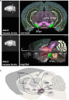
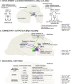
The Substantia Nigra pars reticulata (SNpr) is the major information output site of the basal ganglia network and instrumental for the activation and adjustment of movement, regulation of the behavioral state and response to reward. Due to both overlapping and unique input and output connections, the SNpr might also have signal integration capacity and contribute to action selection. How the SNpr regulates these multiple functions remains incompletely understood. The SNpr is located in the ventral midbrain and is composed primarily of inhibitory GABAergic projection neurons that are heterogeneous in their properties. In addition, the SNpr contains smaller populations of other neurons, including glutamatergic neurons. Here, we discuss regionalization of the SNpr, in particular the division of the SNpr neurons to anterior (aSNpr) and posterior (pSNpr) subtypes, which display differences in many of their features. We hypothesize that unique developmental and molecular characteristics of the SNpr neuron subtypes correlate with both region-specific connections and notable functional specializations of the SNpr. Variation in both the genetic control of the SNpr neuron development as well as signals regulating cell migration and axon guidance may contribute to the functional diversity of the SNpr neurons. Therefore, insights into the various aspects of differentiation of the SNpr neurons can increase our understanding of fundamental brain functions and their defects in neurological and psychiatric disorders, including movement and mood disorders, as well as epilepsy.
Keywords: GABAergic neuron; Substantia Nigra pars reticulata; basal ganglia; movement; neurogenesis; reward; seizure; sleep.
Copyright © 2022 Partanen and Achim.
Conflict of interest statement
The authors declare that the research was conducted in the absence of any commercial or financial relationships that could be construed as a potential conflict of interest.
Figures


Similar articles
-
Activity of pars reticulata neurons of monkey substantia nigra in relation to motor, sensory, and complex events.J Neurophysiol. 1986 Apr;55(4):660-77. doi: 10.1152/jn.1986.55.4.660. J Neurophysiol. 1986. PMID: 3701399
-
Topography of dyskinesias and torticollis evoked by inhibition of substantia nigra pars reticulata.Mov Disord. 2013 Apr;28(4):460-8. doi: 10.1002/mds.25215. Epub 2012 Oct 31. Mov Disord. 2013. PMID: 23115112
-
The modulation of striatonigral and nigrotectal pathways by CB1 signalling in the substantia nigra pars reticulata regulates panic elicited in mice by urutu-cruzeiro lancehead pit vipers.Behav Brain Res. 2021 Mar 5;401:112996. doi: 10.1016/j.bbr.2020.112996. Epub 2020 Nov 7. Behav Brain Res. 2021. PMID: 33171147
-
Topographic and functional neuroanatomical study of GABAergic disinhibitory striatum-nigral inputs and inhibitory nigrocollicular pathways: neural hodology recruiting the substantia nigra, pars reticulata, for the modulation of the neural activity in the inferior colliculus involved with panic-like emotions.J Chem Neuroanat. 2006 Aug;32(1):1-27. doi: 10.1016/j.jchemneu.2006.05.002. Epub 2006 Jul 3. J Chem Neuroanat. 2006. PMID: 16820278 Review.
-
GABAergic control of substantia nigra dopaminergic neurons.Prog Brain Res. 2007;160:189-208. doi: 10.1016/S0079-6123(06)60011-3. Prog Brain Res. 2007. PMID: 17499115 Review.
Cited by
-
Striatonigral distribution of a fluorescent reporter following intracerebral delivery of genome editors.Front Bioeng Biotechnol. 2023 Jul 26;11:1237613. doi: 10.3389/fbioe.2023.1237613. eCollection 2023. Front Bioeng Biotechnol. 2023. PMID: 37564994 Free PMC article.
-
Dopamine depletion weakens direct pathway modulation of SNr neurons.Neurobiol Dis. 2024 Jun 15;196:106512. doi: 10.1016/j.nbd.2024.106512. Epub 2024 Apr 24. Neurobiol Dis. 2024. PMID: 38670278 Free PMC article.
-
Silencing of dentate gyrus inhibits mossy fiber sprouting and prevents epileptogenesis through NDR2 kinase in pentylenetetrazole kindling rat model of TLE.PLoS One. 2023 Apr 12;18(4):e0284359. doi: 10.1371/journal.pone.0284359. eCollection 2023. PLoS One. 2023. PMID: 37043471 Free PMC article.
-
The Brain of the African Wild Dog. VI. The Motor System.J Comp Neurol. 2025 Jul;533(7):e70072. doi: 10.1002/cne.70072. J Comp Neurol. 2025. PMID: 40664361 Free PMC article.
-
Dopamine Dysregulation in Reward and Autism Spectrum Disorder.Brain Sci. 2024 Jul 22;14(7):733. doi: 10.3390/brainsci14070733. Brain Sci. 2024. PMID: 39061473 Free PMC article. Review.
References
-
- Arber S., Costa R. M. (2022). Networking brainstem and basal ganglia circuits for movement. Nat. Rev. Neurosci. 23 342–360. - PubMed
LinkOut - more resources
Full Text Sources
Miscellaneous

