. 2022 Sep 6:13:1000007.
doi: 10.3389/fphys.2022.1000007. eCollection 2022.

Novel TrueVue series of 3D echocardiography: Revealing the pathological morphology of congenital heart disease

Affiliations

Novel TrueVue series of 3D echocardiography: Revealing the pathological morphology of congenital heart disease

Feifei Sun et al. Front Physiol. .

Abstract

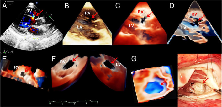
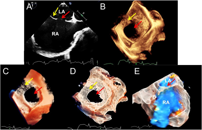
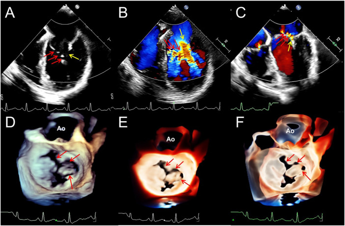
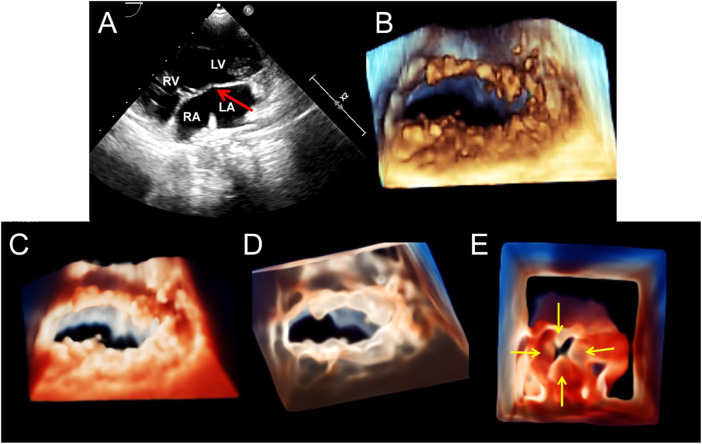
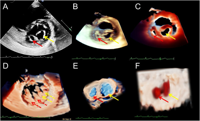
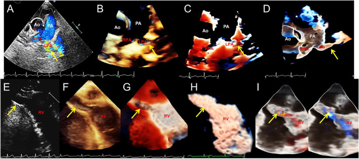
Aims: This study explored the advantages and limitations of novel series of three-dimensional (3D) echocardiographic techniques and summarized their application methods for congenital heart diseases (CHDs). Method and result: Two-dimensional (2D), traditional 3D echocardiography, and TrueVue plus light and/or Glass novel 3D technologies were performed on 62 patients with CHD, and a clinical survey was designed to judge whether the novel 3D images were more helpful for understanding the cardiac condition and guide treatment than traditional 3D images. TrueVue increased the visual resolution and simulated the true texture of cardiac tissue, significantly improving the display ability of abnormal anatomical structures in CHDs. TrueVue Glass displayed the blood channel and the internal structure of cardiac cavity more intuitively, indicating a new observation aspect not shown by conventional echocardiography. The clinical survey results showed that the new 3D imaging methods effectively increased the diagnostic confidence of echocardiographers, enabled surgeons to better understand the details of lesions, promoted efficient communication, and improved the confidence of both doctors and patients in treatment. Conclusion: The combined application of TrueVue, TrueVue Light, and TrueVue Glass more closely simulated real anatomical features, showed more comprehensive and subtle blood flow in the lumen, not only increased the visual effect but also provided more useful diagnostic information, improved the accuracy of evaluation and treatment of CHD when compared to traditional imaging techniques, indicating that this combined application has significant clinical value.

Keywords: congenital abnormalities; echocardiography; heart defects; real-time three-dimensional; ultrasonic imaging.


Conflict of interest statement

The authors declare that the research was conducted in the absence of any commercial or financial relationships that could be construed as a potential conflict of interest.

Figures

FIGURE 1
FIGURE 1
Transthoracic echocardiogram showing ventricular septal defect. (A–D). On long axis section of the left ventricle, the 2D, traditional 3D, TrueVue Light, and TrueVue Glass images indicate the defects of the peri-membranous part of the interventricular septum (5 mm × 10 mm, arrows), respectively. (E). In the same patient as A-D, looking down the defect from the right ventricle directly through a single view. (F). In another patient with a large (22 mm × 14 mm) peri-membranous ventricularseptal defect, the “Dual Volume” imaging mode in TrueVue was used to observe the defects (arrows) directly from the right and left ventricles simultaneously. (G). It is the same patient as in Figure (F), showing the three-dimensional shape of the defect displayed by using TrueVue Glass to simulate the “surgical view”(left, arrow), and the comparison of the doctor’s field of vision during cardiac surgery (right, arrow). In surgery, after lifting the tricuspid valve, the mitral valve can be seen through the septal defect. 2D, two-dimensional; 3D, three-dimensional; LV, left ventricle; RV, right ventricle.
FIGURE 2
FIGURE 2
Transesophageal echocardiography of double atrial-superior and inferior vena cava section to diagnose atrial septal defect. (A–D). The 2D, traditional 3D, TrueVue Light, and TrueVue Glass show the secondary type of atrial septal defect (25 mm × 23 mm, red arrows) and the upper soft, thin rim (yellow arrows), respectively. Figure (B–D) is a direct view of the atrial septum from the left atrium perspective. (E). TrueVue Glass shows the spatial path of the atrial shunt from left to right. 2D, two-dimensional; 3D, three-dimensional; LA, left atrium; RA, right atrium.
FIGURE 3
FIGURE 3
Transesophageal echocardiography reveals multiple mitral valve clefts. (A). 2D echocardiography four-chamber view showing multiple loss of echo in the anterior leaflet of mitral valve (MV) (the sizes of the clefts from A1 to A3 area are 3.1, 4.6, and 3.4 mm respectively, red arrows). The yellow arrow points to the junction between the anterior and posterior leaflets. (B,C). Blood flows into the left ventricle during diastole (red arrows) and regurgitation jets into left atrium during systole (red arrows) from the clefts. Yellow arrows represent the blood flow signals of the normal MV orifices. (D–F). From a surgical perspective, traditional 3D, TrueVue Light, and TrueVue Glass show three irregular clefts (the range of the clefts from A1 to A3 area are 2.5–5.4, 2.5–4.3, and 2.0–3.3 mm, respectively) from the perspective of the left atrium, and all are complete cleft to the annulus. 2D, two-dimensional; 3D, three-dimensional; AO, Aorta; A1, A1 scallop of MV; A3, A3 scallop of MV.
FIGURE 4
FIGURE 4
Transthoracic echocardiography shows complete atrioventricular septal defect. (A). The four-chamber view of a 2D echocardiography shows only one atrioventricular valve (arrow), combined with a large defect of the lower part of the atrial septum and large ventricular septal defect. (B–D). Traditional 3D, TrueVue, and TrueVue Glass show a single atrioventricular annulus and a group of atrioventricular valves open from the atrium view in diastole. (E). TrueVue Glass shows the systolic phase with atrioventricular valve closed as a single annulus and four lobes from the perspective of the atrium, which are left, right, anterior, and posterior bridge lobes (arrows), confirmed to be a complete atrioventricular septal defect deformity. 2D, two-dimensional; 3D, three-dimensional. LA, left atrium; RA, right atrium; LV, left ventricle; RV, right ventricle.
FIGURE 5
FIGURE 5
Transthoracic echocardiography of the short-axis view of the mitral valve (MV) showing the double orifice MV (arrows). (A). 2D display of asymmetric orifices; (B,C). Traditional 3D and TrueVue show the spatial shape of the orifices; (D). TrueVue shows the abnormal multiple papillary muscles and the tendons connected to them from the perspective of the left ventricle; the red dotted arrows represent the additional structure corresponding to the small orifice, and the yellow dotted arrows represents the additional structure corresponding to the large orifice. TrueVue Glass (E) plus color Doppler (F) directly visualizes the two orifices of the MV from the left atrium side (surgical view) and the two blood streams entering the left ventricle during diastole. The red and yellow solid line arrows in (A–C,E,F) indicate two asymmetric orifices of MV, respectively. 2D, two-dimensional; 3D, three-dimensional.
FIGURE 6
FIGURE 6
Transthoracic echocardiography shows pulmonary artery sling and right ventricular diverticulum. (A–D). Transthoracic echocardiography showing the pulmonary artery sling on the short-axis section of the great artery. 2D, traditional 3D, TrueVue Light, and TrueVue Glass show abnormal orientation of the left pulmonary artery, which originate from a lower position, bypassing the rear of the trachea and then reflexed to the left, with local stenosis showing increased blood flow speed (arrows). (E–H). 2D, traditional 3D, TrueVue Light, and TrueVue Glass show the diverticula (the end-systolic volume is 15 mm × 9 mm × 9 mm, and the end-diastolic volume is 24 mm × 14 mm × 14 mm, arrows) at the apex of the RV (arrows). (I). TrueVue Glass with color Doppler shows that blood in the right ventricle enters the diverticulum during diastole (left), and blood flow in the diverticulum returns to the right ventricle during systole (right). 2D, two-dimensional; 3D, three-dimensional; Ao, aorta; PA, pulmonary artery; LPA, left pulmonary artery; RPA, right pulmonary artery; RV, right ventricle.

References

    1. Anwar A. M., Soliman O., Bosch A. E. V. D., Mcghie J. S., Meijboom F. J., ten Cate F. J., et al. (2007). Assessment of pulmonary valve and right ventricular outflow tract with real-time three-dimensional echocardiography. Int. J. Cardiovasc. Imaging 23, 167–175. 10.1007/s10554-006-9142-3 - DOI - PubMed
    1. Charakida M., Pushparajah K., Anderson D., Simpson J. M. (2014). Insights gained from three-dimensional imaging modalities for closure of ventricular septal defects. Circ. Cardiovasc. Imaging 7, 954–961. 10.1161/CIRCIMAGING.114.002502 - DOI - PubMed
    1. Cossor W., Cui V. W., Roberson D. A. (2015). Three-dimensional echocardiographic en face views of ventricular septal defects: Feasibility, accuracy, imaging protocols and reference image collection. J. Am. Soc. Echocardiogr. 28, 1020–1029. 10.1016/j.echo.2015.05.014 - DOI - PubMed
    1. Erkol A., Karagoz A., Ozkan A., Koca F., Yilmaz F., Sonmez K., et al. (2009). Double-orifice mitral valve associated with bicuspid aortic valve: A rare case of incomplete form of shone's complex. Eur. J. Echocardiogr. 10, 801–803. 10.1093/ejechocard/jep067 - DOI - PubMed
    1. Fabricius A., Walther T., Falk V., Mohr F. (2004). Three-dimensional echocardiography for planning of mitral valve surgery: Current applicability? Ann. Thorac. Surg. 78, 575–578. 10.1016/j.athoracsur.2003.10.031 - DOI - PubMed

LinkOut - more resources