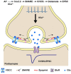
Ca2+ -independent transmission at the central synapse formed between dorsal root ganglion and dorsal horn neurons
- PMID: 36148511
- PMCID: PMC9638852
- DOI: 10.15252/embr.202154507
Ca2+ -independent transmission at the central synapse formed between dorsal root ganglion and dorsal horn neurons
Abstract
A central principle of synaptic transmission is that action potential-induced presynaptic neurotransmitter release occurs exclusively via Ca2+ -dependent secretion (CDS). The discovery and mechanistic investigations of Ca2+ -independent but voltage-dependent secretion (CiVDS) have demonstrated that the action potential per se is sufficient to trigger neurotransmission in the somata of primary sensory and sympathetic neurons in mammals. One key question remains, however, whether CiVDS contributes to central synaptic transmission. Here, we report, in the central transmission from presynaptic (dorsal root ganglion) to postsynaptic (spinal dorsal horn) neurons in vitro, (i) excitatory postsynaptic currents (EPSCs) are mediated by glutamate transmission through both CiVDS (up to 87%) and CDS; (ii) CiVDS-mediated EPSCs are independent of extracellular and intracellular Ca2+ ; (iii) CiVDS is faster than CDS in vesicle recycling with much less short-term depression; (iv) the fusion machinery of CiVDS includes Cav2.2 (voltage sensor) and SNARE (fusion pore). Together, an essential component of activity-induced EPSCs is mediated by CiVDS in a central synapse.
Keywords: Ca2+-dependent secretion; Ca2+-independent but voltage-dependent secretion; dorsal horn; dorsal root ganglion; synaptic transmission.
© 2022 The Authors.
Figures

- A
(a1) Whole‐cell current recording of evoked EPSC signals in response to local electrical field stimulation (Estim, arrows) from a postsynaptic dorsal horn (DH) neuron co‐cultured with presynaptic dorsal root ganglion (DRG) neurons in Ca2+‐free (0Ca2+, black) and 2.5 mM Ca2+ bath (2Ca2+, green); (a2), evoked intracellular Ca2+ signals (dF/F0) in a DRG neuron; inset, micrograph showing the setup for EPSC recording from DH neurons co‐cultured with EGFP‐expressing DRG neurons (scale bar, 20 μm).
- B
(b1) Evoked EPSCs from cultured hippocampal neurons in Ca2+‐free (black) and 2.5 mM Ca2+‐containing solution (green); (b2) evoked intracellular Ca2+ signals (dF/F0) in hippocampal neurons; inset, micrograph showing the setup for EPSC recordings from a hippocampal neuron (scale bar, 20 μm).
- C
As in (A), except that 50 μM BAPTA‐AM was pre‐loaded into the DRG and DH neurons before recording in Ca2+‐free (black) or 2.5 mM Ca2+‐containing solution (green). BAPTA‐AM was loaded for 30 min at 37°C.
- D
Left, quantification of amplitude as in (A) (n = 26 cells for 0Ca2+ and 29 cells for 2Ca2+). Right, quantification of EPSC amplitudes as in (C) (n = 16 cells for 0Ca2+ and 14 cells for 2Ca2+).
- E
Evoked EPSCCiVDS (0Ca2+, black) and total EPSCs (CDS + CiVDS, 2Ca2+, green) induced by paired‐pulse stimulation with a 50‐ms interval from DH neurons co‐cultured with DRG neurons. The 0Ca2+ and 2Ca2+ EPSCs were recorded in two different DH neurons.
- F
Summary plot of paired‐pulse ratios as in (E) with different intervals (in 0Ca2+, n = 7 cells for 10 ms, 13 for 50 ms, 14 for 500 ms, 10 for 5 s, and 8 for 15 s; in 2Ca2+, n = 14 cells for 10 ms, 17 for 50 ms, 22 for 500 ms, 19 for 5 s, and 14 for 15 s). Inset shows the initial plot at an expanded scale.
- G
Representative EPSCs induced by a 10‐Hz stimulus train in DH neurons co‐cultured with DRG neurons in Ca2+‐free (black) or 2.5 mM Ca2+‐containing solution (green). The 0Ca2+ and 2Ca2+ EPSCs were recorded in two different DH neurons.
- H
Summary plots of the EPSC amplitudes as in (G), including CiVDS (0Ca2+, black), CDS + CiVDS (2Ca2+, green), and CDS (2Ca2+ − 0Ca2+, blue) (n = 11 cells).
- I
As in (H), statistics for amplitudes of EPSCCiVDS (0Ca2+, black) and EPSCCDS (“2Ca2+” − “0Ca2+,” blue) evoked by single pulse (first EPSC, left) or 10 pulses (cumulative 10 EPSCs, ΣEPSC, right) during 10‐Hz train stimulation (n = 11 cells).

- A
A representative photograph showing the co‐cultured DRG and DH neurons. The DRG neurons were expressed with Spy‐pHluorin for imaging the synaptic transmission. Scale bar, 10 μm.
- B
Images of a presynaptic bouton marked in (A) showing the Spy‐pHluorin fluorescence at 20 s before (−20 s, pre‐stimulus), 20, 40, and 120 s after electrical stimulation (post‐stimulus) in 0Ca2+ (upper panel) or 2Ca2+ (lower panel) solution.
- C
Averaged fluorescence changes (ΔF/F0) of Spy‐pHluorin in 0Ca2+ (left) or 2Ca2+ solution (right) in response to the same electrical stimulation (20 Hz, 20 s) (n = 45 puncta from six cells for 0Ca2+ and 57 puncta from six cells for 2Ca2+). The shadow in the traces represents the error bars (s.e.m) of each point.
- D–F
The same as in (A–C), but the experiments were performed in cultured hippocampal neurons (n = 70 puncta from three cells for 0Ca2+ and 72 puncta from three cells for 2Ca2+).

- A
Representative evoked EPSCs in DH neurons co‐cultured with DRG neurons before (black) and after (red) applying 50 μM AP5 and 10 μM CNQX in Ca2+‐free (left) or 2.5 mM Ca2+‐containing solution (right). The EPSCs of 0Ca2+ and 2Ca2+ were recorded in two different DH neurons.
- B
Quantification of EPSC amplitudes as in (A) (n = 15 cells for 0Ca2+ and 10 cells for 2Ca2+).
- C
Evoked EPSCs recorded in DH neurons co‐cultured with DRG neurons before (black) and after (red) applying 100 μM cyclothiazide (CTZ) in 0Ca2+ (left) or 2Ca2+ solution (right). The traces are fitted with a single exponential curve (blue). The EPSCs of 0Ca2+ and 2Ca2+ were recorded in two different DH neurons.
- D
Quantification of the decay time (τ) and charge as in (C) (n = 10 cells for 0Ca2+ and 12 cells for 2Ca2+). EPSCs were evoked by local electrical stimulation (Estim) at arrows.

- A
Setup for paired patch‐clamp recording of dorsal root ganglion (DRG) and dorsal horn (DH) neurons in co‐culture. The presynaptic DRG neuron was whole‐cell dialyzed with 10 mM BAPTA under current‐clamp mode, while the postsynaptic DH neuron was in voltage‐clamp mode.
- B
Left, representative dual recordings of presynaptic action potentials (upper) and postsynaptic EPSCs (lower) following current‐step injection (1,000 pA, 5 ms) in a DRG neuron dialyzed with 10 mM BAPTA. Following whole‐cell break‐in and intracellular BAPTA dialysis, double recordings were performed at 0 min/2.5 mM Ca2+ bath (green), and 5 min/0Ca2+ bath (black), Right, statistics of EPSC amplitudes (n = 9 cells).
- C, D
As in (A) and (B), except that EPSCs were recorded from cultured hippocampal neurons (n = 11 cells).
- E
Image showing a DRG neuron infected by ChR2‐mCherry AAV2/9 virus and a co‐cultured DH neuron.
- F
Upper, diagram showing the protocol for infection of DRG neurons by ChR2‐mCherry virus and co‐culture with DH neurons; lower, cartoon of EPSC recording from DH neurons co‐cultured with ChR2‐expressing DRG neurons.
- G
Left, typical EPSCs from DH neurons induced by light stimulation (475 nm, 5 ms, at arrows) of co‐cultured ChR2‐expressing DRG neurons in Ca2+‐free or 2.5 mM Ca2+‐containing solution before (black) and after (red) exposure to 50 μM AP5 and 10 μM CNQX. The EPSCs of 0Ca2+ and 2Ca2+ were recorded in two different DH neurons; right, statistics of EPSC amplitude (n = 5 cells for 0Ca2+ and 14 for 2Ca2+).

- A
Left, diagram showing the protocol for EPSC recording from DH neurons co‐cultured with Syb2‐knockout (cKO) DRG neurons; right, cartoon of EPSC recording from DH neurons co‐cultured with Syb2‐cKO DRG neurons.
- B
Representative western blots (upper) and analysis (lower) of the expression levels of Syb2 in control (Ctrl) and Syb2‐cKO DRG neurons (n = 3 per group). Control DRG neurons are from homozygous floxed Syb2‐null mice infected with AAV5‐CAG‐EGFP virus.
- C
Evoked EPSCs and statistics from DH neurons co‐cultured with control (Ctrl) or Syb2‐cKO DRG cells (cKO) in 0Ca2+ solution (n = 13 cells for Ctrl and 11 for cKO).
- D
Evoked EPSCs and statistics from DH neurons co‐cultured with DRG neurons before (black) and after (red) applying 1 μM ω‐conotoxin‐GVIA (GVIA) in 0Ca2+ bath (n = 9 cells).
- E
Upper, diagram showing the protocol for EPSC recordings from DH neurons co‐cultured with Cav2.2 knockdown (KD) DRG neurons; lower, cartoon of EPSC recording from DH neurons co‐cultured with Cav2.2‐KD DRG neurons.
- F
Left panels, evoked EPSCs from DH neurons co‐cultured with scrambled shRNA control (Ctrl) or Cav2.2‐KD (sh‐1/sh‐2) DRG neurons in 0Ca2+ solution. Right panel, quantification of evoked EPSCs (n = 17 cells for Ctrl, 12 for sh‐1, and 19 for sh‐2). EPSCs were evoked by local electrical stimulation (Estim, at arrows).

References
-
- Augustine GJ, Charlton MP, Smith SJ (1987) Calcium action in synaptic transmitter release. Annu Rev Neurosci 10: 633–693 - PubMed
-
- Chai Z, Wang C, Huang R, Wang Y, Zhang X, Wu Q, Wang Y, Wu X, Zheng L, Zhang C et al (2017) CaV2.2 gates calcium‐independent but voltage‐dependent secretion in mammalian sensory neurons. Neuron 96: 1317–1326 - PubMed
-
- Ellinor PT, Zhang JF, Horne WA, Tsien RW (1994) Structural determinants of the blockade of N‐type calcium channels by a peptide neurotoxin. Nature 372: 272–275 - PubMed
Publication types
MeSH terms
LinkOut - more resources
Full Text Sources
Miscellaneous

