. 2022 Dec;11(23):e2201714.
doi: 10.1002/adhm.202201714. Epub 2022 Oct 17.

Thermoresponsive and Injectable Hydrogel for Tissue Agnostic Regeneration

Affiliations

Thermoresponsive and Injectable Hydrogel for Tissue Agnostic Regeneration

Dax Calder et al. Adv Healthc Mater. 2022 Dec.

Abstract

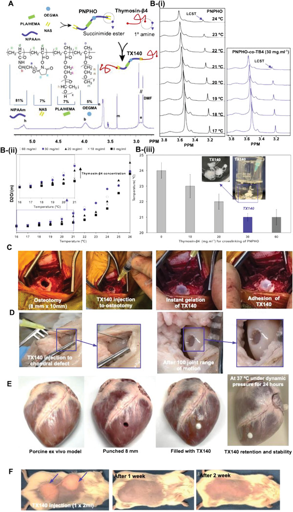
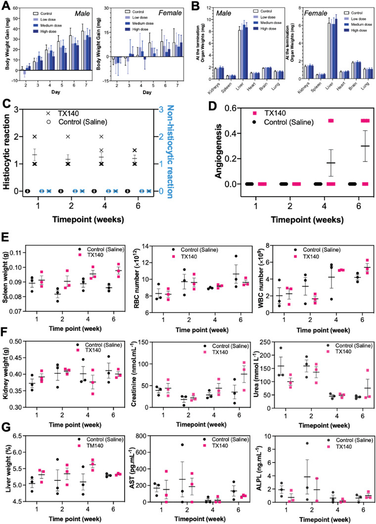
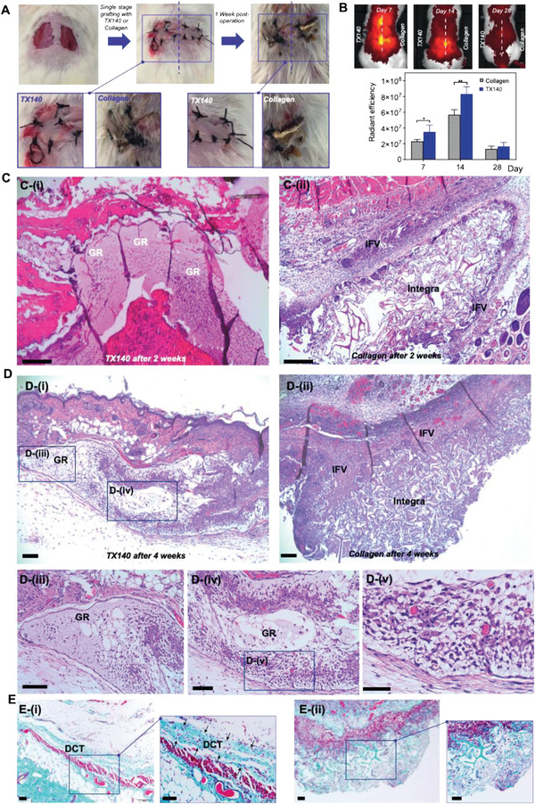
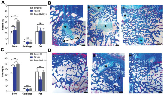
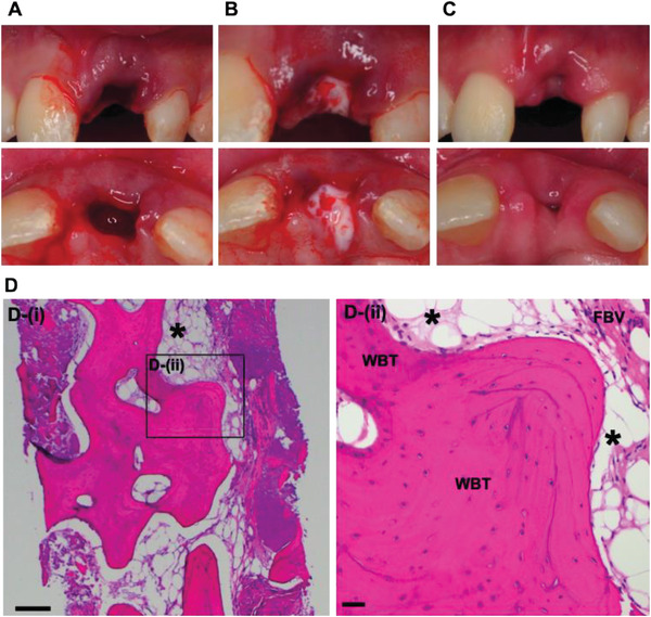
Injectable hydrogels can support the body's innate healing capability by providing a temporary matrix for host cell ingrowth and neovascularization. The clinical adoption of current injectable systems remains low due to their cumbersome preparation requirements, device malfunction, product dislodgment during administration, and uncontrolled biological responses at the treatment site. To address these challenges, a fully synthetic and ready-to-use injectable biomaterial is engineered that forms an adhesive hydrogel that remains at the administration site regardless of defect anatomy. The product elicits a negligible local inflammatory response and fully resorbs into nontoxic components with minimal impact on internal organs. Preclinical animal studies confirm that the engineered hydrogel upregulates the regeneration of both soft and hard tissues by providing a temporary matrix to support host cell ingrowth and neovascularization. In a pilot clinical trial, the engineered hydrogel is successfully administered to a socket site post tooth extraction and forms adhesive hydrogel that stabilizes blood clot and supports soft and hard tissue regeneration. Accordingly, this injectable hydrogel exhibits high therapeutic potential and can be adopted to address multiple unmet needs in different clinical settings.

Keywords: injectable hydrogel; platform technology; regenerative medicine.


Conflict of interest statement

A.F., T.A., S.M., and J.M. are employed by Tetratherix. A.F. and F.D. are co‐inventors in AU2016301103; EP16829490.8; JP2018‐503480; US15/746810; U2016314146; EP344304; JP2018‐529692; US 17/090078; EP 2794701; US9546235. A.F., D.C., and T.A. are co‐inventors in PCT/AU2020/051332 and Chinese national phase patent application (2020800006185.0). D.C. is a clinical advisor to Tetratherix. While the material in the current study has not been published, the main authors have previously invented and disclosed information pertaining to a family of polymers as outlined in granted patents WO 2013/091001; WO 2017/035587; WO 2017/015703; WO 2021/119727. All other authors declare no competing interest.

Figures

Figure 1
Figure 1
Chemical composition of TX140 and its application at different anatomical sites; A) schematic representation of PNPHO along with its chemical composition and 1H‐NMR spectrum of the polymer with the use of Thymosin‐β4 to conjugate with PNPHO. B) The region of interest, (m) peak, in 1H‐NMR spectra of PNPHO polymer solution i) and PNPHO‐co‐Thymosin‐β4 with 30 mg mL−1 at different temperatures ranging from 17 to 24 °C. D2O/(m) of PNPHO‐co‐Thymosin‐β4 solution at different temperature to find LCST of the solutions ii). LCST of the different PNPHO‐co‐Thymosin‐β4 solution with different Thymosin‐β4 concentration (iii, source data in Figure S4 (Supporting Information), and n = 10). C) The hydrogel is injectable and forms a matrix in physiological conditions in a live sheep osteotomy model (n = 6). D) Adhesion and retention of TX140 in an ex vivo bovine cadaveric subchondral defect model without the need for physical containment in a dynamic model after 100 cycles of complete joint motion (n = 12). E) Demonstration of underwater and dynamic stability of the TX140 hydrogels applied to seal an 8 mm puncture in porcine heart tissue (n = 6). The hydrogels remained stable after 24 h soaking in a 37 °C water bath (Movies S1 and S2, Supporting Information). F) live mice subcutaneous injection model that confirms gelation and adhesion of TX140 without any physical containment (n = 24).
Figure 2
Figure 2
Acute toxicity, local, and systemic biocompatibility assessment of TX140. A) Body weight gain of female and male rats by polar and nonpolar extracts of TX140 during a week for acute toxicity assessment (n = 80, 40 female and 40 male animals). B) Organ weights of female and male rats by polar and nonpolar extracts of TX140 at the termination of the acute toxicity assessment (n = 80, 40 female and 40 male animals). C) inflammatory response (histiocytic and nonhistiocytic responses) to TX140 and normal saline at different time points. D) local angiogenesis at TX140 and normal saline injection site at different time points. E) spleen weight, red blood cell (RBC) and white blood cell (WBC) counts of TX140 and normal saline injected animals at different time points. F) kidney weight and kidney biochemistry markers (urea and creatinine) of TX140 and normal saline injected animals at different time points. G) liver weight and liver biochemistry markers of TX140 and normal saline injected animals at different time points (aspartate aminotransferase (AST) and alkaline phosphatase (ALP)). n = 24 in total, 6 at each time point for measurements reported in (C)–(G).
Figure 3
Figure 3
Soft‐tissue healing and angioconductive properties of TX140. A) Intraoperative wounds after harvesting full‐thickness skin grafts and grafting, treated with TX140 (left) and commercially available collagen scaffold as the gold standard (right) at day 0 and day 7 post skin grafting (n = 27). B) Radiant efficiency of TX140 and collagen scaffold in a murine model after 7, 14, and 28 days (n = 6, statistical significance * for p < 0.05 and ** for p < 0.01)). C) H&E stained cross‐sections of TX140 i) and collagen scaffold ii) after 14 days. D) H&E stained cross‐sections of TX140 i) and the collagen scaffold ii) after 28 days. Cross sections of TX140 iii–v). E) MT stained cross‐sections of Integra and TX140 i) and collagen scaffold ii) biopsies 28 days postskin grafting. GR: TX140 hydrogel residues and IFV: inflammatory fibrosis. DCT: dermal connective tissue. The scale bar in all panel is 100 µm. For (C)–(E) n = 27 in total and 12 at each time point.
Figure 4
Figure 4
Hard‐tissue healing in sheep osteotomy model. A) Bone, cartilage, and fat formation in osteotomy sheep model after 6 weeks. B) Masson's Trichrome (MT)‐stained decalcified sections of lesion explants from i) Empty, ii) TX140, and iii) bone graft groups after 6‐weeks postsurgery. C) Bone, cartilage, and fat formation in an osteotomy sheep model after 12 weeks postsurgery D) MT‐stained decalcified sections of lesion explants from i) Empty, ii) TX140, and iii) bone graft groups after 12‐weeks postsurgery. The scale bar in all panels is 1 mm. Asterisks highlight the new bone formation foci of fibrous connective tissue (n = 6).
Figure 5
Figure 5
Clinical study of TX140 post tooth extraction. A) Extraction site with active bleeding, B) in situ gel formation of TX140 in extraction site, adhesion and mixing with blood at the injection site without the need for primary closure C) soft tissue healing and wound closure at TX140 treated site 7 days post operation (n = 10). D) MT stained TX140 treated site 3 months postoperation, product residues (asterisk), woven bone trabecular (WBT) and fibrovascular tissue (FBV). The scale bar in panel (D) is 200 µm (n = 9).

References

    1. Sun Y., Nan D., Jin H., Qu X., Polym. Test. 2020, 81, 106283.
    1. Rizzo F., Kehr N. S., Adv. Healthcare Mater. 2021, 10, 2001341. - PubMed
    1. Busscher H. J., van der Mei H. C., Subbiahdoss G., Jutte P. C., van den Dungen J. J. A. M., Zaat S. A. J., Schultz M. J., Grainger D. W., Sci. Transl. Med. 2012, 4. - PubMed
    1. Ren X., Zhao M., Lash B., Martino M. M., Julier Z., Front. Bioeng. Biotechnol. 2019, 7, 469. - PMC - PubMed
    1. Carragee E. J., Hurwitz E. L., Weiner B. K., Spine J. 2011, 11, 471. - PubMed

Publication types

MeSH terms

LinkOut - more resources