

Totally Organic Hydrogel-Based Self-Closing Cuff Electrode for Vagus Nerve Stimulation
- PMID: 36148587
- PMCID: PMC11468388
- DOI: 10.1002/adhm.202201627
Totally Organic Hydrogel-Based Self-Closing Cuff Electrode for Vagus Nerve Stimulation
Abstract
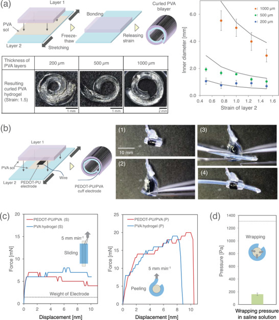
An intrinsically soft organic electrode consisting of poly(3,4-ethylenedioxythiophene)-modified polyurethane (PEDOT-PU) is embedded into a bilayer film of polyvinyl alcohol (PVA) hydrogels for developing a self-closing cuff electrode for neuromodulation. The curled form of the PVA hydrogel is prepared by releasing internal stress in the bilayer structure. The inner diameter of the cuff electrode is set to less than 2 mm for immobilization to the vagus nerve (VN) of humans and pigs. The stability of the immobilization is examined, while the pressure applied to a nerve bundle is at a harmless level (≈200 Pa). Since the electrode is totally organic, MRI measurements can be conducted without image artifacts. The large electric capacitance of the PEDOT-PU (≈27 mF cm-2 ) ensures a safe stimulation of living tissues without Faradaic reactions. The practical performance of the cuff electrode for VN stimulation is demonstrated by observation of bradycardia induction in a pig.
Keywords: MRI compatible implants; cuff electrodes; hydrogels; self-closing; vagus nerve stimulation.
© 2022 The Authors. Advanced Healthcare Materials published by Wiley-VCH GmbH.
Conflict of interest statement
The authors declare no conflict of interest.
Figures


Similar articles
-
Hydrogel-Based Organic Subdural Electrode with High Conformability to Brain Surface.Sci Rep. 2019 Sep 16;9(1):13379. doi: 10.1038/s41598-019-49772-z. Sci Rep. 2019. PMID: 31527626 Free PMC article.
-
Vagus nerve stimulation using an endovascular electrode array.J Neural Eng. 2023 Jul 14;20(4):10.1088/1741-2552/acdb9b. doi: 10.1088/1741-2552/acdb9b. J Neural Eng. 2023. PMID: 37276858 Free PMC article.
-
Flexible IrOx neural electrode for mouse vagus nerve stimulation.Acta Biomater. 2023 Mar 15;159:394-409. doi: 10.1016/j.actbio.2023.01.026. Epub 2023 Jan 17. Acta Biomater. 2023. PMID: 36669547 Free PMC article.
-
Electrochemical and Electrophysiological Performance of Platinum Electrodes Within the Ninety-Nine-Electrode Stimulating Nerve Cuff.Artif Organs. 2015 Oct;39(10):886-96. doi: 10.1111/aor.12625. Artif Organs. 2015. PMID: 26471140 Review.
-
Vagus Nerve and Vagus Nerve Stimulation, a Comprehensive Review: Part I.Headache. 2016 Jan;56(1):71-8. doi: 10.1111/head.12647. Epub 2015 Sep 14. Headache. 2016. PMID: 26364692 Review.
Cited by
-
A biodegradable and flexible neural interface for transdermal optoelectronic modulation and regeneration of peripheral nerves.Nat Commun. 2024 Jun 3;15(1):4721. doi: 10.1038/s41467-024-49166-4. Nat Commun. 2024. PMID: 38830884 Free PMC article.
-
Stretchable Surface Electrode Arrays Using an Alginate/PEDOT:PSS-Based Conductive Hydrogel for Conformal Brain Interfacing.Polymers (Basel). 2022 Dec 25;15(1):84. doi: 10.3390/polym15010084. Polymers (Basel). 2022. PMID: 36616434 Free PMC article.
-
Design considerations for optogenetic applications of soft micro-LED-based device systems across diverse nervous systems.Bioact Mater. 2025 Feb 19;48:217-241. doi: 10.1016/j.bioactmat.2025.02.006. eCollection 2025 Jun. Bioact Mater. 2025. PMID: 40046014 Free PMC article. Review.
-
PEDOT:PSS Wire: A Two-Terminal Synaptic Device for Operation in Electrolyte and Saline Solutions.ACS Appl Mater Interfaces. 2024 Oct 9;16(40):54636-54644. doi: 10.1021/acsami.4c12037. Epub 2024 Sep 30. ACS Appl Mater Interfaces. 2024. PMID: 39344494 Free PMC article.
-
Hydrogels in wearable neural interfaces.Med X. 2024;2(1):23. doi: 10.1007/s44258-024-00040-4. Epub 2024 Dec 9. Med X. 2024. PMID: 39659711 Free PMC article. Review.
References
Publication types
MeSH terms
Substances
LinkOut - more resources
Full Text Sources
Miscellaneous
