. 2022 Dec;232(5-6):115-123.
doi: 10.1007/s00427-022-00696-1. Epub 2022 Sep 23.

Exploring the roles of FGF/MAPK and cVG1/GDF signalling on mesendoderm induction and convergent extension during chick primitive streak formation

Affiliations

Exploring the roles of FGF/MAPK and cVG1/GDF signalling on mesendoderm induction and convergent extension during chick primitive streak formation

Hyung Chul Lee et al. Dev Genes Evol. 2022 Dec.

Abstract

During primitive streak formation in the chick embryo, cells undergo mesendoderm specification and convergent extension at the same time and in the same cells. Previous work has implicated cVG1 (GDF3) as a key factor for induction of primitive streak identity and positioning the primitive streak, whereas FGF signalling was implicated in regulating cell intercalation via regulation of components of the WNT-planar cell polarity (PCP) pathway. FGF has also been reported to be able to induce a primitive streak (but lacking the most axial derivatives such as notochord/prechordal mesendoderm). These signals emanate from different cell populations in the embryo, so how do they interact to ensure that the same cells undergo both cell intercalation and acquire primitive streak identity? Here we begin to address this question by examining in more detail the ability of the two classes of signals in regulating the two developmental events. Using misexpression of inducers and/or exposure to inhibitors and in situ hybridisation, we study how these two signals regulate expression of Brachyury (TBXT) and PRICKLE1 as markers for the primitive streak and the PCP, respectively. We find that both signals can induce both properties, but while FGF seems to be required for induction of the streak by cVG1, it is not necessary for induction of PRICKLE1. The results are consistent with cVG1 being a common regulator for both primitive streak identity and the initiation of convergent extension that leads to streak elongation.

Keywords: Embryonic polarity; Epithelial cell intercalation; Gastrulation; Planar cell polarity; Primitive streak elongation.


Conflict of interest statement

The authors declare no competing interests.

Figures

Fig. 1
Fig. 1
Expression patterns of FGF8, TBXT and PRICKLE1 in the pre-primitive-streak stage embryo, revealed by in situ hybridisation. FGF8 is detected exclusively in the hypoblast (hypo) at EGK stage XII (A), then in both the epiblast (epi) and the lower layers at EGK stage XIII (D). TBXT starts to be expressed in the posterior epiblast at EGK stage XII (B), then is also expressed in the forming mesendoderm at HH stage 2 (E). PRICKLE1 is expressed both in the epiblast and the lower layers of the primitive streak at EGK stage XIV (C and F). The upper (AF) and lower (A’–F’ and A’’–F’’, 20 × and 40 × magnification, respectively) panels correspond, respectively, to a whole mount and a sagittal section at the level of the dashed line. Scale bar, 100 µm for all sections
Fig. 2
Fig. 2
cVG1 misexpression induces PRICKLE1 and TBXT expression. Two cVG1-expressing cell pellets were grafted to the anterior region of the embryo at EGK X-XI, and the expression of PRICKLE1 (AD) and TBXT (EH) was investigated by in situ hybridisation after 6, 9, 12 and 15 h incubation. Both genes begin to be expressed from 9 h after incubation in the epiblast above the grafted cell pellet. The upper (AH) and lower (B’–D’ and F’–H’) panels show the whole embryo and a sagittal section at the level of the dotted lines. Arrows: location of the cVG1-expressing cell pellet. Scale bars, 100 µm for all sections. The number of embryos with ectopic expression and total number of embryos in (AH) were 0/6, 5/6, 5/6, 4/5, 0/4, 5/5, 6/6 and 5/5
Fig. 3
Fig. 3
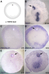
FGF8 misexpression induces PRICKLE1 and TBXT. (A) Experimental design. An FGF8-soaked bead was grafted to the anterior region of the embryo at EGK X-XI. (B) After overnight culture, an ectopic primitive streak, expressing TBXT (arrow), was induced near the bead. (CD) An FGF8-soaked bead induces PRICKLE1 by 6 h after grafting (arrow, D). (EF) FGF8-beads also induce weak TBXT expression as soon as 3 h after grafting; this later intensifies (arrows, E and F). Number of embryos with ectopic expression and total number of embryos in (BF): 4/6, 0/5, 3/5, 4/5 and 6/6
Fig. 4
Fig. 4
Effect of SU5402, a FGF inhibitor, on the induction of TBXT and PRICKLE1. (A and F) Experimental design. SU5402-soaked or control (DMSO) beads were grafted either into the posterior area pellucida (four beads, A), or into the anterior area pellucida at EGK X-XI together with a cVG1-expressing cell pellet (two beads, F). Then, after 9-h incubation, expression of TBXT or PRICKLE1 was investigated by in situ hybridisation. (BE) SU5402-soaked beads inhibit TBXT expression but not PRICKLE1 in the posterior area pellucida (prospective primitive streak cells). (GJ) SU5402-soaked beads inhibit induction of TBXT but not PRICKLE1 by a cVG1-pellet in the anterior area pellucida. Arrows, induced expression. Number of embryos with expression near the beads and total number of embryos: 0/4, 2/2, 4/4 and 3/3, respectively, for BE, and 0/6, 4/4, 2/3 and 6/6, respectively, for GJ. Note that in some embryos, some of the beads become detached during fixation or in situ hybridisation
Fig. 5
Fig. 5
Effect of U0126, a MEK1/2 inhibitor, on the induction of TBXT and PRICKLE1. (A and F) Experimental design. U0126-soaked or control (U0124) beads were grafted into either the posterior area pellucida (four beads, A), or into the anterior area pellucida at EGK X-XI together with a cVG1-expressing cell pellet (two beads, F). Then, after 9-h incubation, expression of TBXT or PRICKLE1 was investigated by in situ hybridisation. (BE) U0126-soaked beads, but not control (U0124) beads, inhibit TBXT expression but not PRICKLE1 posteriorly. (GJ) U0126-soaked beads inhibit induction of TBXT but not PRICKLE1 by cVG1 anteriorly. Arrows, induced expression. Number of embryos with expression near the beads and total number of embryos: 2/12, 8/9, 11/12 and 5/8, respectively, for BE, and 0/4, 3/4, 4/5 and 5/5, respectively, for GJ. Note that as in Fig. 4, some of the beads have become detached during processing of the embryos
Fig. 6
Fig. 6
Inhibition of TBXT and PRICKLE1 by BMP4 misexpression. (A) Experimental design. A BMP4-expressing cell pellet is grafted to the posterior region of the embryo at EGK X-XI, and the expression of PRICKLE1 (after 9-h incubation) (BC) or TBXT (after overnight incubation) (DE) was investigated by in situ hybridisation. BMP4 misexpression splits the primitive streak forming region, sometimes generating a double primitive streak, one on either side of the grafted cell pellet (B, D). Dotted circles, location of the cell pellet. Number of embryos showing inhibitory effect and total number of embryos: 7/7, 0/5, 5/6 and 0/5, respectively, for BE

Similar articles

References

    1. Bertocchini F, Stern CD. The hypoblast of the chick embryo positions the primitive streak by antagonizing nodal signaling. Dev Cell. 2002;3:735–744. doi: 10.1016/S1534-5807(02)00318-0. - DOI - PubMed
    1. Bertocchini F, Stern CD. Gata2 provides an early anterior bias and uncovers a global positioning system for polarity in the amniote embryo. Development. 2012;139:4232–4238. doi: 10.1242/dev.081901. - DOI - PMC - PubMed
    1. Bertocchini F, Skromne I, Wolpert L, Stern CD. Determination of embryonic polarity in a regulative system: evidence for endogenous inhibitors acting sequentially during primitive streak formation in the chick embryo. Development. 2004;131:3381–3390. doi: 10.1242/dev.01178. - DOI - PubMed
    1. Candia AF, Watabe T, Hawley SH, Onichtchouk D, Zhang Y, Derynck R, Niehrs C, Cho KW. Cellular interpretation of multiple TGF-beta signals: intracellular antagonism between activin/BVg1 and BMP-2/4 signaling mediated by Smads. Development. 1997;124:4467–4480. doi: 10.1242/dev.124.22.4467. - DOI - PubMed
    1. Demagny H, De Robertis EM. Point mutations in the tumor suppressor Smad4/DPC4 enhance its phosphorylation by GSK3 and reversibly inactivate TGF-beta signaling. Mol Cell Oncol. 2016;3:e1025181. doi: 10.1080/23723556.2015.1025181. - DOI - PMC - PubMed

Publication types

LinkOut - more resources