. 2022 Oct;38(10):971-980.
doi: 10.1002/kjm2.12587. Epub 2022 Sep 23.

Long non-coding RNA MALAT1 promotes Th2 differentiation by regulating microRNA-135b-5p/GATA-3 axis in children with allergic rhinitis

Affiliations

Long non-coding RNA MALAT1 promotes Th2 differentiation by regulating microRNA-135b-5p/GATA-3 axis in children with allergic rhinitis

Xiong-Hui Wu et al. Kaohsiung J Med Sci. 2022 Oct.

Abstract

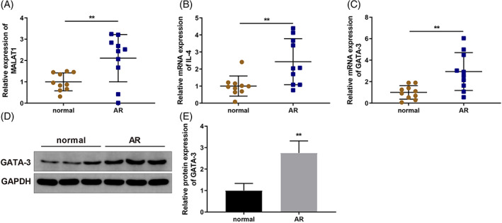
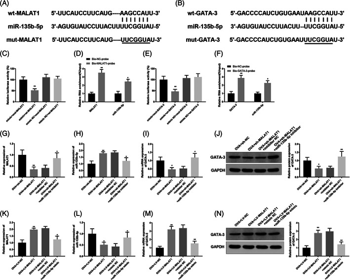
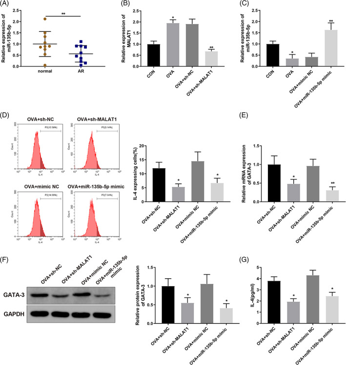
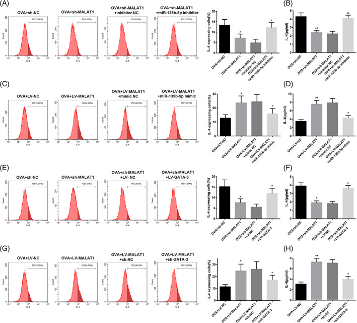
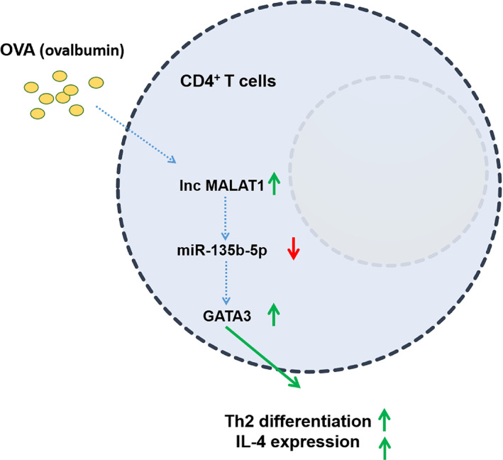
Allergic rhinitis (AR) threatens patient survival. CD4+ T cells play key roles in AR progression. Long non-coding RNAs (lncRNAs) are key regulators of cell differentiation. Therefore, we investigated the molecular mechanism of the lncRNA metastasis-associated lung adenocarcinoma transcript 1 (MALAT1) in AR. Expression levels of MALAT1, microRNA (miR)-135b-5p, interleukin-4 (IL-4), and GATA-binding protein 3 (GATA-3) in the nasal mucosa of AR patients were quantified. CD4+ T cells were isolated from the peripheral blood of healthy volunteers and treated with ovalbumin (OVA) and Th2 inducers. After MALAT1 and miR-135b-5p levels changed in CD4+ T cells, the proportion of IL-4-expressing cells and the levels of IL-4 and GATA-3 in OVA-induced CD4+ T cells were determined. Binding relationships among MALAT1, miR-135b-5p, and GATA-3 were predicted and verified. Rescue experiments were performed to confirm the role of the MALAT1/miR-135b-5p/GATA-3 axis in Th2 differentiation of CD4+ T cells. MALAT1, IL-4, and GATA-3 expression was upregulated, whereas miR-135b-5p expression was downregulated, in patients with AR. MALAT1 knockdown or miR-135b-5p overexpression in CD4+ T cells notably decreased the proportion of IL-4-expressing cells and downregulated GATA-3 and IL-4 expression in OVA-induced CD4+ T cells. MALAT1 and GATA-3 exhibited competitive binding toward miR-135b-5p. MALAT1 facilitated CD4+ T cell Th2 differentiation via the miR-135b-5p/GATA-3 axis. MALAT1 facilitated AR development by facilitating CD4+ T cell Th2 differentiation via the miR-135b-5p/GATA-3 axis. This study may provide guidance for clinical treatment of AR.

Keywords: GATA binding protein 3; T-helper 2 differentiation; allergic rhinitis; long non-coding RNA MALAT1; microRNA-135b-5p.


Conflict of interest statement

All authors declare no conflict of interest.

Figures

FIGURE 1
FIGURE 1
MALAT1 and GATA‐3 expression levels are upregulated in AR patients. Nasal mucosa tissue samples of 10 AR patients and 10 normal volunteers were collected. (A) MALAT1 expression in nasal mucosal tissues was quantified via qRT‐PCR; (B) IL‐4 expression in nasal mucosal tissues was quantified via qRT‐PCR; (C‐E) GATA‐3 expression in nasal mucosal tissues was quantified via qRT‐PCR (C) and WB (D‐E). N = 10, **p < 0.01, compared to the normal group. AR, allergic rhinitis; GATA‐3, GATA‐binding protein 3; MALAT1, metastasis‐associated lung adenocarcinoma transcript 1; qRT‐PCR, quantitative reverse transcription‐polymerase chain reaction; WB, western blotting
FIGURE 2
FIGURE 2
MALAT1 competes with GATA‐3 to bind to miR‐135b‐5p. (A) Binding site between miR‐135b‐5p and MALAT1 3'UTR; (B) binding site between miR‐135b‐5p and GATA‐3 3'UTR; (C) dual‐luciferase reporter gene assay was used to confirm the interaction between MALAT1 and miR‐135b‐5p, **p < 0.01, compared to the OVA + mimic NC + wt‐MALAT1 group; (D) RNA pull‐down assay was used to verify the interaction between MALAT1 and miR‐135b‐5p, *p < 0.05, **p < 0.01, compared to the bio‐NC‐probe group; (E) binding between miR‐135b‐5p and GATA‐3 was verified using dual luciferase reporter gene assay, **p < 0.01, compared to the OVA + mimic NC + wt‐GATA‐3 group; (F) RNA pull‐down assay was used to verify the interaction between miR‐135b‐5p and GATA‐3, *p < 0.05, **p < 0.01, compared to the bio‐NC‐probe group. CD4+ T cells were transfected with sh‐MALAT1 or LV‐MALAT1 lentiviral vectors, sh‐MALAT1 plus miR‐135b‐5p inhibitor, or LV‐MALAT1 plus miR‐135b‐5p mimic and then induced with OVA and Th2 inducers. (G–N) qRT‐PCR was used to quantify MALAT1 (G, K), miR‐135b‐5p (H, L) or GATA‐3 (I, M) expression levels, and WB was utilized to quantify GATA‐3 expression levels (J, N) in CD4+ T cells, *p < 0.05, **p < 0.01, compared to the OVA + sh‐NC/OVA + LV‐NC group or OVA + sh‐MALAT1 + inhibitor NC/OVA + LV‐MALAT1 + mimic NC group. Each experiment was independently repeated three times. AR, allergic rhinitis; GATA‐3, GATA‐binding protein 3; IL, interleukin; lncRNA, long non‐coding RNA; MALAT1, metastasis‐associated lung adenocarcinoma transcript 1; miR, microRNA; OVA, ovalbumin; qRT‐PCR, quantitative reverse transcription‐polymerase chain reaction; WB, western blotting
FIGURE 3
FIGURE 3
MALAT1 knockdown or miR‐135b‐5p overexpression inhibits OVA‐induced Th2 differentiation of CD4+ T cells. (A) miR‐135b‐5p expression levels in the collected nasal mucosa tissue samples were quantified via qRT‐PCR and compared to normal group. CD4+ T cells were transfected with sh‐MALAT1 lentiviral vector or miR‐135b‐5p mimic and induced with OVA and Th2 inducers. (B) qRT‐PCR was performed to quantify MALAT1 expression levels in OVA‐induced CD4+ T cells, compared to CON group or OVA + sh‐NC/OVA + mimic NC group; (C) qRT‐PCR was used to quantify miR‐135b‐5p expression levels in OVA‐induced CD4+ T cells; (D) Flow cytometry was used to determine the percentage of IL‐4‐expressing cells; (E, F) qRT‐PCR (E) and WB (F) were used to quantify GATA‐3 expression levels in OVA‐induced CD4+ T cells; (G) ELISA was used to quantify IL‐4 levels in the supernatant of OVA‐induced CD4+ T cells. *p < 0.05, **p < 0.01, compared to the OVA + sh‐NC group or the OVA + mimic NC group. Each experiment was independently repeated three times. AR, allergic rhinitis; ELISA, enzyme‐linked immunosorbent assay; lncRNA, long non‐coding RNA; MALAT1, metastasis‐associated lung adenocarcinoma transcript 1; miR, microRNA; IL, interleukin; GATA‐3, GATA‐binding protein 3; OVA, ovalbumin; qRT‐PCR, quantitative reverse transcription‐polymerase chain reaction; WB, western blotting
FIGURE 4
FIGURE 4
MALAT1 promotes OVA‐induced Th2 differentiation of CD4+ T cells via the miR‐135b‐5p/GATA‐3 axis. CD4+ T cells were transfected with sh‐MALAT1 lentiviral vector or co‐transfected with sh‐MALAT1 lentiviral vector and miR‐135b‐5p inhibitor, followed by the induction using OVA and Th2 inducers. (A) Percentage of IL‐4‐expressing cells was determined using flow cytometry. (B) IL‐4 level in the supernatant of OVA‐induced CD4+ T cells was measured using ELISA, *p < 0.05, **p < 0.01, compared to the OVA + sh‐NC group or the OVA + sh‐MALAT1 + inhibitor NC group. CD4+ T cells were transfected with LV‐MALAT1 or co‐transfected with LV‐MALAT1 and miR‐135b‐5p mimic and induced for Th2 differentiation; (C) percentage of IL‐4‐expressing cells was determined using flow cytometry; (D) IL‐4 level in the supernatant of OVA‐induced CD4+ T cells was measured using ELISA, *p < 0.05, **p < 0.01, compared to the OVA + LV‐NC group or the OVA + LV‐MALAT1 + mimic NC group. CD4+ T cells were transfected with sh‐MALAT1 or co‐transfected with sh‐MALAT1 and LV‐GATA‐3 and induced using OVA and Th2 inducers; (E) Percentage of IL‐4‐expressing cells was determined using flow cytometry; (F) IL‐4 level in the supernatant of OVA‐induced CD4+ T cells was measured using ELISA, *p < 0.05, compared to the OVA + sh‐NC group or the OVA + sh‐MALAT1 + LV‐NC group. CD4+ T cells were transfected with LV‐MALAT1 or co‐transfected with LV‐MALAT1 and sh‐GATA‐3 and induced using OVA and Th2 inducers. (G) Percentage of IL‐4‐expressing cells was determined using flow cytometry; (H) IL‐4 level in the supernatant of OVA‐induced CD4+ T cells was measured using ELISA, *p < 0.05, **p < 0.01, compared to the OVA + LV‐NC group or the OVA + LV‐MALAT1 + sh‐NC group. Each experiment was independently repeated three times. AR, allergic rhinitis; ELISA, enzyme‐linked immunosorbent assay; GATA‐3, GATA‐binding protein 3; IL, interleukin; lncRNA, long non‐coding RNA; MALAT1, metastasis‐associated lung adenocarcinoma transcript 1; miR, microRNA; OVA, ovalbumin; qRT‐PCR, quantitative reverse transcription‐polymerase chain reaction; WB, western blotting
FIGURE 5
FIGURE 5
A schematic diagram of long noncoding RNA MALAT1 promoting ovalbumin‐induced Th2 differentiation and IL‐4 expression in CD4+ T cells by the miR‐135b‐5p/GATA‐3 axis. Ovalbumin induction upregulated lncRNA MALAT1 expression and downregulated miR‐135b‐5p expression, which upregulated GATA‐3 expression, thereby promoting Th2 differentiation and IL‐4 expression in CD4+ T cells. AR, allergic rhinitis; GATA‐3, GATA‐binding protein 3; IL, interleukin; lncRNA, long non‐coding RNA; MALAT1, metastasis‐associated lung adenocarcinoma transcript 1; miR, microRNA; OVA, ovalbumin

Similar articles

Cited by

References

    1. Kalmarzi RN, Ataee P, Fathollahpour A, Behzadifar M, Moradi M, Sharifian F, et al. The prevalence of allergic rhinitis among Iranian children: a systematic review and meta‐analysis. Endocr Metab Immune Disord Drug Targets. 2020;20(2):189–97. - PubMed
    1. Testa D, Di Bari M, Nunziata M, Cristofaro G, Massaro G, Marcuccio G, et al. Allergic rhinitis and asthma assessment of risk factors in pediatric patients: a systematic review. Int J Pediatr Otorhinolaryngol. 2020;129:109759. - PubMed
    1. Loekmanwidjaja J, Carneiro ACF, Nishinaka MLT, Munhoes DA, Benezoli G, Wandalsen GF, et al. Sleep disorders in children with moderate to severe persistent allergic rhinitis. Braz J Otorhinolaryngol. 2018;84(2):178–84. - PMC - PubMed
    1. Kakli HA, Riley TD. Allergic rhinitis. Prim Care. 2016;43(3):465–75. - PubMed
    1. Ridolo E, Martignago I, Masieri S. Mechanisms of allergic diseases in otorhinolaryngology. J Biol Regul Homeost Agents. 2018;32(1 Suppl. 1):9–12. - PubMed