Review
. 2022 Nov 1:192:130-140.
doi: 10.1016/j.freeradbiomed.2022.09.015. Epub 2022 Sep 21.

α-Syn overexpression, NRF2 suppression, and enhanced ferroptosis create a vicious cycle of neuronal loss in Parkinson's disease

Affiliations
Review

α-Syn overexpression, NRF2 suppression, and enhanced ferroptosis create a vicious cycle of neuronal loss in Parkinson's disease

Anandhan A et al. Free Radic Biol Med. .

Abstract

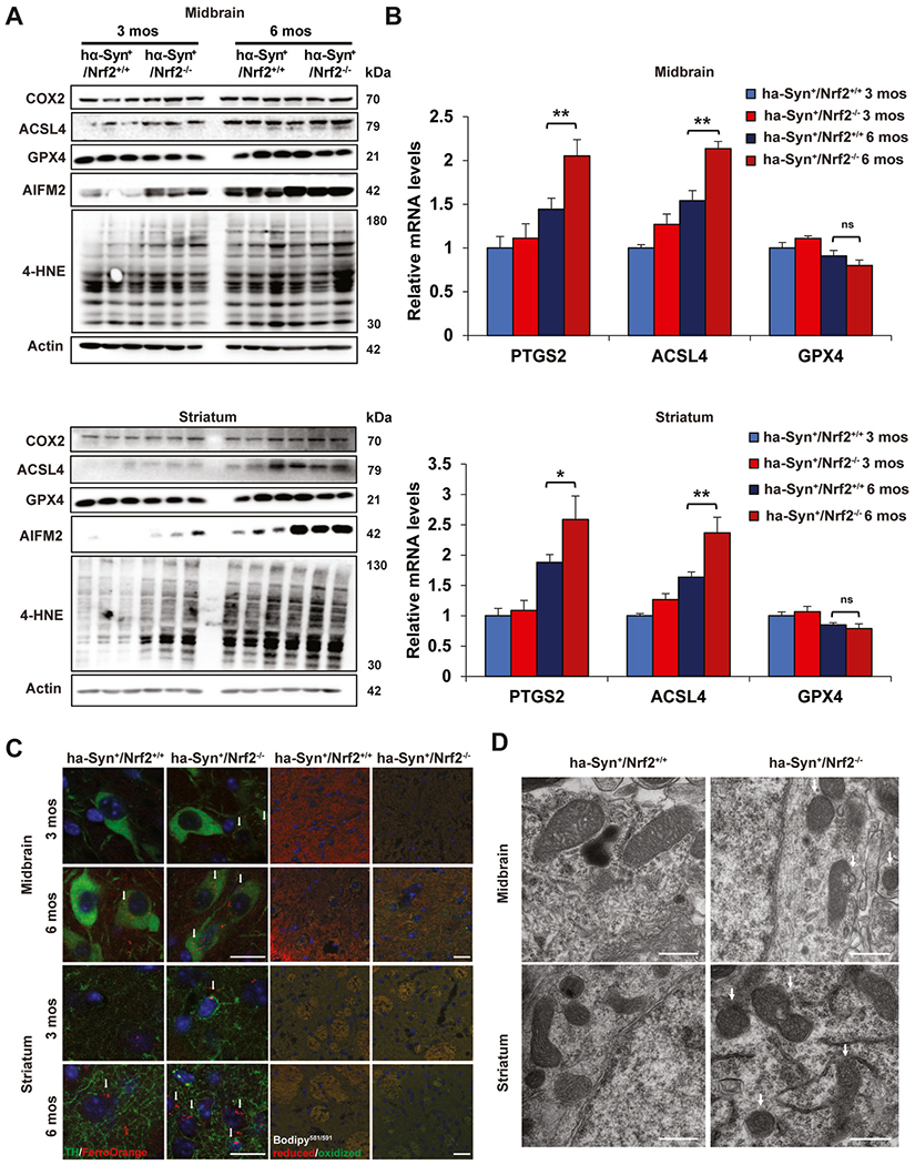
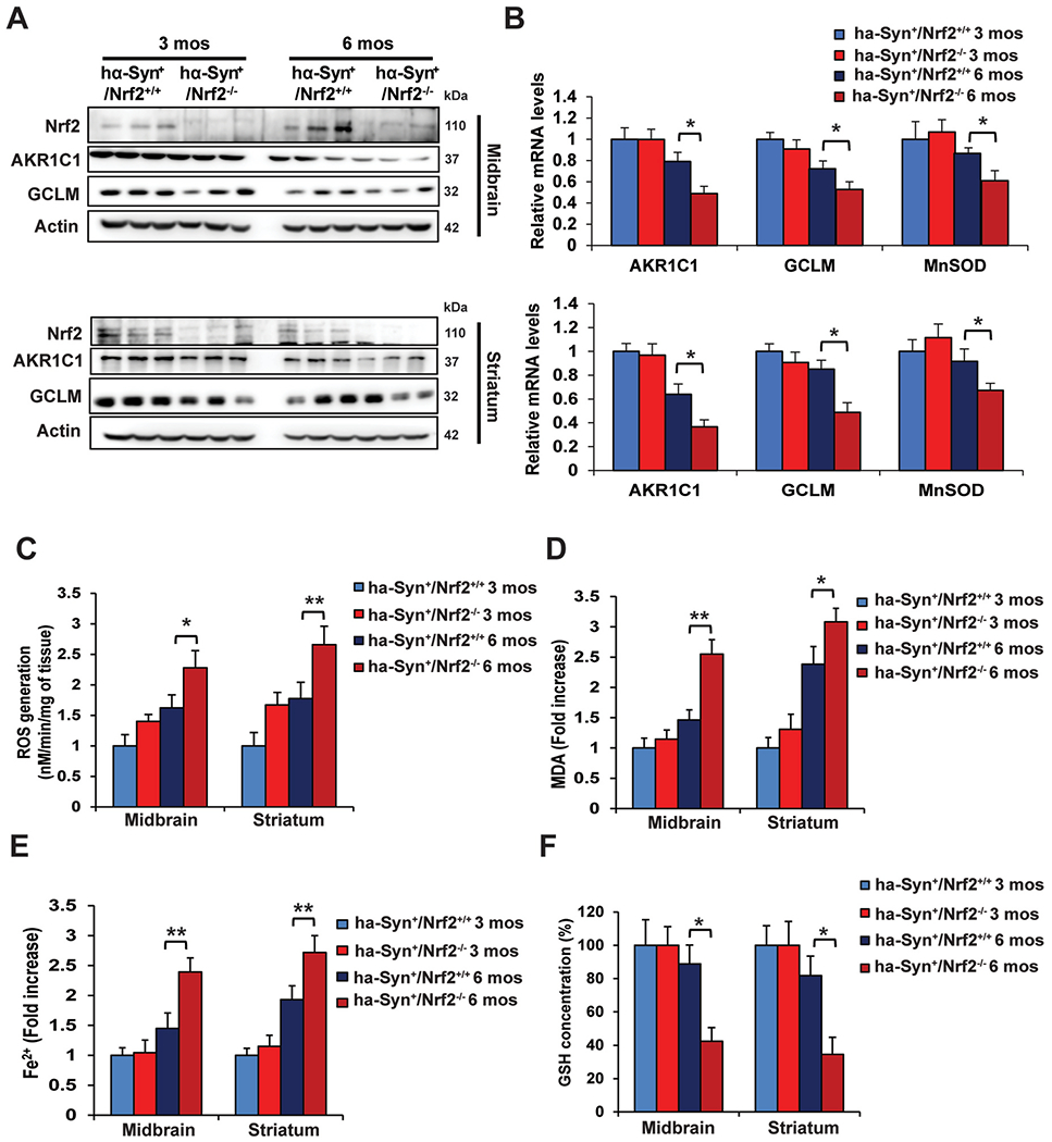
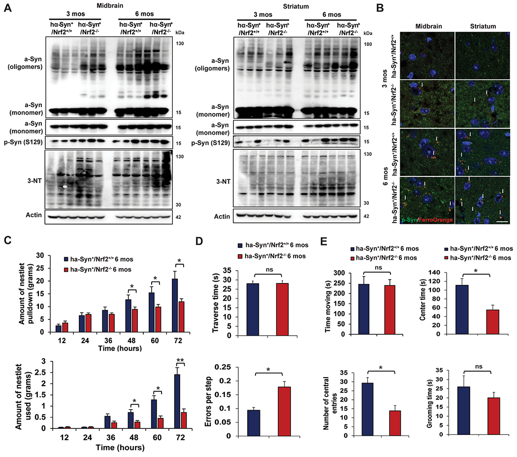
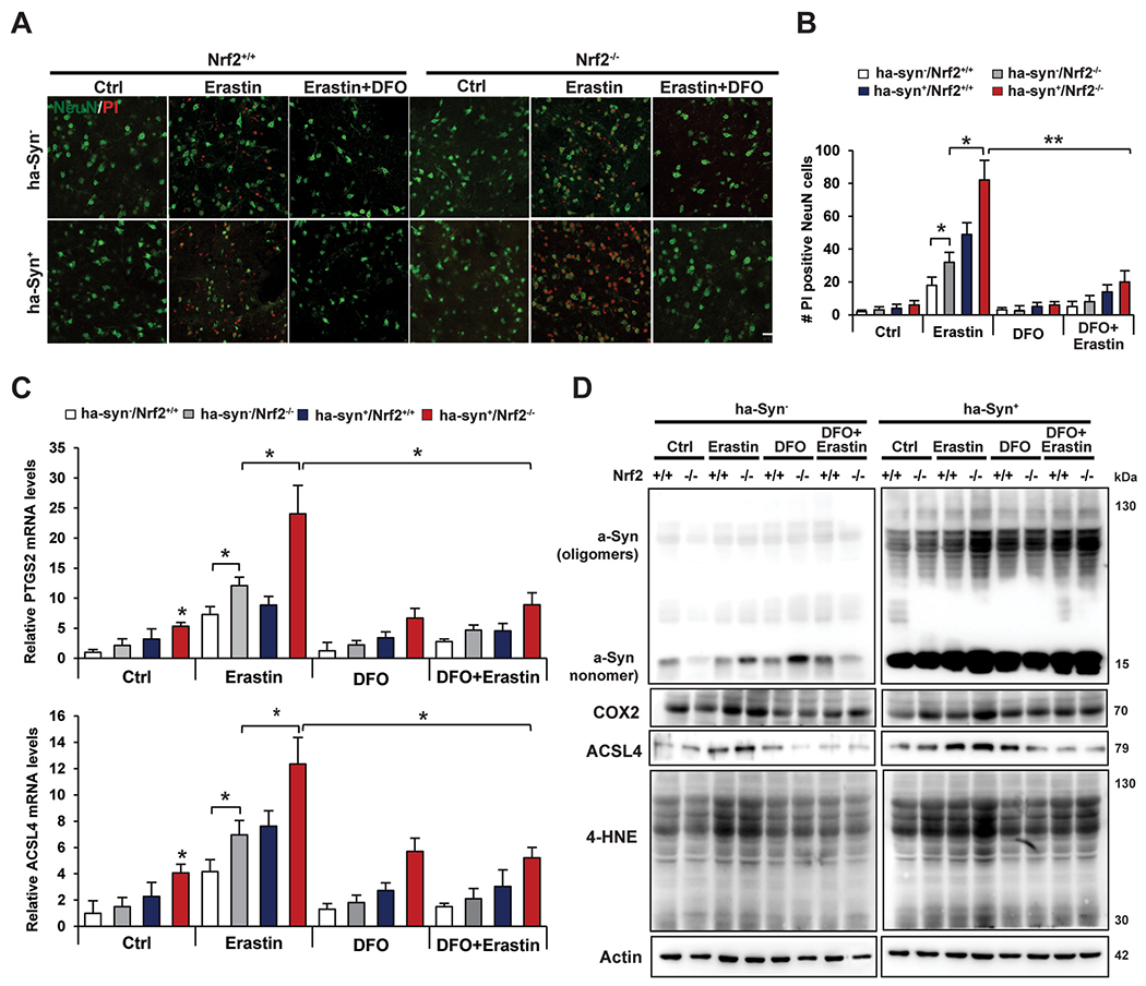
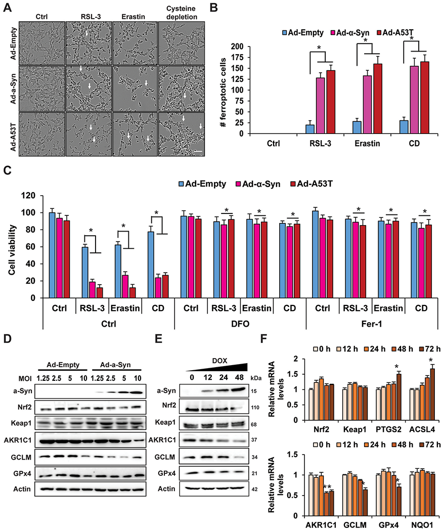
Parkinson's disease (PD) is the second most common neurodegenerative disorder, affecting millions each year. Most PD cases (∼90%) are sporadic, resulting from the age-dependent accumulation of pathogenic effects. One key pathological hallmark of PD progression is the accumulation of alpha-synuclein (α-syn), which has been shown to negatively affect neuronal function and viability. Here, using 3- and 6-month-old Nrf2+/+ and Nrf2-/- mice overexpressing human α-syn (PD model), we show that loss of NRF2 increases markers of ferroptosis across PD-relevant brain regions. Increased ferroptosis was associated with an age- and genotype-dependent increase in α-syn pathology and behavioral deficits. Finally, we demonstrate that α-syn overexpression sensitizes neuronal cells and ex vivo brain slices to ferroptosis induction, which may be due to α-syn decreasing NRF2 protein levels. Altogether, these results indicate that NRF2 is a critical anti-ferroptotic mediator of neuronal survival, and that the vicious cycle of α-syn overexpression and NRF2 suppression, leading to enhanced neuronal ferroptotic cell death, could represent a targetable and currently untapped means of preventing PD onset and progression.

Keywords: Nrf2; Parkinson's disease; aging; alpha-synuclein; ferroptosis.


Figures

Fig. 1.
Fig. 1.. Loss of Nrf2 increases markers of ferroptosis in the PD brain.
The midbrain (MB) and striatum (ST) of 3 and 6 month old hα-Syn+:Nrf2+/+ and hα-Syn+:Nrf2−/− mice were collected and assessed for key markers of ferroptosis. (A) Immunoblot analysis of Cox2, Acsl4, Gpx4, xCT, Aifm2, and 4-HNE adduct protein levels, β-actin was used as an internal loading control. n = 3 mice per group. (B) qRT-PCR of Ptgs2, Acsl4, and Gpx4 mRNA levels. n = 5 mice per group (C) Immunofluorescent staining of tyrosine hydroxylase (green) and FerroOrange (orange; left panels), or C11-BODIPY581/591 (red = reduced, green = oxidized; right panels). Scale bar = 20 μm. (D) TEM micrographs of the MB and ST of 6-month-old hα-Syn+:Nrf2+/+ and hα-Syn+:Nrf2−/− mice. Arrows indicate condensed mitochondria. Scale bar = 500 nm *p < 0.05, **p < 0.01, One-way ANOVA with Tukey’s post-hoc test. (For interpretation of the references to color in this figure legend, the reader is referred to the Web version of this article.)
Fig. 2.
Fig. 2.. 6-month-old hα-Syn+:Nrf2−/− mice exhibit increased free iron, ROS, and lipid peroxidation.
The midbrain (MB) and striatum (ST) of 3 and 6 month old hα-Syn+:Nrf2+/+ and hα-Syn+ :Nrf2−/− mice were collected and assessed for changes related to loss of NRF2. (A) Immunoblot analysis of Nrf2, and two of its known target genes, Akr1c1 and Gclm. β-actin was used as an internal loading control. n = 3 mice per group. (B) qRT-PCR analysis of Akr1c1, Gclm, and Sod2 (MnSOD). n = 5 mice per group. (C) ROS generation measured by electron paramagnetic resonance (EPR) spectroscopy. n = 6 mice per group. (D) Malondialdehyde (MDA) formation measured by TBARs assay. n = 6 mice per group. (E) Free ferrous iron (Fe2+) measured via Ferene-S based colorimetry. n = 6 mice per group. (F) Total glutathione (GSH) levels measured via Quantichrom assay. n = 6 mice per group. *p < 0.05, **p < 0.01, One-way ANOVA with Tukey’s post-hoc test.
Fig. 3.
Fig. 3.. 6-month-old hα-Syn+ :Nrf2−/− mice exhibit greater α-syn pathology and behavioral deficits.
The midbrain (MB) and striatum (ST) of 3 and 6 month old hα-Syn+:Nrf2+/+ and hα-Syn+ :Nrf2−/− mice were collected and assessed for changes in α-syn accumulation/phosphorylation and indicators of pro-ferroptotic stress. (A) Immunoblot analysis of monomeric and oligomeric α-syn, phosphorylated α-syn (S129), and 3-nitrotyrosine (3-NT). β-actin was used as an internal loading control. n = 3 mice per group. (B) Immunofluorescent staining of phosphorylated α-syn (green) and FerroOrange (orange). Scale bar = 50 μm. 6 month old hα-Syn+:Nrf2+/+ (n = 16) and hα-Syn+ :Nrf2−/− (n = 16) mice were subjected to a battery of behavioral tests. (C) Nestlet pulled down (top panel) and nestlet used (bottom panel). (D) Narrow beam traverse time (top panel) and foot slips/errors per step (bottom panel). (E) Time spent moving (top left panel), center time (top right panel), and number of central entries (bottom left panel) during an open field test, and grooming time (bottom right panel). For (C), *p < 0.05, **p < 0.01 Two-way repeated measure ANOVA. For (D–E), *p < 0.05, Unpaired Student’s t-test. (For interpretation of the references to color in this figure legend, the reader is referred to the Web version of this article.)
Fig. 4.
Fig. 4.. Loss of Nrf2 increases susceptibility to erastin-induced ferroptosis ex vivo.
Brain slices were prepared from 6 month old hα-Syn:Nrf2+/+, hμ-Syn:Nrf2−/−, hα-Syn+ :Nrf2+/+, and hα-Syn+ :Nrf2−/− mice and were either left untreated or treated with 10 μM deferoxamine (DFO) for 1 h, followed by treatment with 10 μM erastin for 16 h. (A) Immunofluorescent staining of NeuN (green; neuronal marker) and propidium iodide (PI, red; marker of cells undergoing ferroptosis). (B) qRT-PCR of Ptgs2 (top panel) and Acsl4 (bottom panel) mRNA levels. n = 3 replicates (C) Immunoblot analysis of monomeric and oligomeric α-syn, Cox2, Acsl4, and 4-HNE adduct protein levels. β-actin was used as an internal loading control. n = 3 replicates *p < 0.05, **p < 0.01, Two-way ANOVA Holm-Sidak post hoc test. All cell experiments were repeated three times to ensure reproducibility. (For interpretation of the references to color in this figure legend, the reader is referred to the Web version of this article.)
Fig. 5.
Fig. 5.. α-syn overexpression enhances susceptibility to ferroptosis and suppresses NRF2 in a neuronal cell type.
SH-SY5Y neuroblastoma cells were differentiated for 7 days, transduced with adenovirus (Ad) encoding WT or A53T α-syn for 24 h, then treated with 0.5 μM RSL-3, 1 μM erastin, or cystine depletion (CD) for 24 h. (A) Representative images of ferroptotic cell morphology (ballooning) taken using the Incucyte Bioimaging system. Scale bar = 100 μm. (B) Number of ferroptotic cells from (A). n = 3 replicates. 500 Neu-N positive cells were counted per group. (C) Cells were treated the same as (A–B) in the presence or absence of 100 μM DFO or 10 μM ferrostatin-1 (Fer-1) and cell viability was measured via MTT assay. n = 3 replicates. (D) Immunoblot analysis of monomeric α-syn, NRF2, KEAP1, AKR1C1, xCT, GLCM, and GPX4 protein levels from SH-SY5Y cells transduced with increasing MOIs of adenovirus encoding either empty vector or WT α-syn for 24 h. Doxycycline-inducible α-syn SH-SY5Y cells were induced with doxycycline for the indicated time points. n = 3 replicates. (E) Immunoblot analysis of monomeric α-syn, NRF2, KEAP1, AKR1C1, xCT, GLCM, and GPX4 protein levels. n = 3 replicates. (F) qRT-PCR of NFE2L2/NRF2, KEAP1, PTGS2, ACSL4, AKR1C1, GCLM, GPX4, and NQO1. n = 3 replicates. (B and D) *p < 0.05, **p < 0.01, One-way ANOVA with Tukey’s post-hoc test. (C) *p < 0.05, Two-way ANOVA Holm-Sidak post hoc test. All cell experiments were repeated three times to ensure reproducibility.

References

    1. Yang W, Hamilton JL, Kopil C, Beck JC, Tanner CM, Albin RL, Ray Dorsey E, Dahodwala N, Cintina I, Hogan P, Thompson T, Current and projected future economic burden of Parkinson’s disease in the U.S, NPJ Parkinsons Dis 6 (2020) 15. - PMC - PubMed
    1. Meade RM, Fairlie DP, Mason JM, Alpha-synuclein structure and Parkinson’s disease - lessons and emerging principles, Mol. Neurodegener 14 (1) (2019) 29. - PMC - PubMed
    1. Fields CR, Bengoa-Vergniory N, Wade-Martins R, Targeting alpha-synuclein as a therapy for Parkinson’s disease, Front. Mol. Neurosci 12 (2019) 299. - PMC - PubMed
    1. Dodson M, de la Vega MR, Cholanians AB, Schmidlin CJ, Chapman E, Zhang DD, Modulating NRF2 in disease: timing is everything, Annu. Rev. Pharmacol. Toxicol 59 (2019) 555–575. - PMC - PubMed
    1. Dodson M, Castro-Portuguez R, Zhang DD, NRF2 plays a critical role in mitigating lipid peroxidation and ferroptosis, Redox Biol. (2019), 101107. - PMC - PubMed

Publication types