Review
. 2022 Oct:288:105897.
doi: 10.1016/j.tvjl.2022.105897. Epub 2022 Sep 20.

Papillomaviral skin diseases of humans, dogs, cats and horses: A comparative review. Part 1: Papillomavirus biology and hyperplastic lesions

Affiliations
Review

Papillomaviral skin diseases of humans, dogs, cats and horses: A comparative review. Part 1: Papillomavirus biology and hyperplastic lesions

John S Munday et al. Vet J. 2022 Oct.

Abstract

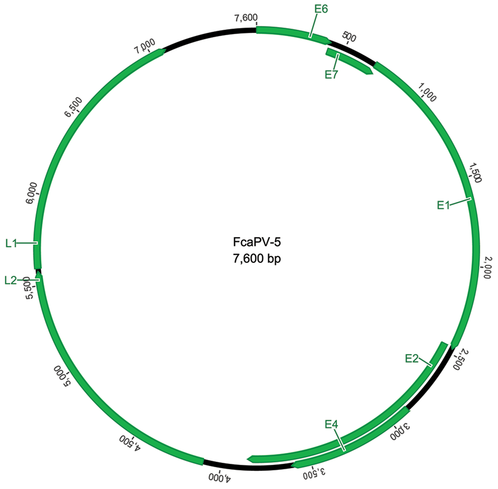
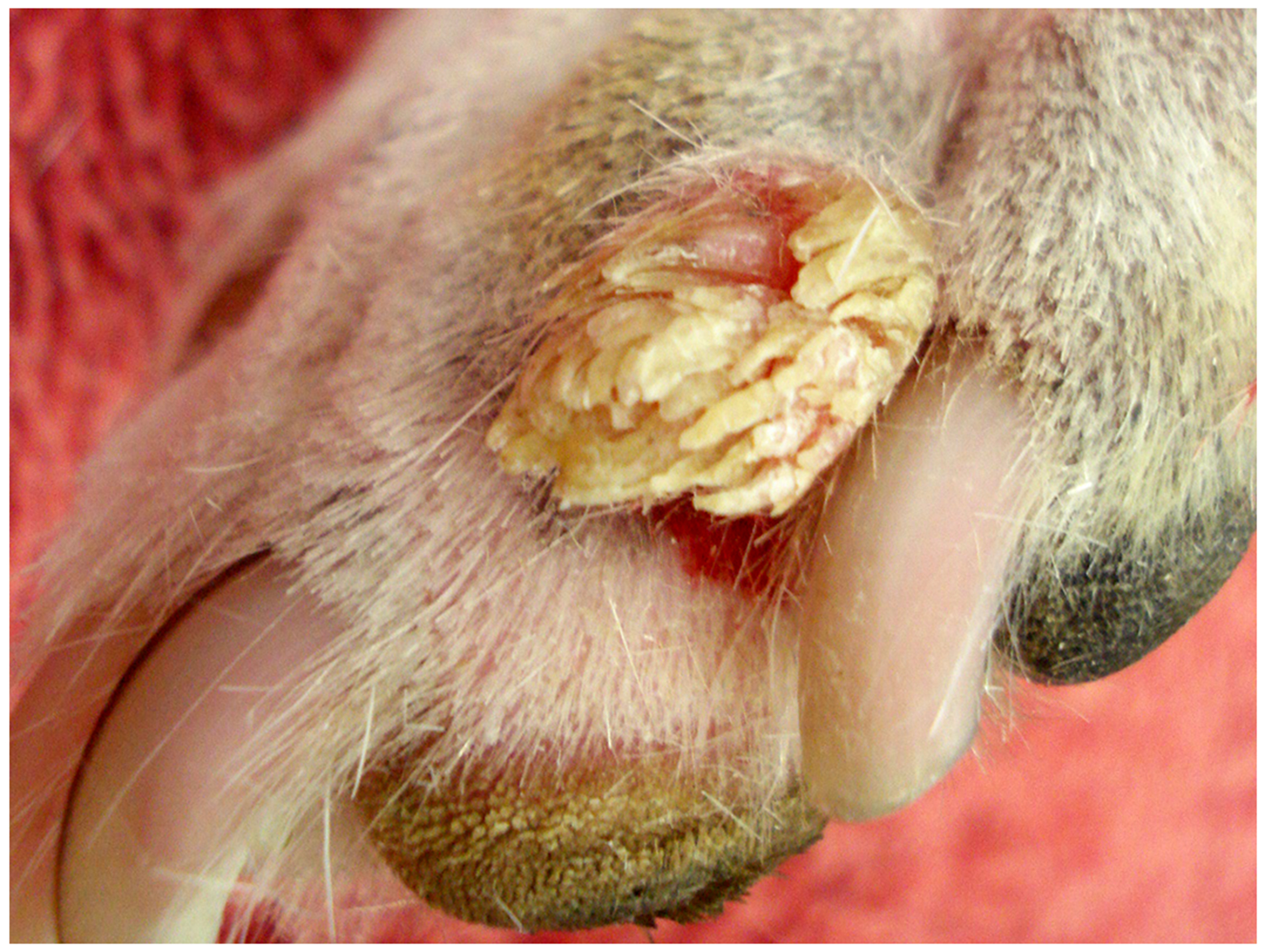
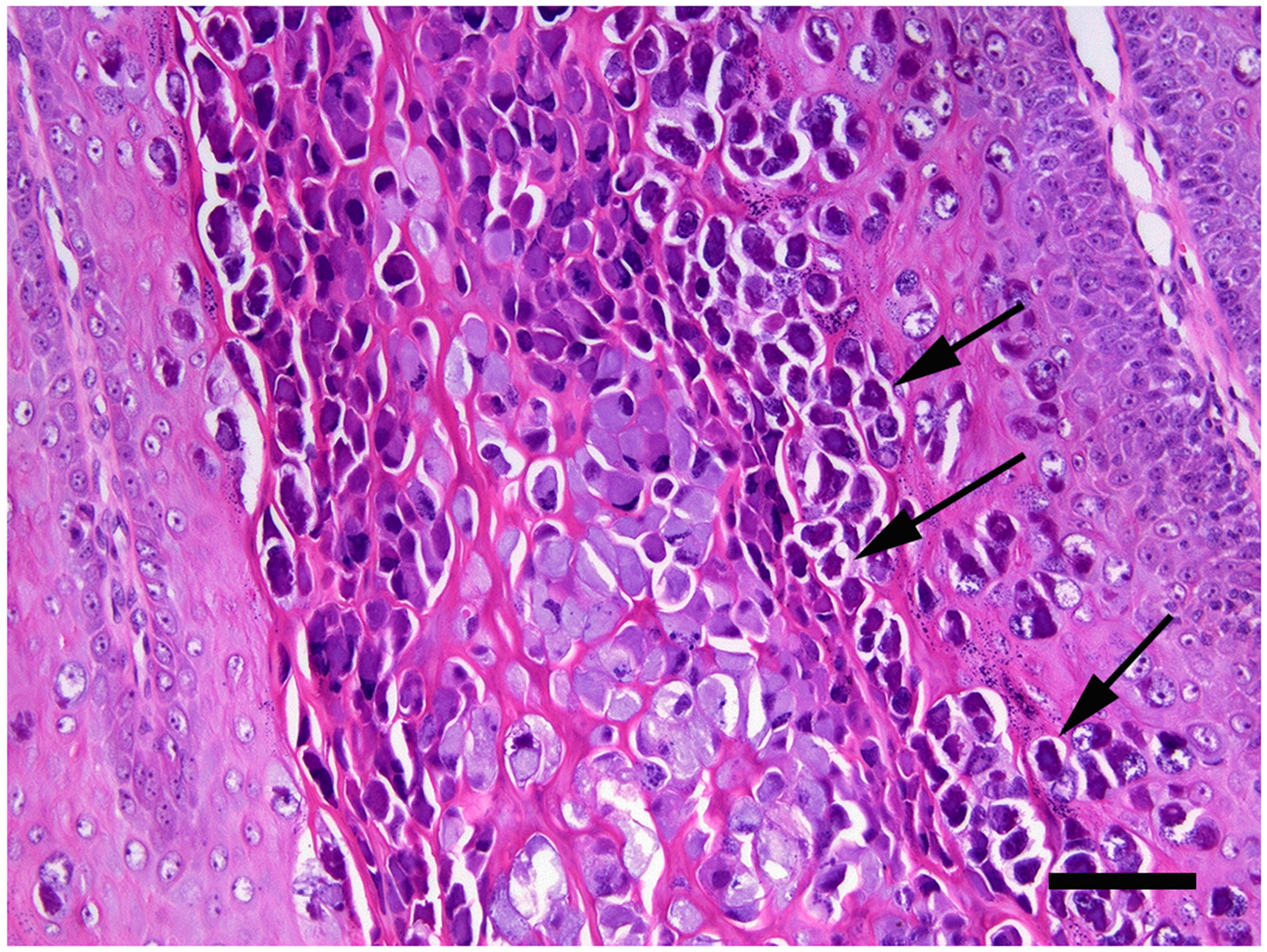
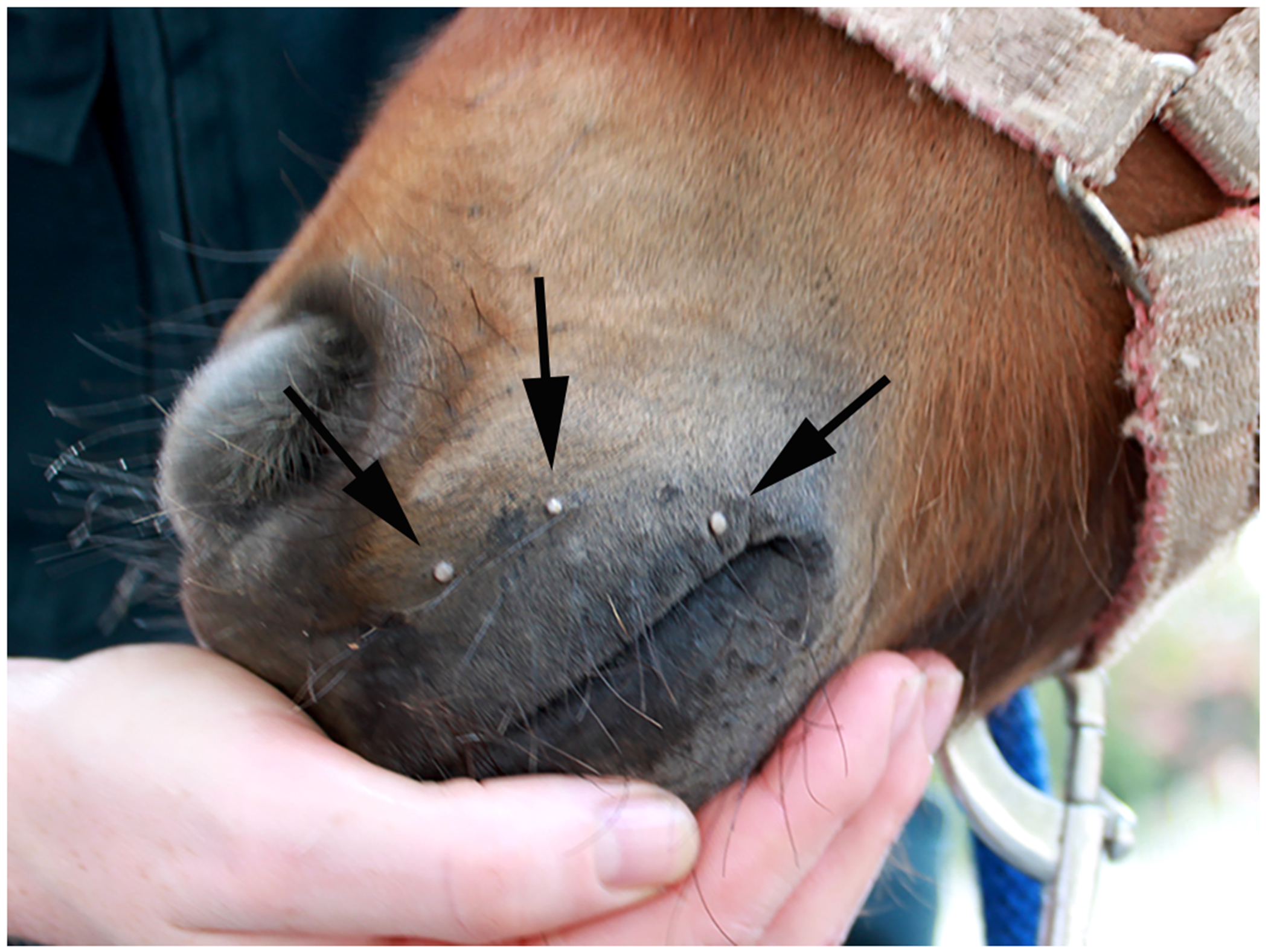
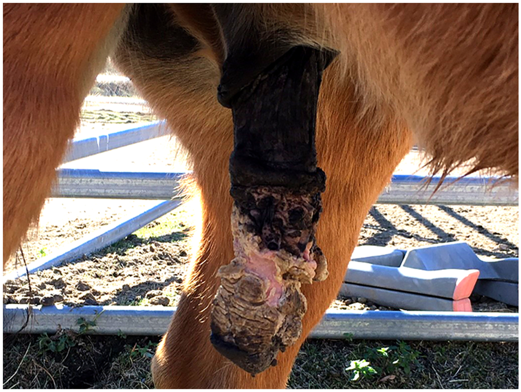
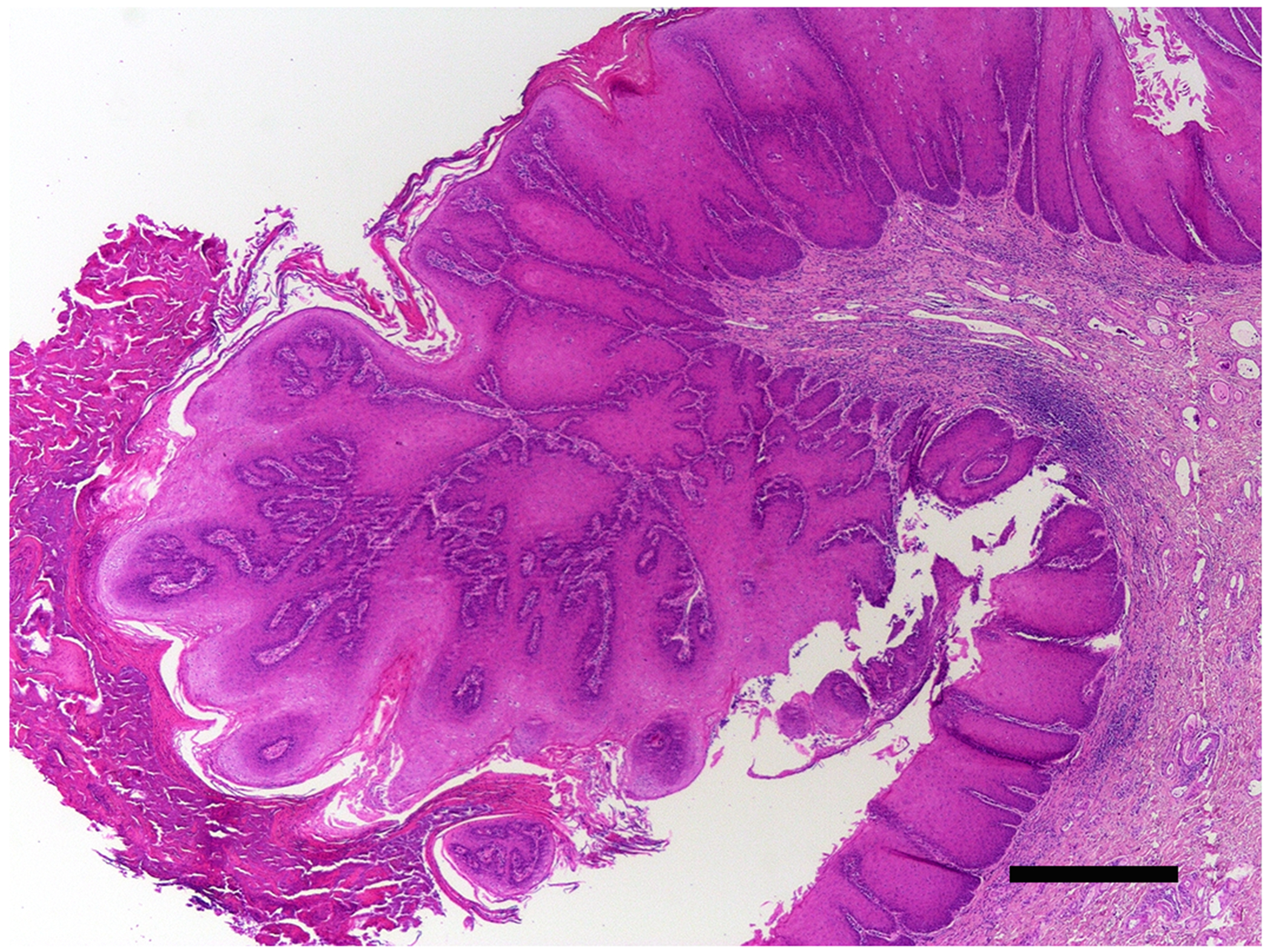
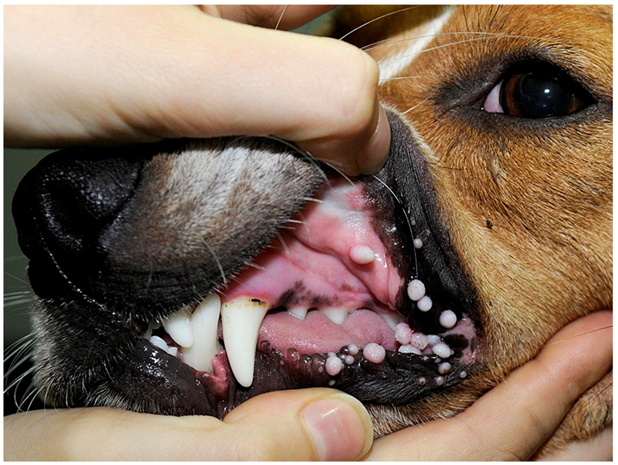
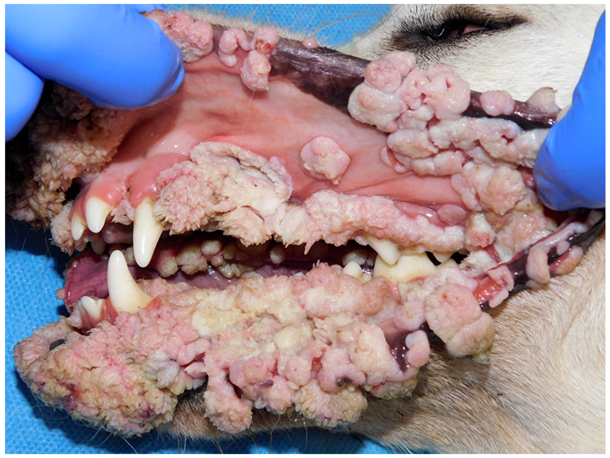
Papillomaviruses (PVs) cause disease in humans, dogs, cats, and horses. While there are some differences, many aspects of the pathogenesis, presentation, and treatment of these diseases are similar between the four species. In this review, the PV-induced diseases of humans are compared to the similar diseases that develop in the companion animal species. By comparing with the human diseases, it is possible to make assumptions about some of the less common and less well-studied diseases in the veterinary species. In the first part of this review, the PV lifecycle is discussed along with the classification of PVs and the immune response to PV infection. The hyperplastic diseases caused by PVs are then discussed; including PV-induced cutaneous, anogenital, and oral warts within the four species.

Keywords: Hyperplasia; Oncogenic viruses; Papillomas; Treatment; Warts.


Figures

Figure 1.
Figure 1.
Schematic genomic organization of Felis catus papillomavirus type 5 showing the circular arrangement of the papillomaviral DNA and the organization of the 5 early (E) and 2 late (L) open reading frames.
Figure 2.
Figure 2.
Cutaneous wart, dog. The wart appears as an exophytic vegetative mass on the toe of this dog. The frond-like appearance of the surface is due to the folding of the epidermis caused by papillomavirus-induced epidermal hyperplasia (Image courtesy of Dr Scott Martin).
Figure 3.
Figure 3.
Cutaneous wart, dog. A high proportion of cells within the thickened, folded epidermis contain marked papillomaviral-induced cellular changes. These include cells with cytoplasm that is expanded by large quantities of amorphous basophilic material and cells that have a dark nucleus with an irregular nuclear membrane surrounded by a clear cytoplasmic halo (koilocytes; arrows). Scale bar = 30μm. Haematoxylin and eosin.
Figure 4.
Figure 4.
Cutaneous warts, horse. The warts appear as multiple small exophytic lesions on the muzzle of this young horse. These warts resolved spontaneously and did not appear to cause discomfort (Image courtesy of Dr Emma Gordon).
Figure 5.
Figure 5.
Penile papillomatosis, horse. The penis is covered by numerous warts and plaques. Some of the warts are covered in a thick layer of keratin that has resulted in a ‘horn-like’ appearance. These warts were stable for around two years. Progression to squamous cell carcinoma then occurred necessitating euthanasia of the horse (Image courtesy of Dr Richard Malik).
Figure 6.
Figure 6.
Penile wart, horse. The epithelium is markedly thickened throughout the section. However, an area of folding has resulted in an exophytic papillomatous mass that is covered by increased quantities of keratin. PCR amplified Equulis caballus papillomavirus type 2 DNA from the lesion. Scale bar = 0.5mm. Haematoxylin and eosin.
Figure 7.
Figure 7.
Oral warts, dog. The warts are exophytic and either appear smooth or have a more roughened surface. The warts in this dog remained small and, as expected for the vast majority of cases, resolved spontaneously (Image courtesy of Dr Anne Quain).
Figure 8.
Figure 8.
Oral warts, dog. The warts in this dog were first observed over a year ago and have continued to grow since this time. Additionally, new warts are continuing to develop in the mouth of this dog. Warts recurred after surgical excision. This dog most likely has an immune disorder that prevents the development of a normal cell-mediated response to papillomavirus infection (Image courtesy of Dr Margherita Gracis).

Similar articles

Cited by

References

    1. Altamura G, Tommasino M, Borzacchiello G, 2020. Cutaneous vs. mucosal tropism: The papillomavirus paradigm comes to an “and”. Frontiers of Microbiology 11, 588663. - PMC - PubMed
    1. Antonsson A, Forslund O, Ekberg H, Sterner G, Hansson BG, 2000. The ubiquity and impressive genomic diversity of human skin papillomaviruses suggest a commensalic nature of these viruses. Journal of Virology 74, 11636–11641. - PMC - PubMed
    1. Antonsson A, Hansson BG, 2002. Healthy skin of many animal species harbors papillomaviruses which are closely related to their human counterparts. Journal of Virology 76, 12537–12542. - PMC - PubMed
    1. Antonsson A, Erfurt C, Hazard K, Holmgren V, Simon M, Kataoka A, Hossain S, Håkangård C, Hansson BG, 2003a. Prevalence and type spectrum of human papillomaviruses in healthy skin samples collected in three continents. Journal of General Virology 84, 1881–1886. - PubMed
    1. Antonsson A, Karanfilovska S, Lindqvist PG, Hansson BG, 2003b. General acquisition of human papillomavirus infections of skin occurs in early infancy. Journal of Clinical Microbiology 41, 2509–2514. - PMC - PubMed