Review
. 2023 Jan 24:18:181-203.
doi: 10.1146/annurev-pathmechdis-031521-034739. Epub 2022 Sep 23.

Pathogenesis of Zika Virus Infection

Affiliations
Review

Pathogenesis of Zika Virus Infection

Maria I Giraldo et al. Annu Rev Pathol. .

Abstract

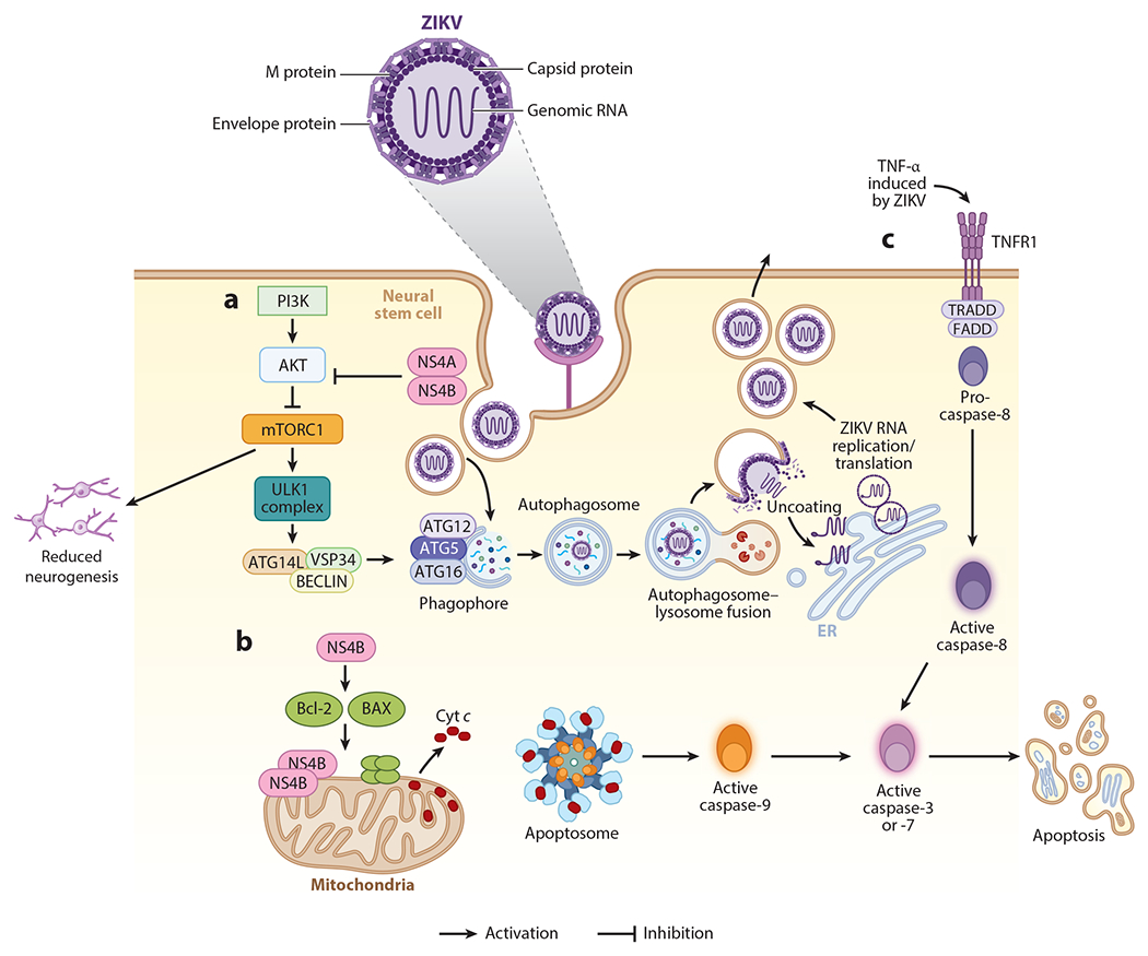
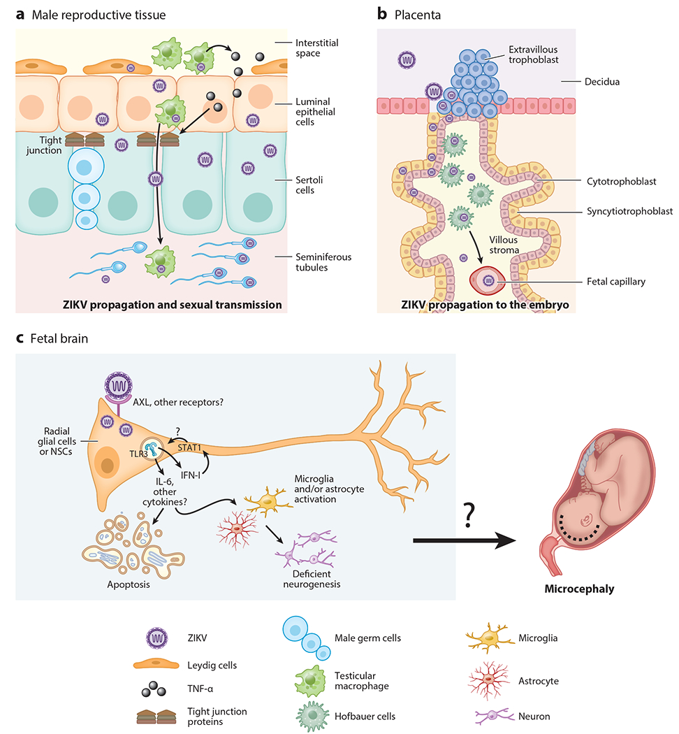
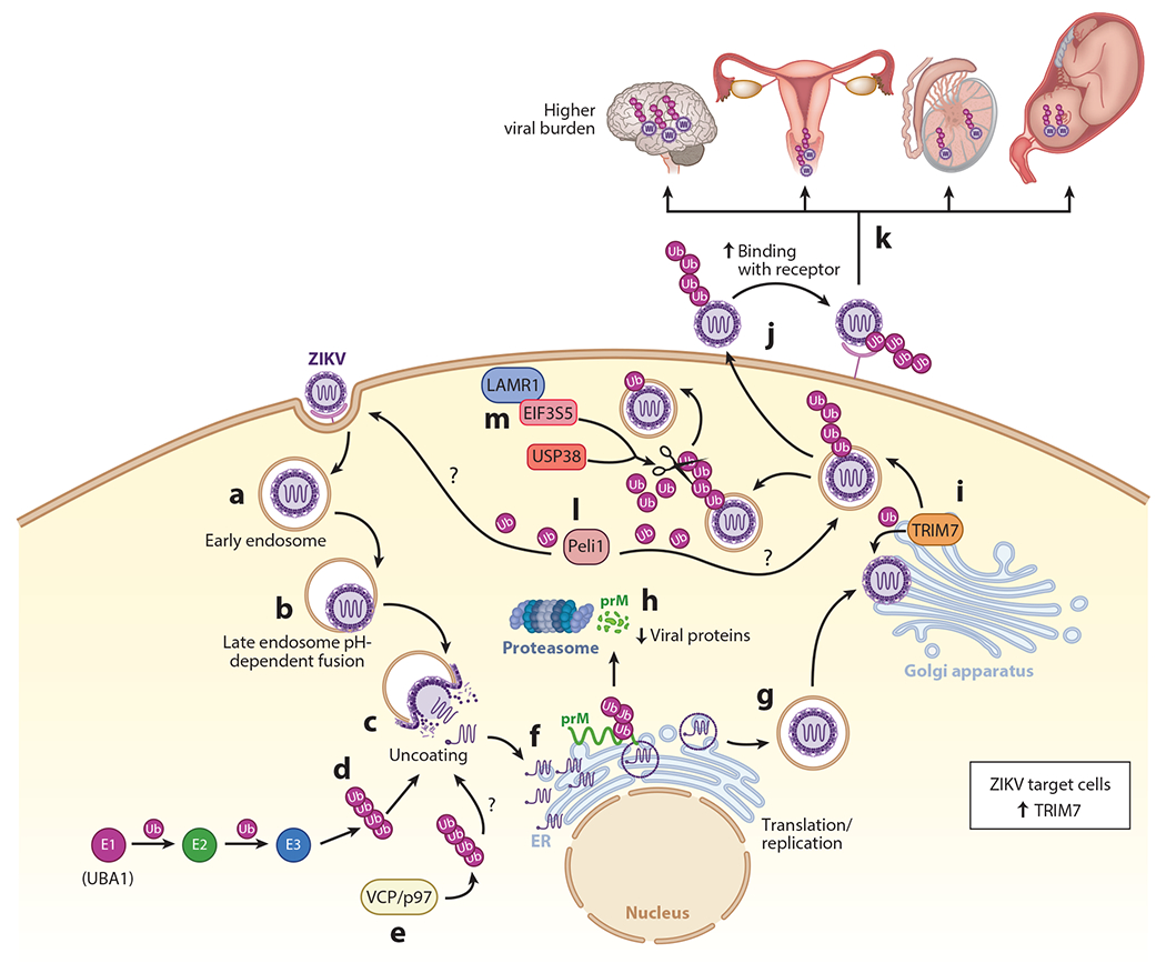
Zika virus (ZIKV) is an emerging virus from the Flaviviridae family that is transmitted to humans by mosquito vectors and represents an important health problem. Infections in pregnant women are of major concern because of potential devastating consequences during pregnancy and have been associated with microcephaly in newborns. ZIKV has a unique ability to use the host machinery to promote viral replication in a tissue-specific manner, resulting in characteristic pathological disorders. Recent studies have proposed that the host ubiquitin system acts as a major determinant of ZIKV tropism by providing the virus with an enhanced ability to enter new cells. In addition, ZIKV has developed mechanisms to evade the host immune response, thereby allowing the establishment of viral persistence and enhancing viral pathogenesis. We discuss recent reports on the mechanisms used by ZIKV to replicate efficiently, and we highlight potential new areas of research for the development of therapeutic approaches.

Keywords: TRIM7; Zika virus; antagonism of immune responses; pathogenesis; tropism; ubiquitin.


Figures

Figure 1
Figure 1
Intrinsic immunity and mechanisms of antagonism by Zika virus (ZIKV). (a) ZIKV proteins NS4A and NS4B can block the activation of AKT and subsequently lead to the inactivation of mTORC1, allowing activation of the ULK complex by AMP-activated protein kinase and the initiation of the autophagosome formation through ATG16. ZIKV is internalized via endosomes and autophagosomes. The autophagosome fuses with the lysosome, leading to the acidification of the autophagolysosome and activation of proteases to degrade the viral components. This process could be beneficial for ZIKV because acidification allows fusion of the ZIKV envelope with the endosomal membrane, allowing the viral RNA to reach the cytoplasm. The inhibition of the AKT-mTOR pathway has an adverse effect in the proliferation of the infected cells, with catastrophic consequences in the formation of the neuronal network and tissue development. (b) ZIKV NS4B protein can localize near the mitochondria membrane and activate the Bcl-1-associated X protein (BAX) to form a complex with Bcl-2,which interacts with the mitochondrial voltage-dependent anion channel, leading to loss in membrane potential and release of cytochrome c (Cyt c). This in turn leads to the activation of the intrinsic apoptosis pathway, with the activation of caspase-9 and caspase-3. (c) The activation of the extrinsic pathway can be a bystander effect, since it is mediated by the activation of cell death receptors FAS and TNFR1, and it is induced by external signals such as tumor necrosis factor alpha (TNF-α), leading to the activation of caspase-8 and caspase-3 to induce cell apoptosis.
Figure 2
Figure 2
Innate immune activation and evasion mediated by Zika virus (ZIKV). (a) After infecting a cell, ZIKV RNA is recognized by multiple cell pattern recognition receptors, such as endosomal Toll-like receptors (TLRs) and cytosolic RIG-I and MDA5. Viral recognition triggers downstream signaling pathways that culminate in the activation and translocation to the nucleus of transcription factors IRF3 [interferon (IFN)-regulatory factor 3] and IRF7 to induce the transcription of type-I IFNs (IFN-α and IFN-β). (b) The production of IFNs is important to reduce the viral spread to surrounding cells, as they can signal in an autocrine or paracrine manner through the IFN receptor (IFNAR1 and IFNAR2). IFN signaling involves the JAK1 and TYK2 kinases that phosphorylate STAT1 and STAT2, which then together with IRF9 form the ISGF3 complex. ISGF3 translocates to the nucleus to promote transcription of IFN-stimulated genes (ISGs), which many have antiviral activity. (c) ZIKV antagonizes the antiviral response. The viral protein NS3 binds to 14-3-3ε and prevents the translocation of RIG-I and MDA5 to the mitochondria for interaction with MAVS. NS4A binds to MAVS, preventing the association with RIG-I and MDA5. NS1, NS2A, NS2B, and NS4B interact with TBK1, reducing its phosphorylation, and inhibit the IFN-I production pathway. NS2B3 targets STING and blocks the signaling through TBK1. NS5 and NS2A act later in the pathway, inhibiting IRF3 and preventing its translocation to the nucleus and the induction of IFN-I. In the IFN-I signaling pathway, NS2B3 binds to JAK1 and induces its degradation, preventing the activation of STAT proteins. NS4B inhibits the pathway by inhibiting the phosphorylation of STAT1, while NS5 binds to STAT2 and promotes its proteasomal degradation. All of these mechanisms prevent induction of ISGs and diminish the antiviral response.
Figure 3
Figure 3
Zika virus (ZIKV) pathogenesis and its target tissues. ZIKV infects and persists in the target tissues, causing its characteristic pathology. (a) ZIKV can travel through the male reproductive tract, within infected immune cells. Once the virus reaches the blood testicular barrier, it infects Leydig cells and activates resident macrophages. This leads to the induction of inflammatory mediators such as tumor necrosis factor alpha (TNF-α) that can activate the epithelial barrier and promote the disruption of the tight junctions. ZIKV can also infects Sertoli cells, spermatogonia, primary spermatocytes, and spermatozoa and can infect and replicate in the sperm. The virus can be sexually transmitted via these cell types. (b) In pregnant women, ZIKV can infect the placenta, where it breaks the decidua and chorionic villi and also infects endothelial cells, Hofbauer cells, and cytotrophoblasts, among others. ZIKV can cross the maternal-fetal blood barrier and infect the embryo. (c) In the fetus, ZIKV efficiently infects neural stem cells (NSCs) during the first trimester of gestation. AXL has been proposed as one important receptor; however, this has not been confirmed in vivo, and a dominant receptor for ZIKV has not been found yet. Proinflammatory cytokines, including interleukin 6 (IL-6) and type I interferons (IFN-I), are induced via Toll-like receptor 3 (TLR3) and other pattern recognition receptors upon ZIKV infection and can promote tissue damage by causing apoptosis of neural stem cells and reducing neurogenesis. Together, these can contribute to pathology, potentially including microcephaly.
Figure 4
Figure 4
The role of the ubiquitin system in Zika virus (ZIKV) pathogenesis and tissue tropism. (a) After ZIKV attachment to cell receptors, the virus enters via endosomes. (b) Acidification in the endosome allows fusion of the envelope with the endosomal membrane. (c) The virus is then released in the cytoplasm after uncoating. (d) This step can be blocked with inhibitors of the E1-activating enzyme (UBA1) required for ubiquitination (Ub) of proteins, suggesting uncoating requires an ubiquitination step. How exactly ubiquitination mediates the uncoating step is unknown. (e) The ATPase valosin-containing protein p97 (VCP/p97) has been proposed to be involved in uncoating and speculated to do so by extracting an ubiquitinated protein during the uncoating process. (f) After uncoating, the viral RNA is replicated and transcribed in the endoplasmic reticulum (ER). The polypeptide is cleaved into the different viral proteins. (g) After virus assembly, which starts in the ER, the immature virion travels through the Golgi, where precursor membrane (prM) is cleaved to mature the virion and it continues to exit the cell. (h) prM protein of ZIKV is ubiquitinated on K6, resulting in proteasomal degradation. (i) The envelope protein of ZIKV is ubiquitinated by tripartite motif containing 7 (TRIM7) on K38 and K281, presumably around the Golgi or Golgi-associated membranes. (j) ZIKV-infectious virions released outside the cell containing a proportion of the ubiquitinated envelope can attach to receptors more efficiently, and provide a determinant of tissue tropism, by enhancing virus entry. (k) This further leads to increased viral loads in target tissues (brain, reproductive tissues, and embryo). (l) The E3-ubiquitin ligase Pellino 1 (Peli1) promotes entry and replication, but the viral target protein of ubiquitination is not known. (m) The host deubiquitinates ubiquitin-specific peptidase 38 (USP38) and eukaryotic translation initiation factor 3 subunit 5 (EIF3S5), which interact with laminin receptor 1 (LAMR1) to reduce the ubiquitination levels of the envelope protein and restrict ZIKV replication.

Similar articles

Cited by

References

    1. Sirohi D, Chen Z, Sun L, Klose T, Pierson TC, et al. 2016. The 3.8 Å resolution cryo-EM structure of Zika virus. Science 352:467–70 - PMC - PubMed
    1. Gutierrez-Bugallo G, Piedra LA, Rodriguez M, Bisset JA, Lourenco-de-Oliveira R, et al. 2019. Vector-borne transmission and evolution of Zika virus. Nat. Ecol. Evol 3:561–69 - PMC - PubMed
    1. Pletnev AG, Maximova OA, Liu G, Kenney H, Nagata BM, et al. 2021. Epididymal epithelium propels early sexual transmission of Zika virus in the absence of interferon signaling. Nat. Commun 12:2469. - PMC - PubMed
    1. Miner JJ. 2017. Congenital Zika virus infection: more than just microcephaly. Sci. Transl. Med 9:8195 - PubMed
    1. Dick GW, Kitchen SF, Haddow AJ. 1952. Zika virus. I. Isolations and serological specificity. Trans. R. Soc. Trop. Med. Hyg 46:509–20 - PubMed

Publication types