Clinical Trial
. 2022 Nov;32(11):995-1007.
doi: 10.1038/s41422-022-00721-y. Epub 2022 Sep 23.

Genetically modified CD7-targeting allogeneic CAR-T cell therapy with enhanced efficacy for relapsed/refractory CD7-positive hematological malignancies: a phase I clinical study

Affiliations
Clinical Trial

Genetically modified CD7-targeting allogeneic CAR-T cell therapy with enhanced efficacy for relapsed/refractory CD7-positive hematological malignancies: a phase I clinical study

Yongxian Hu et al. Cell Res. 2022 Nov.

Abstract

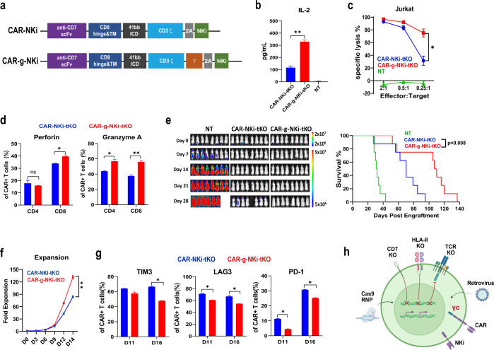
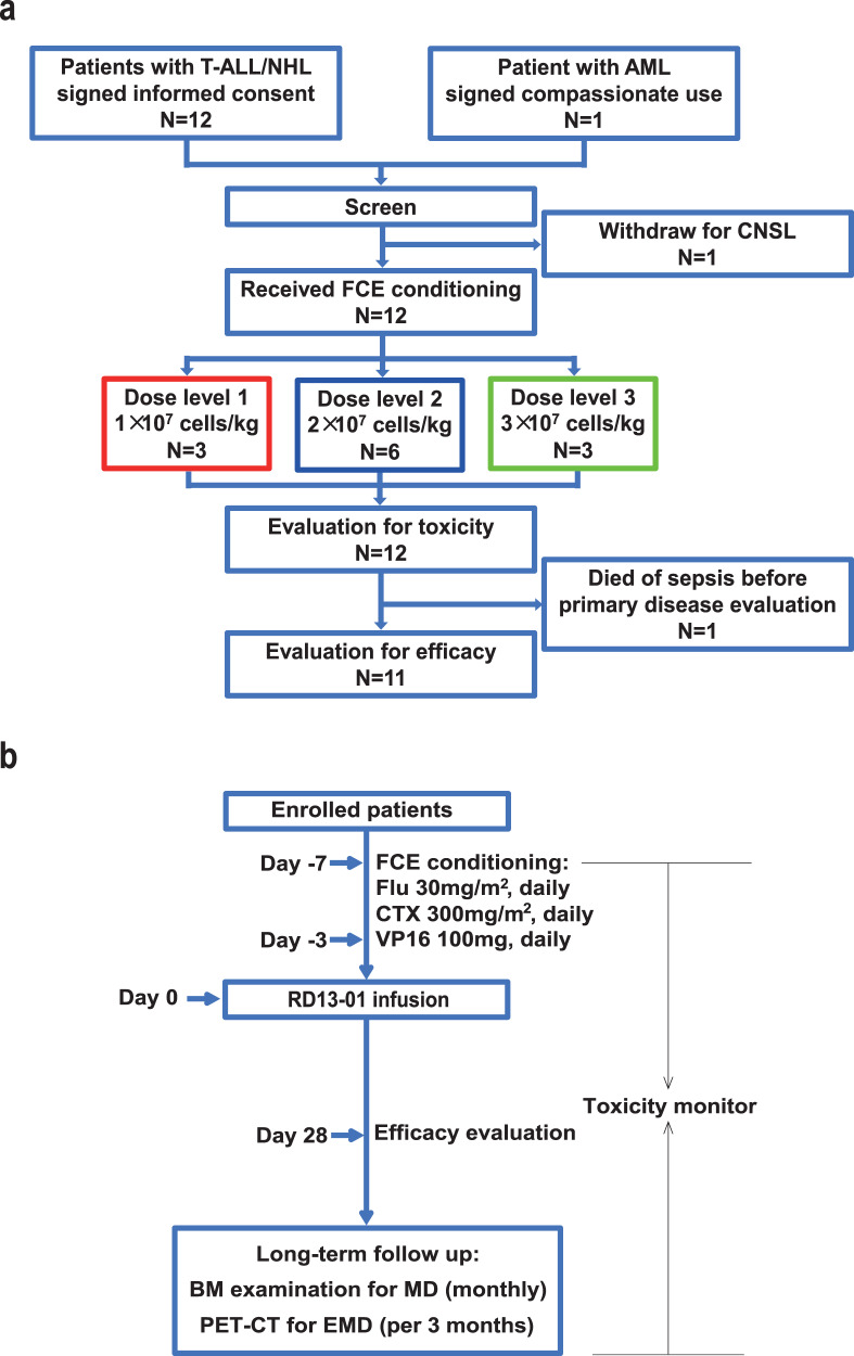
Chimeric antigen receptor (CAR)-T cell therapy against T cell malignancies faces major challenges including fratricide between CAR-T cells and product contamination from the blasts. Allogeneic CAR-T cells, generated from healthy donor T cells, can provide ready-to-use, blast-free therapeutic products, but their application could be complicated by graft-versus-host disease (GvHD) and host rejection. Here we developed healthy donor-derived, CD7-targeting CAR-T cells (RD13-01) with genetic modifications to resist fratricide, GvHD and allogeneic rejection, as well as to potentiate antitumor function. A phase I clinical trial (NCT04538599) was conducted with twelve patients recruited (eleven with T cell leukemia/lymphoma, and one with CD7-expressing acute myeloid leukemia). All patients achieved pre-set end points and eleven proceeded to efficacy evaluation. No dose-limiting toxicity, GvHD, immune effector cell-associated neurotoxicity or severe cytokine release syndrome (grade ≥ 3) were observed. 28 days post infusion, 81.8% of patients (9/11) showed objective responses and the complete response rate was 63.6% (7/11, including the patient with AML). 3 of the responding patients were bridged to allogeneic hematopoietic stem cell transplantation. With a median follow-up of 10.5 months, 4 patients remained in complete remission. Cytomegalovirus (CMV) and/or Epstein-Barr virus (EBV) reactivation was observed in several patients, and one died from EBV-associated diffuse large B-cell lymphoma (DLBCL). Expansion of CD7-negative normal T cells was detected post infusion. In summary, we present the first report of a Phase I clinical trial using healthy donor-derived CD7-targeting allogeneic CAR-T cells to treat CD7+ hematological malignancies. Our results demonstrated the encouraging safety and efficacy profiles of the RD13-01 allogeneic CAR-T cells for CD7+ tumors.


Conflict of interest statement

Y.Z., W.G., G.C., L.H., T.G., X.J., X.Zheng, S.Y., Xiaolong L., X.Zhang, M.C., Xiuju L., M.G., K.W., X.H., Y.W., and J.R. are employees of Nanjing Bioheng Biotech Co. All other authors declare no competing interests.

Figures

Fig. 1
Fig. 1. Construction of rejection-resistant “off-the-shelf” CD7-targeting CAR-T cells.
a CAR expression and efficiency of TCR/CD3 and CD7 disruption on CAR-dKO T cells, measured by flow cytometry. b, c Tumor control ability of CAR-dKO T cells evaluated in NCG mice bearing Jurkat tumors. Mice were injected with 1 × 106 Jurkat tumor cells intravenously (i.v.) and treated with a single injection of 10 × 106 non-transduced (NT) T cells (n = 4) or CAR-dKO T cells (n = 8) 72 h after tumor inoculation. Bioluminescent images obtained at the indicated time points after tumor injection (b). Kaplan–Meier curves of mice treated with NT or CAR-dKO T cells (c), with P value calculated using a Log-rank test. d Top, composition of CD7+ and CD7 subsets in CD4+/CD8+ T cells from healthy donors (n = 5) and patients with T-ALL (n = 14). Bottom, elimination of HLA-II expression by disrupting RFX5, generating CAR-tKO T cells. e Expansion of allogeneic CD4+ T cells in the presence of CAR-dKO or CAR-tKO T cells in a mixed lymphocyte reaction (MLR) assay. f E-cadherin expression on CAR-tKO and CAR-NKi-tKO T cells. g NK cell cytotoxicity against CAR-tKO and CAR-NKi-tKO cells was detected using a luciferase-based killing assay at different NK:CAR-T ratios after 24 h of co-culture (n = 3). h Illustration of CAR-T cell development from “CAR-dKO” to “CAR-tKO” to “CAR-NKi-tKO”. Data are presented as means ± SD; All comparisons (except for Kaplan–Meier curves) were determined using student’s t-tests; *P < 0.05; **P < 0.01; ns not significant.
Fig. 2
Fig. 2. Incorporation of γc improves CAR-T cell function.
a Schematic illustration of γc-tethered CAR design. b Secretion of IL-2 by CAR-NKi-tKO and CAR-g-NKi-tKO T cells following 8 h of stimulation using Jurkat tumor cells (n = 3). c Cytotoxicity of CAR-NKi-tKO and CAR-g-NKi-tKO T cells against Jurkat cells at indicated Effector: Target (CAR-T: Tumor) ratios after 8 h of co-culture. d Secretion of cytotoxic molecules in CD4+ and CD8+ subsets of CAR-NKi-tKO and CAR-g-NKi-tKO T cells following 8 h of stimulation using Jurkat tumor cells (n = 3). e NCG mice were injected with 1 × 106 Jurkat tumor cells (i.v.) and treated with a single injection of 10 × 106 NT, CAR-NKi-tKO or CAR-g-NKi-tKO T cells. Left, bioluminescent images obtained at the indicated time points after tumor injection; right, Kaplan–Meier curves of mice treated with NT, CAR-NKi-tKO or CAR-g-NKi-tKO T cells, with P value calculated using a Log-rank test. f Ex vivo expansion of CAR-NKi-tKO or CAR-g-NKi-tKO T cells during production. g Expression of TIM3, LAG3, and PD-1 on CAR-NKi-tKO or CAR-g-NKi-tKO T cells at indicated time points during production (n = 3). h Illustration of RD13-01 CAR-T cell degisn. Data are presented as means ± SD; All comparisons (except for Kaplan–Meier curves) were determined using student’s t-tests; *P < 0.05; **P < 0.01; ns not significant.
Fig. 3
Fig. 3. Clinical procedure of treatment with RD13-01 CAR-T cells.
a CONSORT diagram of the clinical trial. AML, acute myeloid leukemia; CNSL, central neural system lymphoma; FCE, fludarabine cyclophosphamide and etoposide. b Diagram of clinical treatment protocol: the day of RD13-01 infusion was set as day 0; the lympho-depleting condition regimen consisted of fludarabine (30 mg/m2), cyclophosphamide (300 mg/m2) and etoposide (100 mg/day) on days ‒7 to ‒3.
Fig. 4
Fig. 4. Safety and efficacy profiles of RD13-01 CAR-T cells.
a Graph of incidences of CRS and ICANS in all patients treated. b Serum concentrations of IFN-γ, IL-6 and IL-10 in patients who developed CRS after RD13-01 infusion. Samples were taken 2 h after CAR-T infusion (“before CRS”), and at the times of initiation, peak and restoration of CRS-related symptoms. Data are presented as means ± SD. c Swimmer plot (n = 12) showing patient responses. Each bar represents an individual patient. Responses were determined on day 28 (arrowhead) and are indicated by different colors (blue, CR; green, PR; orange, PD; gray, NE). Bars with solid arrows represent patients in an ongoing follow-up. d CD7+ malignant cells in bone marrow measured using flow cytometry (left) and microscopic images (100× and 1000×, bar indicates 10 μm) of bone marrow samples (right) of Patient 5, who achieved a complete response at day 28 after RD13-01 infusion. e Computed tomography scans showed a complete response of extramedullary leukemia in Patient 9. Pre-treatment (baseline) tumor lesion was indicated by the red arrow.
Fig. 5
Fig. 5. RD13-01 expansion and endogenous T cell dynamics.
a, b Flow cytometry (a) and qPCR (b) detections of CAR-T cells in the peripheral blood of patients who achieved responses (Responders) or no response (Non-responders) at different time points post CAR-T infusion. c Stacked pie charts showing subset compositions of normal lymphocytes from responders. Plotted are median values. DNT, double negative T cell. d Fold changes of different types of endogenous lymphocytes at indicated time points compared to day 1 post-infusion. Plotted are median values for responders. e, f Surface antigen expression on normal lymphocytes (defined as the CD45-bright population) from Patient 5 at different times points. CD3 and CD7 expression on normal lymphocytes before CAR-T cell infusion (e); CD3, CD7 and CAR expression on normal lymphocytes on day 15 and 42 after infusion (f). CAR was gated from the CD3 population, and CD4/CD8 were gated from the CD3+ population. g MLR assay was performed by mixing PBMCs from Patient 5 (day 42 post infusion) with naïve T cells from the same donor origin as RD13-01 CAR-T cells, or naïve T cells from an unrelated donor (PBMC: naive T cell = 4:1), for 72 h. Percentages of patient-originated CD7CD8+ T cells expressing various activation markers (OX40, CD137, CD25, CD40, CD69) were determined by flow cytometry to indicate allogeneic T cell activation against the CAR-T cell donor. Data are presented as means ± SD. All comparisons were calculated using Student’s t-tests. *P < 0.05; **P < 0.01; ns not significant.

References

    1. Marks DI, Rowntree C. Management of adults with T-cell lymphoblastic leukemia. Blood. 2017;129:1134–1142. doi: 10.1182/blood-2016-07-692608. - DOI - PubMed
    1. Rodriguez-Zuniga MJM, Cortez-Franco F, Qujiano-Gomero E. Adult T-cell leukemia/lymphoma. Review of the literature. Actas Dermosifiliogr (Engl Ed) 2018;109:399–407. - PubMed
    1. Brown CE, Mackall CL. CAR T cell therapy: inroads to response and resistance. Nat. Rev. Immunol. 2019;19:73–74. doi: 10.1038/s41577-018-0119-y. - DOI - PubMed
    1. MacKay M, et al. The therapeutic landscape for cells engineered with chimeric antigen receptors. Nat. Biotechnol. 2020;38:233–244. doi: 10.1038/s41587-019-0329-2. - DOI - PubMed
    1. Neelapu SS, et al. Axicabtagene ciloleucel CAR T-cell therapy in refractory large B-cell lymphoma. N. Engl. J. Med. 2017;377:2531–2544. doi: 10.1056/NEJMoa1707447. - DOI - PMC - PubMed

Publication types

MeSH terms

Substances

Associated data