. 2022 Sep 23;12(1):15858.
doi: 10.1038/s41598-022-20271-y.

WNK1/HSN2 mediates neurite outgrowth and differentiation via a OSR1/GSK3β-LHX8 pathway

Affiliations

WNK1/HSN2 mediates neurite outgrowth and differentiation via a OSR1/GSK3β-LHX8 pathway

Masahiro Shimizu et al. Sci Rep. .

Abstract

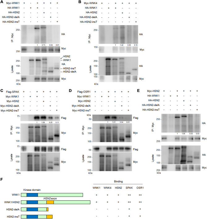
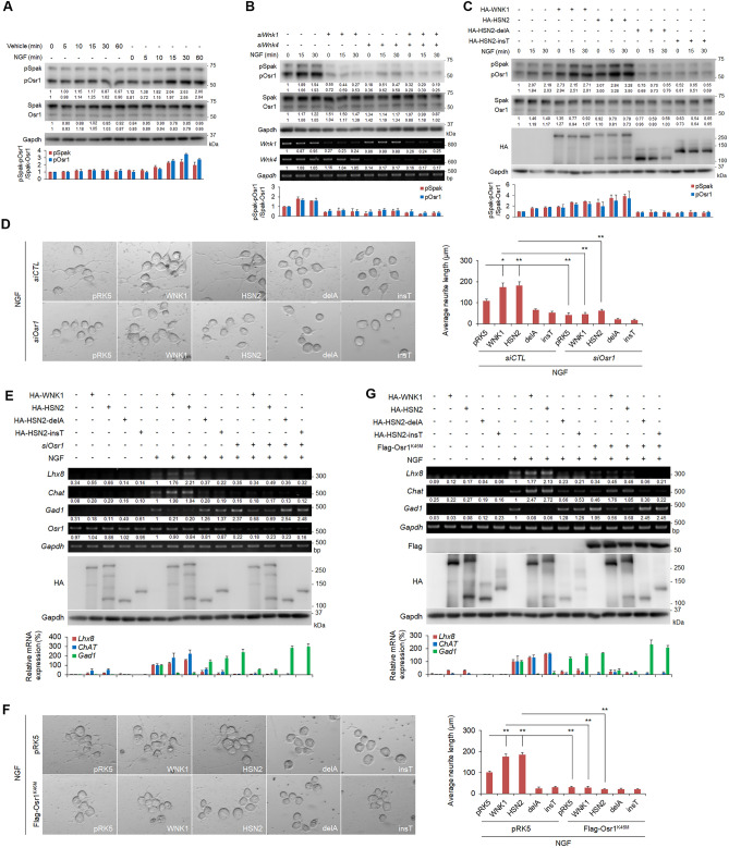
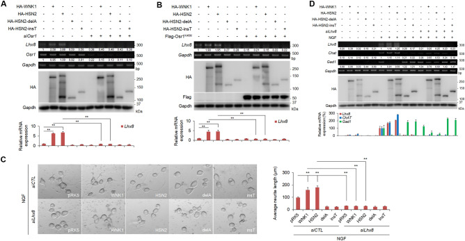
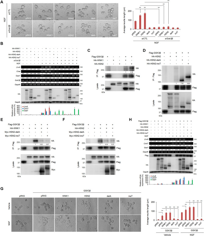
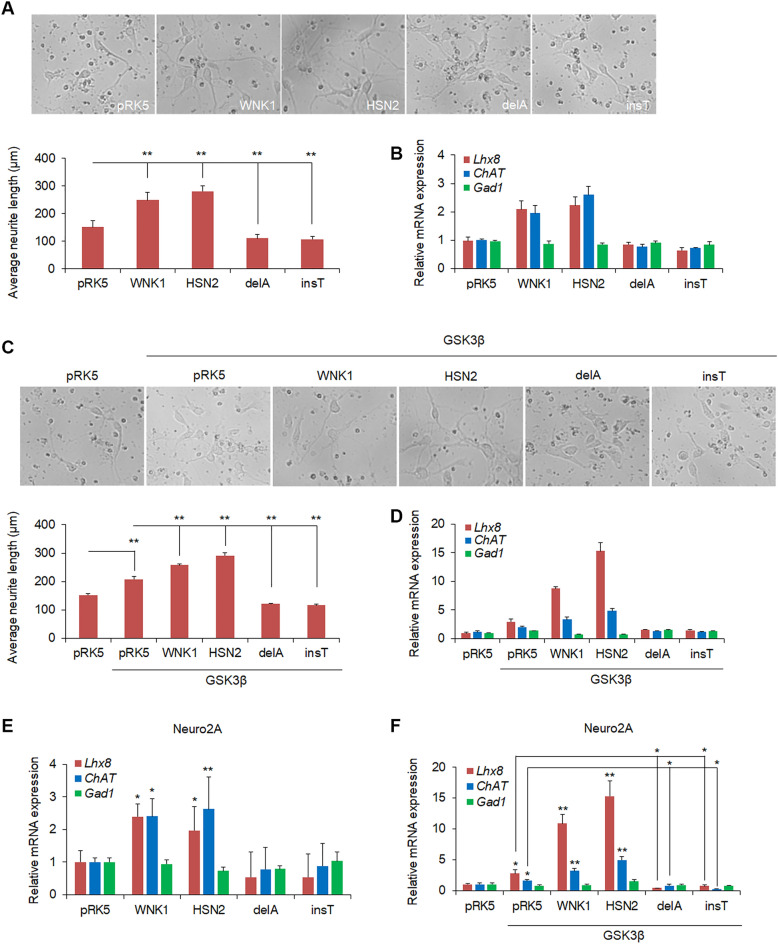
With no lysine kinase 1 (WNK1) phosphorylates and activates STE20/SPS1-related proline-alanine-rich protein kinase (SPAK) and oxidative stress responsive kinase 1 (OSR1) to regulate ion homeostasis in the kidney. Mutations in WNK1 result in dysregulation of the WNK1-SPAK/OSR1 pathway and cause pseudohypoaldosteronism type II (PHAII), a form of hypertension. WNK1 is also involved in the autosomal recessive neuropathy, hereditary sensory and autonomic neuropathy type II (HSANII). Mutations in a neural-specific splice variant of WNK1 (HSN2) cause HSANII. However, the mechanisms underlying HSN2 regulation in neurons and effects of HSN2 mutants remain unclear. Here, we found that HSN2 regulated neurite outgrowth through OSR1 activation and glycogen synthase kinase 3β (GSK3β). Moreover, HSN2-OSR1 and HSN2-GSK3β signalling induced expression of LIM homeobox 8 (Lhx8), which is a key regulator of cholinergic neural function. The HSN2-OSR1/GSK3β-LHX8 pathway is therefore important for neurite outgrowth. Consistently, HSN2 mutants reported in HSANII patients suppressed SPAK and OSR1 activation and LHX8 induction. Interestingly, HSN2 mutants also suppressed neurite outgrowth by preventing interaction of between wild-type HSN2 and GSK3β. These results indicate that HSN2 mutants cause dysregulation of neurite outgrowth via GSK3β in the HSN2 and/or WNK1 pathways.


Conflict of interest statement

The authors declare no competing interests.

Figures

Figure 1
Figure 1
HSN2, but not HSN2 mutants, binds to WNK1, WNK4, SPAK, OSR1 and HSN2. (AE) Interaction between WNK1 (A), WNK4 (B), SPAK (C), OSR1 (D) or HSN2 (E) and WNK1, HSN2, HSN2-delA and HSN2-insT was analysed in HEK293T cells by immunoprecipitation assays. Cells were transiently transfected with indicated vectors and 48 h later lysates were harvested and immunoprecipitated with an anti-Myc antibody for 4 h. Immunoprecipitates were subjected to immunoblotting with the indicated antibodies. (F) The schematic diagram of WNK1, HSN2 and its mutants was shown in left. The information of interaction levels of indicated proteins was shown in right.
Figure 2
Figure 2
HSN2 mutants, HSN2-delA and HSN2-insT, exhibit loss of neurite outgrowth function. (A) Neurite outgrowth from Neuro2A cells treated with NGF (100 ng/ml) or vehicle-containing FBS-free medium for 24 h. Cells were transiently transfected with pRK5, HA-WNK1, HA-HSN2, HA-HSN2-delA or HA-HSN2-insT vectors 24 h prior to treatment. (B) RT-PCR and qPCR analysis of mRNA levels of neural marker genes, Lhx8, ChAT and Gad1, in Neuro2A cells indicated in (A). The value of each gene in empty vector-transfected, vehicle-treated cells was set to 1 and the value in empty vector-transfected, NGF-treated cells was set to 100. (C) Neurite elongation of Neuro2A cells 24 h after transfection of HA-WNK1 or HA-HSN2 with pRK5, HA-HSN2-delA or HA-HSN2-insT vectors. (D) Neurite outgrowth of Neuro2A cells treated with 100 ng/ml NGF-containing FBS-free medium for 24 h. Cells were transiently transfected with control siRNA (siCTL) or siWnk1 and siWnk4 24 h prior to treatment. (E) RT-PCR and qPCR analysis of mRNA levels of Lhx8, ChAT and Gad1 in Neuro2A cells indicated in (C). The value of each gene from vehicle-treated cells under siCTL treatment and from NGF-treated cells under siCTL treatment was set to 1 and 100, respectively.
Figure 3
Figure 3
NGF activates SPAK/OSR1 through WNK1 and HSN2. (A) Immunoblot analysis of phosphorylated SPAK and OSR1 in Neuro2A cells treated with NGF (100 ng/ml) or vehicle-containing FBS-free medium for 0–60 min. (B) Phosphorylation of SPAK and OSR1 in Neuro2A cells treated with 100 ng/ml NGF-containing FBS-free medium for the indicated times. Cells were transfected with control siRNA (siCTL) or siWnk1 and siWnk4 24 h prior to the treatment. (C) Phosphorylation of SPAK and OSR1 in Neuro2A cells treated with 100 ng/ml NGF-containing FBS-free medium for the indicated times. Cells were transiently transfected with pRK5, HA-WNK1, HA-HSN2, HA-HSN2-delA or HA-HSN2-insT vectors 24 h prior to the treatment. (D) Neurite outgrowth of Neuro2A cells treated with 100 ng/ml NGF-containing FBS-free medium for 24 h. Cells were transiently transfected with siCTL or siOsr1 and indicated vectors 24 h prior to the treatment. (E) RT-PCR and qPCR analysis of mRNA levels of Lhx8, ChAT and Gad1 in Neuro2A cells indicated in (D). The value of each gene from vehicle-treated cells under siCTL treatment and from NGF-treated cells under siCTL treatment was set to 1 and 100, respectively. (F) Neurite outgrowth of Neuro2A cells treated with 100 ng/ml NGF-containing FBS-free medium for 24 h. Cells were transiently transfected with empty vector or kinase-negative OSR1 (OSR1K46M) 24 h prior to treatment. (G) RT-PCR and qPCR analysis of mRNA levels of Lhx8, ChAT and Gad1 in Neuro2A cells indicated in (F). The value of each gene from empty vector-transfected, vehicle-treated cells and from empty vector-transfected, NGF-treated cells was set to 1 and 100, respectively. The band intensities of pSpak and pOsr1 in (AC) by normalising intensities to the Spak and Osr1 signal, respectively. The measured values were shown in the graph under each immunoblot data.
Figure 4
Figure 4
The HSN2-OSR1-LHX8 pathway is important for NGF-induced neurite outgrowth. (A,B) RT-PCR and qPCR analysis of Lhx8 mRNA levels in Neuro2A cells 24 h after transfection with pRK5, HA-WNK1, HA-HSN2, HA-HSN2-delA or HA-HSN2-insT vectors and control siRNA (siCTL) or siOsr1 (A) and Flag-OSR1K46M (B). **P < 0.01. (C) Neurite outgrowth of Neuro2A cells treated with 100 ng/ml NGF-containing FBS-free medium for 24 h. Cells were transiently transfected with siCTL or siLhx8 and indicated vectors 24 h prior to treatment. (D) RT-PCR and qPCR analysis of mRNA levels of Lhx8, ChAT and Gad1 in Neuro2A cells indicated in (C). The value of each gene from vehicle-treated cells under siCTL treatment and from NGF-treated cells under siCTL treatment was set to 1 and 100, respectively.
Figure 5
Figure 5
HSN2 mutants suppress GSK3β functions in neurite outgrowth. (A) Neurite outgrowth of Neuro2A cells treated with 100 ng/ml NGF-containing FBS-free medium for 24 h. Cells were transiently transfected with control siRNA (siCTL) or siGsk3β and indicated vectors 24 h prior to treatment. (B) RT-PCR and qPCR analysis of mRNA levels of Lhx8, ChAT and Gad1 in Neuro2A cells indicated in (A). The value of each gene from vehicle-treated cells under siCTL treatment and from NGF-treated cells under siCTL treatment was set to 1 and 100, respectively. (C,D) Binding analysis of WNK1 and HSN2 to GSK3β (C), and of HSN2-delA and HSN2-insT mutants to GSK3β (D). Cells were transiently transfected with indicated vectors and 48 h later lysates were immunoprecipitated with a Flag antibody for 4 h. Immunoprecipitates were subjected to immunoblotting assays with the indicated antibodies. (E,F) Interaction between WNK1 (E) or HSN2 (F) and GSK3β with and without HSN2-delA and HSN2-insT mutants was analysed in HEK293T cells by immunoprecipitation assays. Cells were transiently transfected with indicated vectors and 48 h later lysates were immunoprecipitated with a Flag antibody for 4 h. Immunoprecipitates were subjected to immunoblotting assays with the indicated antibodies. (G) Neurite elongation of Neuro2A cells treated with 100 ng/ml NGF-containing FBS-free medium for 24 h. Cells were transiently transfected with the indicated vectors 24 h prior to treatment. (H) RT-PCR and qPCR analysis of mRNA levels of Lhx8, ChAT and Gad1 in Neuro2A cells indicated in (G). The value of each gene from control vector-transfected and vehicle- or NGF-treated cells was set to 1 and 100, respectively.
Figure 6
Figure 6
The function of HSN2 mutants and GSK3β in mouse primary neuron. (A) Neurite outgrowth of mouse primary cortical neural cells expressing indicated plasmids. Average neurite length was quantified by ImageJ software with NeuronJ plugin. (B) qPCR analysis of mRNA levels of Lhx8, ChAT and Gad1 in primary neural cells indicated in (A). (C) Neurite elongation and average neurite length of mouse primary cortical neural cells expressing indicated plasmids. (D) The expression levels of Lhx8, ChAT and Gad1 mRNA in primary neural cells indicated in (C). (E,F) The expression levels of Lhx8, ChAT and Gad1 mRNA in non-treated Neuro2A cells expressing indicated plasmids. *P < 0.05, **P < 0.01.

Similar articles

Cited by

References

    1. Xu BE, et al. WNK1, a novel mammalian serine/threonine protein kinase lacking the catalytic lysine in subdomain II. J. Biol. Chem. 2000;275:16795–16801. - PubMed
    1. Veríssimo F, Jordan P. WNK kinases, a novel protein kinase subfamily in multi-cellular organisms. Oncogene. 2001;20:5562–5569. - PubMed
    1. Wilson FH, et al. Human hypertension caused by mutations in WNK kinases. Science. 2001;293:1107–1112. - PubMed
    1. Piechotta K, Lu J, Delpire E. Cation chloride cotransporters interact with the stress-related kinases Ste20-related proline-alanine-rich kinase (SPAK) and oxidative stress response 1 (OSR1) J. Biol. Chem. 2002;277:50812–50819. - PubMed
    1. Vitari AC, Deak M, Morrice NA, Alessi DR. The WNK1 and WNK4 protein kinases that are mutated in Gordon’s hypertension syndrome phosphorylate and activate SPAK and OSR1 protein kinases. Biochem. J. 2005;391:17–24. - PMC - PubMed

Publication types

MeSH terms