. 2022 Sep 24;7(1):112.
doi: 10.1038/s41541-022-00530-9.

Dithranol as novel co-adjuvant for non-invasive dermal vaccination

Affiliations

Dithranol as novel co-adjuvant for non-invasive dermal vaccination

Julian Sohl et al. NPJ Vaccines. .

Erratum in

Abstract

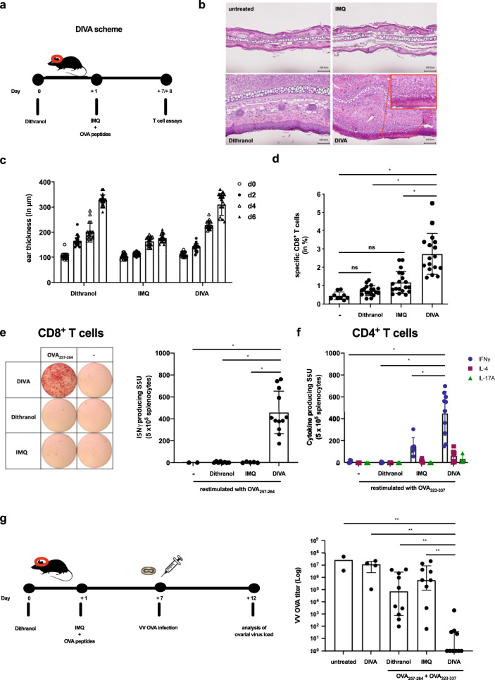
Transcutaneous immunization (TCI) utilizing the TLR7 agonist imiquimod (IMQ-TCI) induces T cell-driven protective immunity upon application onto intact skin. In our present work, we combine the anti-psoriatic agent dithranol with IMQ-TCI to boost vaccination efficacy (Dithranol/IMQ-based transcutaneous vaccination (DIVA)). Using ovalbumin-derived peptides as model antigens in mice, DIVA induced superior cytolytic CD8+ T cells and CD4+ T cells with a TH1 cytokine profile in the priming as well as in the memory phase. Regarding the underlying mechanisms, dithranol induced an oxidant-dependent, monocyte-attracting inflammatory milieu in the skin boosting TLR7-dependent activation of dendritic cells and macrophages leading to superior T cell priming and protective immunity in vaccinia virus infection. In conclusion, we introduce the non-invasive vaccination method DIVA to induce strong primary and memory T cell responses upon a single local treatment. This work provides relevant insights in cutaneous vaccination approaches, paving the way for clinical development in humans.


Conflict of interest statement

J.S., M.S., A.-K.H., and M.P.R. are inventors of a patent application submitted by the UMC Mainz (EP 18204287.9). The others authors declare that they have no competing interests.

Figures

Fig. 1
Fig. 1. Dithranol and IMQ-TCI induce local inflammation and specific T cell-mediated immune responses.
a Schematic overview of the DIVA protocol for the primary immune response. C57B6/J wildtype mice were immunized on both ears with dithranol (day 0) and IMQ together with OVA257–264 & OVA323–337 (stirred in officinal cremor basalis, day 1), treated with the single agents or left untreated (untreated control). b Seven days post treatment, HE-stained ear skin samples were examined for inflammatory signs and c the ear thickness was determined from day 0 until day 6 (n = 4–9). d The frequency of OVA257–264-specific CD8+ T cells in the blood was assessed (n = 6–19) on day 7 by flow cytometry (Supplementary Fig. 1a). e Splenocytes were restimulated ex vivo with OVA257–264 and IFN-γ-producing cells were quantified on day 8. f CD4+ T cell responses were characterized by ex vivo restimulation of splenocytes with OVA323–337 followed by IFN-γ, IL-4, and IL-17A ELISpot assays on day 8. Bars represent mean and SD of data collected from at least two independent experiments (n = 9). g Female C57B6/J wildtype mice were immunized as depicted and infected with an ovalbumin-expressing vaccinia virus on day 7. Five days after the infection, ovaries were collected and homogenized for BSC-40 plaque assays to determine VV-OVA titers (log scale). Bars represent mean and SD of data collected from at least two independent experiments. *p < 0.05 by one-way ANOVA with Bonferroni’s post hoc test. **p < 0.05 by Kruskal–Wallis test and Dunn’s post hoc test as we assumed a non-Gaussian distribution of VV-OVA titers.
Fig. 2
Fig. 2. DIVA generates memory CD8+ and CD4+ T cell responses.
a Schematic overview of the DIVA protocol for the generation of a memory immune response. C57B6/J wildtype mice were immunized on both ears with dithranol (day 0) and IMQ together with OVA257–264 & OVA323–337 (stirred in officinal cremor basalis, day 1) on 2 consecutive days, treated with the single agents or left untreated (untreated control). b The frequency of OVA257–264 specific CD8+ T cells in the blood was assessed on day 7, 14, and 35 (n = 9–19). c On day 36, the in vivo cytolytic activity was estimated 20 h after transfer of peptide-loaded, CFSE-labeled target cells (n = 4–8) (Supplementary Fig. 1c). d Splenocytes were ex vivo restimulated with either OVA257–264 or OVA323–337 peptide and IFN-γ-producing cells were analyzed by ELISpot assays on day 36. Bars represent mean and SD of data collected from at least two independent experiments. e, f The frequency of OVA257–264-specific CD8+ T cells (e) and IFN-γ producing cells (f) after restimulation with OVA257–264 or OVA323–337 were quantified in splenocytes on day 35 after DIVA with or without CD4 T cell depletion (500 µg GK1.5 i. p. as indicated). *p < 0.05 by one-way ANOVA with Bonferroni’s post hoc test, flow cytometry gating strategy in Supplementary Fig. 1a.
Fig. 3
Fig. 3. Dithranol induces localized CD8+ and CD4+ T cell memory responses in the skin.
C57B6/J wildtype mice were immunized as depicted in Fig. 2a. a Representative flow cytometry plots and quantification of dithranol, IMQ-TCI or DI-TCI treated murine ears following digestion with collagenase type IV & quantification of CD45+ leukocytes and TCR+ CD8+ T cells on day 36 following TCI are depicted. b Ex vivo IFN-γ ELISpot assays of 2 × 105 murine ear skin-resident leukocytes following restimulation with either OVA257–264 or OVA323–337 peptide and OVA257–264-specific CD8+ T cells by tetramer staining on day 36. Bars represent mean and SD of data collected from at least two independent experiments. *Significant difference with p < 0.05 by one-way ANOVA with Bonferroni’s post hoc test. Gating strategy in Supplementary Fig. 1b.
Fig. 4
Fig. 4. DIVA-induced T cell activation requires TLR7 in DCs and partly in macrophages.
Both CD11cCre × TLR7flox/flox and LysMCre × TLR7flox/flox (knock-out mice or respective littermates) were transcutaneously immunized as depicted in Fig. 2a. On day 35, the frequency of OVA257–264 specific CD8+ T cells was assessed in the blood of a CD11cCre × TLR7flox/flox or b LysMCre × TLR7flox/flox and the respective littermates (flow cytometry gating in Supplementary Fig. 1a). On day 36, splenocytes of a CD11cCre × TLR7flox/flox or b LysMCre × TLR7flox/flox and the respective littermate were ex vivo restimulated with either OVA257–264 or OVA323–337 peptide and IFN-γ-producing cells were analyzed by ELISpot assays. Bars represent mean and SD of data collected from at least two independent experiments. *Significant difference with p < 0.05 by one-way ANOVA with Bonferroni’s posttest.
Fig. 5
Fig. 5. RNA-seq of whole skin reveals dithranol, but not IMQ, as inducer of monocyte-specific inflammatory gene signature.
a Bulk RNA-seq-based heatmap of differentially expressed (DE) genes (adjusted p value < 0.01 and log2FC ≥ 2) between IMI-Sol (IMQ) or dithranol-treated wildtype mice (n = 3) from murine ear skin cells. Monocyte related genes are highlighted in the blow-up section. The differential expression intensity is colored for each row from min (blue) to max (red). b Box plots with expression values of monocytic genes are shown for each of the genes, that are highlighted in a. In b, *p < 0.05 was evaluated by two-tailed unpaired T test followed by Welch´s correction. The complete data set of this experiment is available under GEO accession number GSE189000.
Fig. 6
Fig. 6. Dithranol induces early monocyte recruitment into the skin and accumulation of monocyte-derived DCs and macrophages in the draining lymph nodes.
a Mice were treated once with dithranol or IMQ on both ears. After 24 h, the ears were digested with collagenase type IV in a GentleMACS dissociator. The percentages of monocytes (MHCII CD11b+ Ly6C+ Ly6G CCR2+), granulocytes (MHCII- CD11b+ Ly6C+ Ly6G+ CCR2), monocyte-derived DCs (MHCII+ CD11b+ Ly6C+ CD64+) and macrophages (MHCII+ CD11b+ Ly6C CD64+) in the living lineage-negative leukocytes in the skin were determined by flow cytometry (Supplementary Fig. 1e). b Mice were treated once with dithranol, IMQ or DIVA as before. After 72 h, auricular lymph nodes were harvested from treated mice and were then digested with collagenase type IV for 60 min on a heat shaker. The total number of monocyte-derived DCs (MHCII+ CD11b+ Ly6C+ CD64+) and macrophages (MHCII+ CD11b+ Ly6C CD64+) and the particular expression of co-stimulatory molecules in the auricular lymph nodes were determined by a Scil Veterinary Excellence cell counter and flow cytometry (Supplementary Fig. 1f). Bars represent mean and SD of data collected from at least two independent experiments. *Significant difference with p < 0.05 by T test (a) or one-way ANOVA with Bonferroni’s posttest (b).
Fig. 7
Fig. 7. Dithranol-induced monocyte recruitment is mediated by oxidative stress and is indispensable for the CD8+ T cell responses following DIVA.
a Mice were treated with dithranol or IMQ for 24 h. When indicated, the radical scavenger α-tocopherol was administered (30 mg, i.p.). Treated murine ears were then digested with collagenase type IV in a GentleMACS dissociator. The total number of monocytes (MHCII CD11b+ Ly6C+ Ly6G) was estimated by a scil veterinary excellence cell counter and flow cytometry (Supplementary Fig. 1d) (n = 4–9). b C57B6/J mice were transcutaneously treated as depicted in the schematic overview. When indicated, the radical scavenger α-Tocopherol was administered (30 mg, i.p.) and c on day 7 the frequency of OVA257–264-specific CD8+ T cells was determined (Supplementary Fig. 1a) and d on day 8 the specific lysis of antigen-loaded target cells by OVA257–264-specific CD8+ T cells was estimated (Supplementary Fig. 1c). e C57B6/J mice were treated as depicted in the schematic overview. When indicated, the CCR2-specific, depleting antibody MC-21 or an isotype control antibody MC-67 (20 µg, i.p.) were administered daily until day 4. f The frequency of OVA257–264-specific CD8+ T cells in the blood was assessed (n = 6–19) on day 7 by flow cytometry (Supplementary Fig. 1a). g Blood cells were restimulated ex vivo with OVA257–264 and IFN-γ producing cells were quantified on day 8. Bars represent mean and SD of data collected from at least two independent experiments. *Significant difference with p < 0.05 by t Test (a) or one-way ANOVA with Bonferroni’s posttest (b).

References

    1. Plotkin S. History of vaccination. Proc. Natl Acad. Sci. USA. 2014;111:12283–12287. - PMC - PubMed
    1. Lurie, N., Saville, M., Hatchett, R. & Halton, J. Developing Covid-19 vaccines at pandemic speed. N. Engl. J. Med.10.1056/nejmp2005630 (2020). - PubMed
    1. Klein, N. P. et al. Surveillance for adverse events after COVID-19 mRNA vaccination. JAMA326, 1390–1399 (2021). - PMC - PubMed
    1. Rappuoli R, Santoni A, Mantovani A. Vaccines: an achievement of civilization, a human right, our health insurance for the future. J. Exp. Med. 2018;551:jem.20182160–273. - PMC - PubMed
    1. Marin-Acevedo JA, Soyano AE, Dholaria B, Knutson KL, Lou Y. Cancer immunotherapy beyond immune checkpoint inhibitors. J. Hematol. Oncol. 2018;11:8. - PMC - PubMed