Review
. 2022 Sep 24;20(1):421.
doi: 10.1186/s12951-022-01631-2.

Application of MOF-based nanotherapeutics in light-mediated cancer diagnosis and therapy

Affiliations
Review

Application of MOF-based nanotherapeutics in light-mediated cancer diagnosis and therapy

Dan Zhao et al. J Nanobiotechnology. .

Abstract

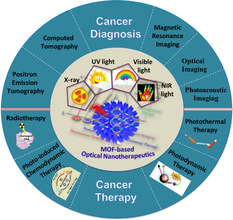
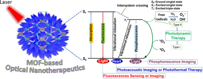
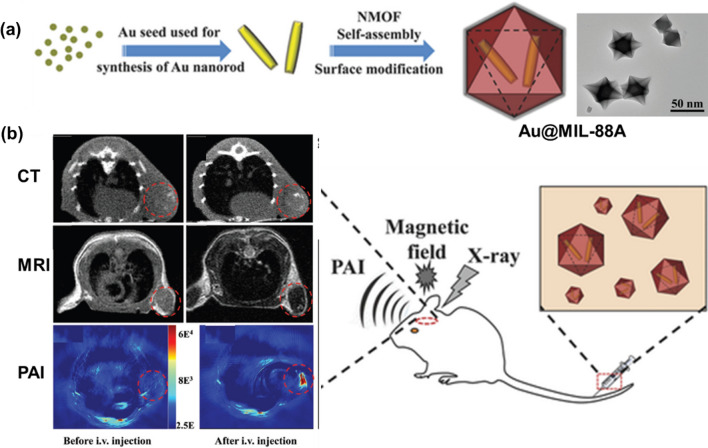
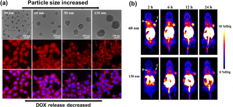
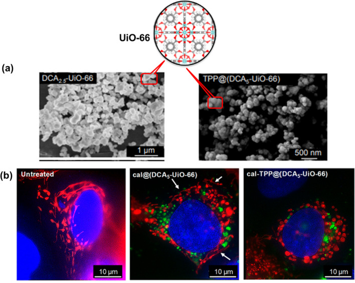
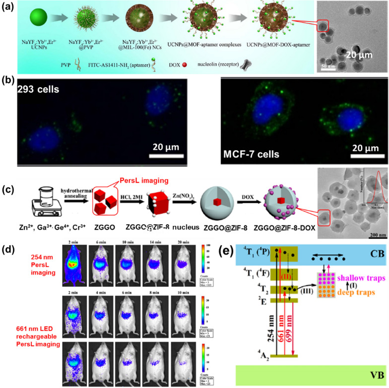
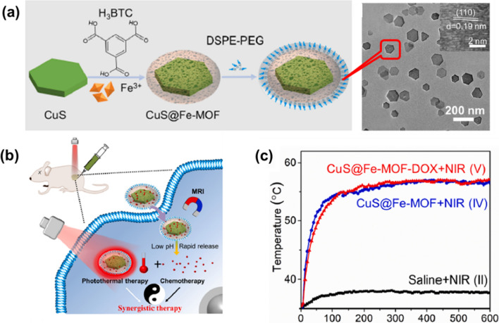
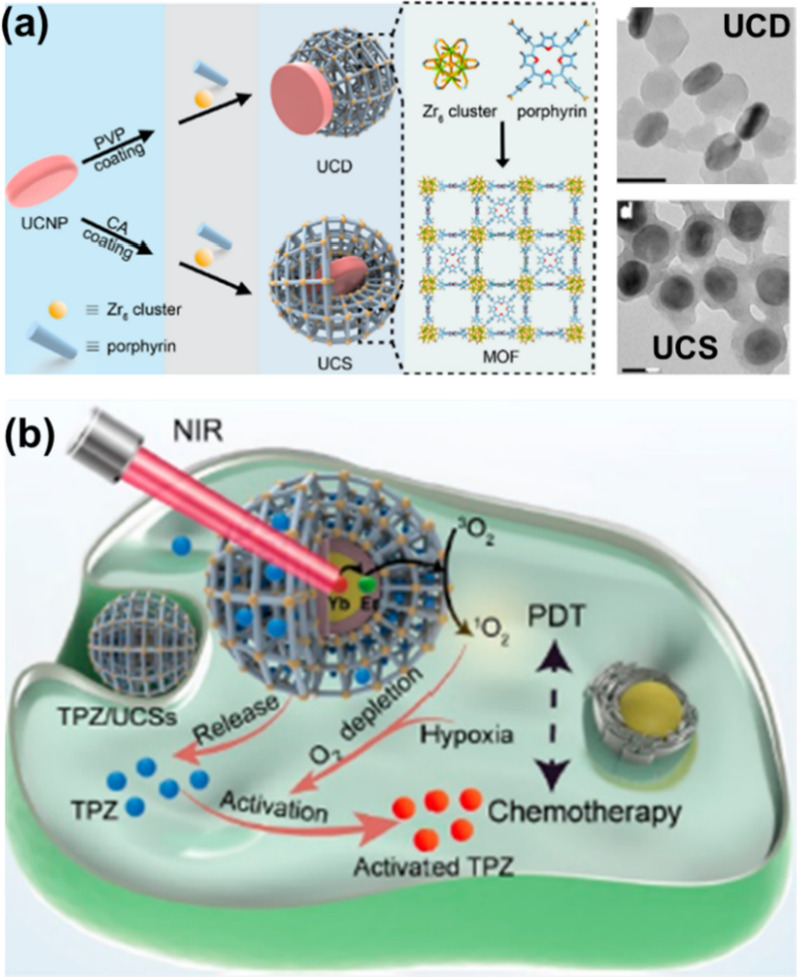
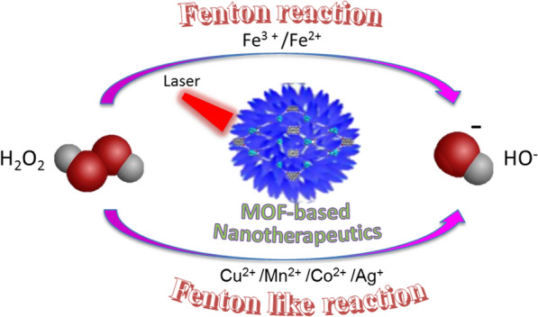
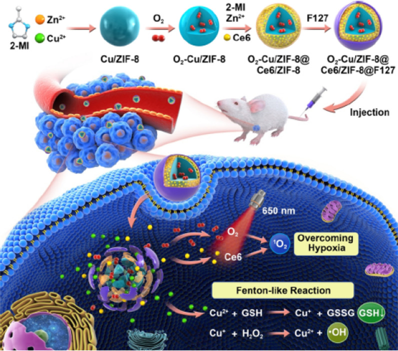
Light-mediated nanotherapeutics have recently emerged as promising strategies to precisely control the activation of therapeutic reagents and imaging probe both in vitro and in vivo, largely ascribed to their unique properties, including minimally invasive capabilities and high spatiotemporal resolution. Nanoscale metal-organic frameworks (NMOFs), a new family of hybrid materials consisting of metal attachment sites and bridging ligands, have been explored as a new platform for enhanced cancer diagnosis and therapy due to their tunable size, modifiable surface, good biocompatibility, high agent loading and, most significantly, their ability to be preferentially deposited in tumors through enhanced permeability and retention (EPR). Especially the light-driven NMOF-based therapeutic platform, which not only allow for increased laser penetration depth and enhanced targeting, but also enable imaging-guided or combined treatments. This review provides up-to-date developments of NMOF-based therapeutic platforms for cancer treatment with emphasis on light-triggered therapeutic strategies and introduces their advances in cancer diagnosis and therapy in recent years.

Keywords: Cancer diagnosis; Light-mediated; Metal–organic frameworks; Nanotherapeutics; Therapy.


Conflict of interest statement

The authors declare no competing interests.

Figures

Fig. 1
Fig. 1
Main scope of this perspective regarding the use of MOF-based nanotherapeutics for light-mediated cancer diagnosis and therapy
Fig. 2
Fig. 2
Schematic diagrams of the photophysical processes of MOF-based optical nanotherapeutics
Fig. 3
Fig. 3
a Synthetic schematic and SEM photograph of Mn3+-TCPP NMOF; b mechanism of Mn3+-TCPP NMOF unlocking triggered by GSH; c in vivo fluorescence (left) and MRI (right) signals after intravenous injection with Mn3+-TCPP NMOF. Reproduced with permission [47]. Copyright 2019, American Chemical Society
Fig. 4
Fig. 4
a Fabrication Process and TEM photograph of FePt-MOFs-tLyp-1; b in vivo T2-weighted MR imaging (axial plane) of a 4T1 tumor-bearing mouse at different time intervals after an intravenous injection with FePt-MOF-tLyp-1; c in vivo CT imaging (axial plane) of a 4T1 tumor-bearing mouse at different time intervals after intravenous injection with FePt-MOF-tLyp-1. Tumor tissue was indicated with red pan. Reproduced with permission [53]. Copyright 2020, American Chemical Society
Fig. 5
Fig. 5
a Fabrication process and TEM photograph of Au@MIL-88(Fe); b application to multimodality imaging-based tumor diagnosis. Reproduced with permission [63]. Copyright 2016, Wiley
Fig. 6
Fig. 6
a Size-controlled TEM images and the DOX fluorescence intensity in nucleus decreased with the increasing size of DOX@AZIF-8; b DOX@AZIF-8 of different sizes exhibited significant difference in the tumor accumulation and anticancer efficacy. Reproduced with permission [69]. Copyright 2018, American Chemical Society
Fig. 7
Fig. 7
a Structure of UiO-66 and SEM images of synthesized DCA2.5-UiO-66 and TPP@(DCA2.5-UiO-66) samples; b images of untreated cells and cells treated with cal@(DCA5-UiO-66) and cal-TPP@(DCA5-UiO-66) for 8 h; mitochondria are colored in red, MOFs in green, and nuclei in blue; white arrows indicate stringy mitochondria. Reproduced with permission [82]. Copyright 2020, American Chemical Society
Fig. 8
Fig. 8
a Fabrication process and TEM photograph of UCNPs@MOF-DOX-AS1411; b images of 293 cells and MCF-7 cells incubated with UCNPs@MOF-DOX-AS1411 NCs for 1 h; c fabrication process and TEM photograph of ZGGO@ZIF-8-DOX; d in vivo NIR PersL imaging in a mouse was radiated 254 nm and 661 nm treated with ZGGO@ZIF-8 (0.2 mL, 1 mg/mL in PBS); e energy-level diagram for Cr3+-activated ZGGO. Reproduced with permission [83]. Copyright 2015, Springer Nature; [87]; Copyright 2019, American Chemical Society
Fig. 9
Fig. 9
a Synthesis procedure and TEM image of Cu-THQNPs; b photoenergy to heat conversion mechanism of Cu-THQNPs; c schematic diagram of the behavior of Cu-THQNPs upon the 1064 nm laser irradiation in vivo. Reproduced with permission [96]. Copyright 2018, American Chemical Society
Fig. 10
Fig. 10
a The one-pot synthesis process of Bi2S3/FeS2@BSA-FA; b Schematic illustration of Bi2S3/FeS2@BSA-FA for MR/CT imaging and PTT. Reproduced with permission [103]. Copyright 2019, Elsevier
Fig. 11
Fig. 11
a Synthesis procedure and structure of 3D porous Zr-PDI; b illustration of photothermal conversion performance of Zr-PDI*−; c schematic diagram of the PET process between TEA and Zr-PDI, excited state Zr-PDI* is reductively quenched by TEA to afford Zr-PDI*−. Reproduced with permission [111]. Copyright 2018, Springer Nature
Fig. 12
Fig. 12
a Synthesis procedure and TEM image of ZnO-CNP-TRGL; b Photothermal images for control media and ZnO-CNP-TRGL (25, 50, 100, and 1000 nm) suspensions (50 µg mL−1) with different irradiation times (0–5 min) at 2 W cm−2; c Illustration images for the size transformation of ZnO-CNP-TRGL from hydrophilic dispersion and hydrophobic micrometer aggregation; LCST is lower critical solution temperature. Reproduced with permission [112]. Copyright 2019, Wiley
Fig. 13
Fig. 13
a Synthesis scheme and TEM image of CuS@Fe-MOF; b Scheme of PAT process; c Temperature elevation curves in the tumor site. Reproduced with permission [115]. Copyright 2019, Elsevier
Fig. 14
Fig. 14
a Schematic illustration and TEM images of the synthesis of UCD and UCS by the surface engineering of UCNPs; b schematic diagram of the treatment process of TPZ/UCSs. Reproduced with permission [124]. Copyright 2020, American Chemical Society
Fig. 15
Fig. 15
Mechanism of photo-driven Fenton or Fenton like reaction in photo-induced CDT by using MOF-based nanoplatform as therapeutic agent
Fig. 16
Fig. 16
Schematic illustration of the fabrication process of the TME-responsive OCZCF nanoplatform for enhanced PDT and CDT through GSH depletion and O2 replenishment. Reproduced with permission [136]. Copyright 2020, American Chemical Society
Fig. 17
Fig. 17
a Schematic illustration and TEM images of the synthesis of 99mTc-Hf-TCPP-PEG. b in vivo SPECT images of 4T1 tumor-bearing mice after intravenous injection with 99mTc-Hf-TCPP-PEG NCPs. c Quantification of SPECT signals in the liver, kidney, tumor, and muscle of 4T1 tumor bearing mice for mice at different time points after intravenous injection with 99mTc-Hf-TCPP-PEG NCPs. Reproduced with permission [139]. Copyright 2018, American Chemical Society
Fig. 18
Fig. 18
Schematic illustration of the fabrication process of the Zr-MOF-QU nanoplatform for enhanced RDT and CDT through 1,4-benzenedicarboxylic acid depletion and relieving hypoxia in the tumor microenvironment. Reproduced with permission [142]. Copyright 2019, American Chemical Society

References

    1. Allemani C, Matsuda T, Di Carlo V, Harewood R, Matz M, Nikšić M, Bonaventure A, Valkov M, Johnson CJ, Estève J, Ogunbiyi OJ, Silva JAE, Chen WQ, Eser S, Engholm G, Stiller AG, Monnereau A, Woods RR, Visser O, Lim GH, Aitken J, Weir HK, Coleman MP. Global surveillance of trends in cancer survival 2000–14 (CONCORD-3): analysis of individual records for 37513025 patients diagnosed with one of 18 cancers from 322 population-based registries in 71 countries. Lancet. 2018;391:1023–1075. doi: 10.1016/S0140-6736(17)33326-3. - DOI - PMC - PubMed
    1. Bray F, Ferlay J, Soerjomataram I, Siegel RL, Torre LA, Jemal A. Global cancer statistics 2018: globocan estimates of incidence and mortality worldwide for 36 cancers in 185 countries. CA Cancer J Clin. 2018;68:394–424. doi: 10.3322/caac.21492. - DOI - PubMed
    1. Thirajan M, Chen YH, Joshi P, Pandey RK. The role of porphyrin chemistry in tumor imaging and photodynamic therapy. Chem Soc Rev. 2011;40:340–362. doi: 10.1039/B915149B. - DOI - PubMed
    1. Wu H, Guo T, Nan J, Yang L, Liao G, Park HJ, Li J. Hyaluronic-acid-coated chitosan nanoparticles for insulin oral delivery: fabrication, characterization, and hypoglycemic ability. Macromol Biosci. 2022;22:2100493. doi: 10.1002/mabi.202100493. - DOI - PubMed
    1. Ai XZ, Mu J, Xing BG. Recent advances of light-mediated. Theranostics. 2016;6:2439–2457. doi: 10.7150/thno.16088. - DOI - PMC - PubMed